





































Large Animal Equine Bovine Ovine Ultrasound Handheld
Large-animal ultrasound puts a handheld scanner to work on horses, cattle, sheep, and pigs, used mainly to read a pregnancy, a breeding cycle, or the soundness of a working horse’s legs. The vet brings the machine to the animal, in a barn, a crush, or an open field, and reads the picture standing beside a cow or kneeling at a horse’s tendon. The work runs on a field probe built to survive mud, rain, and the long reach inside a large animal.
Reproduction is the bulk of the work
The greater part of large-animal ultrasound is breeding work. A dairy farm scans its cows to confirm a pregnancy weeks earlier than any other test allows, to find the open cows that need rebreeding, and to time the herd’s calving. A stud reads a mare through her cycle to breed her on the right day. A sheep flock is scanned to count the lambs each ewe carries. The money a farm makes turns on its breeding. The scan watches that breeding from the first weeks.
The cost of a slow breeder runs the economics of a farm. A cow open past her time eats through feed and gives no calf and no fresh lactation, a loss counted by the day on a dairy. The scan finds her in the first month, in time to rebreed her before another cycle is lost. A whole herd read in a morning turns breeding from a guess into a managed calendar.
The questions are the same across the species. Whether she is pregnant, and how far along. How many young she carries. Whether she is cycling, and when she will breed. The scan answers each in minutes, in the barn, on the animal. The answer comes the same day, in the time it takes to run the animal through. The farm acts on it before the cow leaves the crush.
The reproductive scan of each species fills a page of its own, set out in full elsewhere in this library. The work here is the shape of it across the farm, the place each scan takes in the year a breeding operation runs. A farm runs its calendar around these scans, the breeding, the pregnancy checks, the lamb counts falling on set weeks the vet and the farmer plan together.
The horse
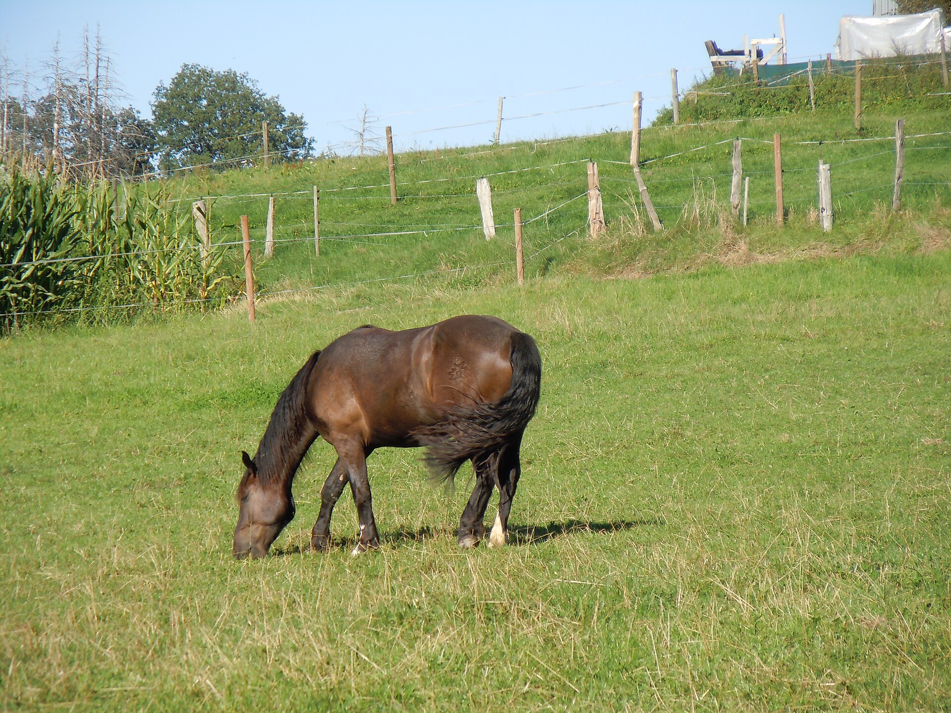
The horse is scanned for its breeding and for its legs. A mare is read through her cycle and her pregnancy, reached by a long arm inside the rectum. The places a vet scans a mare and a stallion run from the ovaries and the uterus of the mare to the testes of the stallion. The mare work fills a stud’s spring, every mare on the books read through her cycle to catch the few days she will hold to a covering.
The breeding mare carries real value the scan is there to guard. It confirms she is in foal, finds the twins a mare cannot carry safely, and dates the pregnancy to plan the foaling. A twin caught in the first weeks is reduced to a single foal the mare can carry to term, the scan turning a lost pregnancy into a live one. A mare is read again through the pregnancy to follow the foal, the heartbeat found by a few weeks in, the sex told from around two months, the growth checked against the months still to run.
The legs are the other half of the horse’s scan. A performance horse lives on its tendons and ligaments. A strain in one of them ends a season or a career. The tendons and ligaments of a horse’s leg read clearly on ultrasound, a tear showing as a dark hole punched through the bright parallel lines of the tendon’s fibres. A vet measures the hole across its width and along its length, putting a number on the damage the first day and a fresh number every few weeks to track it filling back in. The scan turns the long months of a tendon’s healing into a set of pictures a trainer can read, the fibres knitting back into line, the dark gap shrinking pull by pull. A horse brought back to work too soon tears the same tendon again. The scan tells the vet when the fibres have healed enough to carry the load. The probe reads the superficial flexor, the deep flexor, and the suspensory, the three that carry a galloping horse and take the strain that breaks them down.
The joints come into the same scan. The joints of a horse show their fluid, their lining, and the surfaces of the cartilage, the swelling of a joint read for the trouble behind it. A lameness worked up on the farm starts with the probe on the leg, the picture pointing to the tendon, the joint, or the bone beneath. The fluid in a swollen joint reads on the scan for blood, for pus, or for the clear excess of a strain, the cause read before a needle goes in.
The horse work asks a lot of a handheld. The animal is large, the structures sit deep, the patient can kick, and the scan often runs in a stable with poor light. A rugged cordless machine with a screen readable in daylight is the tool that suits the barn and the field, the one probe reaching from a tendon close under the skin to a foal deep in the mare. The stallion is read as well, his testes scanned for the lumps and the changes that cut a breeding career short, the soundness of a sire counting for as much to a stud as the soundness of the mares.
The cow
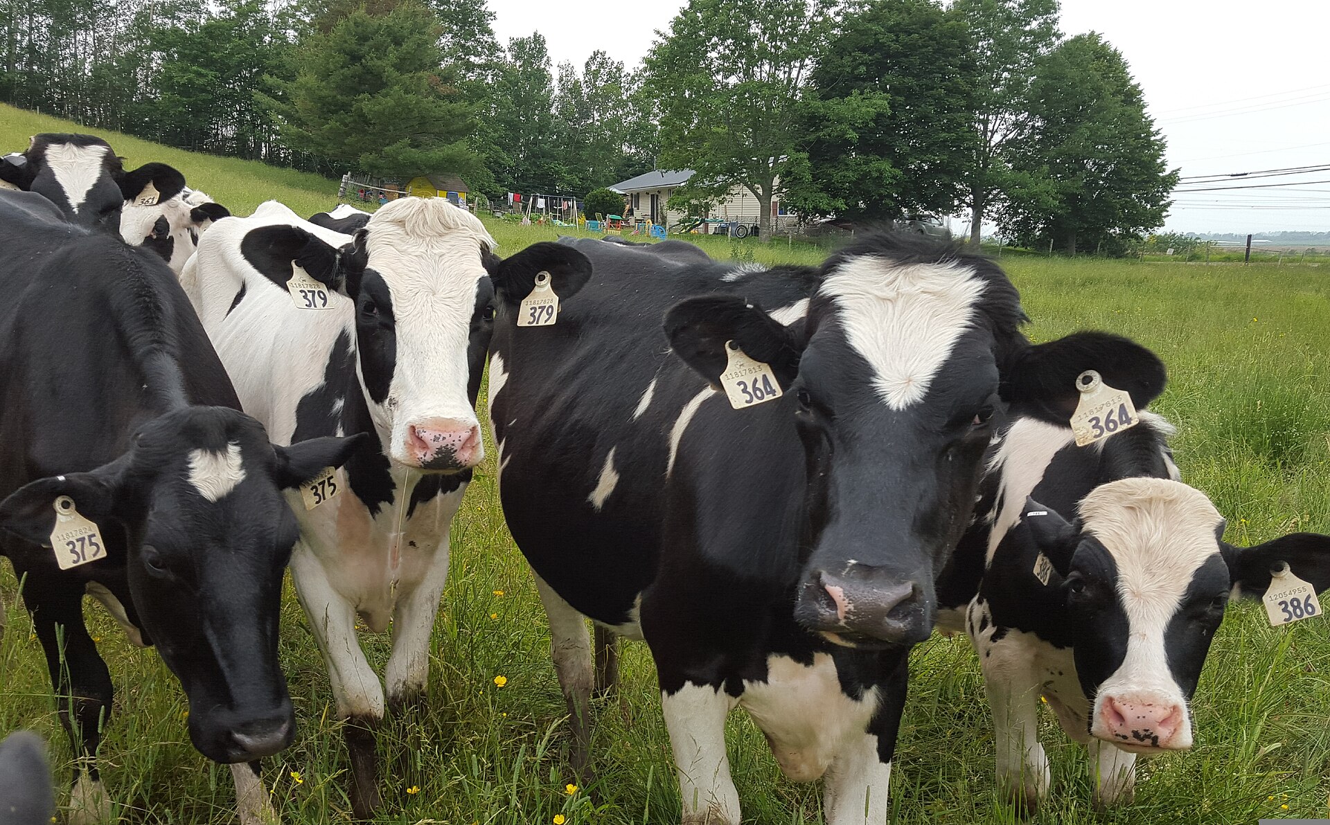
The cow is scanned more than any other farm animal. A dairy or beef herd lives on a tight breeding cycle the scan keeps on time. The vet reads each cow for a pregnancy, for the stage of her cycle, and for the troubles that keep her open. A dairy measures itself on how fast its cows get back in calf after calving. The scan keeps an eye on that number across the whole herd. A herd that calves on time fills the tank with milk and the shed with calves, the whole business resting on the cows getting back in calf.
The pregnancy scan is the everyday job. The transrectal scan that reads a cow in calf finds a calf from around a month after breeding, far sooner than waiting for the cow to show. A cow found open is rebred without losing a cycle, the scan saving the weeks a missed pregnancy would cost a dairy. The same scan ages the calf by its size and its parts, telling the farm when each cow is due and grouping the herd by its calving dates.
The sex of the calf is read on the scan as well. From around the second month a trained eye finds the parts that tell a bull calf from a heifer, the dairy planning its replacements and its beef crosses from the answer. A herd sexed in the womb is sorted long before it hits the ground. The early answer lets a dairy plan the year’s replacements from the start, the heifer calves marked for the milking string from before they are born.
The ovaries tell the rest of the breeding story. The follicles and corpus luteum of a cow read on the scan show where she sits in her cycle, the tool that drives the timed breeding programmes a modern dairy runs on. A cow that has stopped cycling is found and treated, the scan reading an ovary a hand can only guess at. The follicle waves of a cow’s cycle run on a schedule the scan can follow, each wave of growing follicles read and timed. A modern dairy hangs its breeding on this reading, dropping hormones and insemination onto the exact days the ovary calls for.
The udder is scanned when it turns hard or sore. The udder of a cow with mastitis reads for the abscess, the blocked quarter, or the damage left by infection, the picture guiding the choice to treat or to cull. A quarter lost to scarring is read on the screen, the farm spared the cost of treating an udder that will never milk again. The udder scan reads a hard quarter for the difference between an abscess and a solid mass, the picture deciding the cow’s fate between the treatment pen and the cull list.
The arm inside the animal
The cow and the mare are scanned from the inside. The vet’s arm goes into the rectum with a long straight probe in the hand, and reads the uterus and the ovaries through the rectal wall. The picture is as close as ultrasound gets, the probe lying right against the organs it reads. The technique is the one a vet learns first in cattle work, the hand placing the probe and reading the uterus by feel at the same time. A practised arm runs through a row of cows in a crush, a scan and a verdict for each in under a minute.
The probe for this work is a long straight one. A rectal linear probe reaches the length of the arm and lays a wide window against the uterus, the same probe used in the cow, the mare, and the larger farm animals. The handheld carries this probe on a short cord or builds it into a wireless wand the arm takes in whole. A wireless probe spares the vet a cable trailing through the muck of a crush, the picture thrown to a screen on the arm or to a tablet propped at the rail. The whole rig goes on with a glove and comes off for cleaning between farms. The same long probe serves a horse stud and a dairy alike, the one tool a large-animal vet reaches for first on any reproductive call.
A scan beats a guess in the crush
A month after breeding, the scan reads a cow’s pregnancy for certain, long before a hand laid on her could call it.
The flock and the herd
Sheep and pigs are scanned in large numbers, each animal read in well under a minute. A flock or a herd is run past the probe one after another. The scan earns its place by the speed it brings to many animals at once. A scanning contractor sets up a race and a crush at the farm, the sheep flowing through in a steady line, a thousand head read in a day by one practised hand. The cost per ewe falls to a few pence at that pace.
A ewe is scanned to count her lambs. The number of lambs a ewe carries sets how she is fed through the winter, a single lamb on one ration and triplets on another, the scan paying for itself in saved feed and live lambs. The flock is split into feeding groups by the count, each ewe fed for the number she carries, the heavy-bearing ones marked for extra rations in the weeks before lambing.
A sow is scanned to confirm her litter. The pregnancy of a sow is read a few weeks after mating, the empty sows found and culled or rebred before they eat through a month of feed for nothing. The scan keeps a breeding herd full of productive animals. A sow scanned a few weeks after mating holds her place in the batch only if she is carrying, the barren ones pulled before they cost the unit a wasted cycle. A breeding unit run on batches lives on finding the empty sows early. A unit that runs five hundred sows on a tight farrowing schedule cannot carry an empty animal in a pen. The scan finds the empty sows for less than a wasted cycle costs.
Built for mud and rain
The farm is hard on a machine. The scan runs in a barn thick with dust, in a crush splashed with muck, in a field under rain. The handheld lives in a coat pocket or a truck, carried out to the animal in all weather. A handheld used in a crush is kicked, knocked against a rail, and dropped in the muck on a long day. The build that survives the farm is the one still working in five years. A farm vet buys a machine the way a farmer buys a tractor, for the years of hard use it will give before it owes nothing.
A sealed body takes the muck and the wash that follow a farm job. The probe and the screen are wiped down after every muddy session, hosed at the end of a day in the crush, and built to shrug off the water that would kill a machine made for a clean clinic. The cleaning matters for more than the machine. A probe carried from farm to farm is scrubbed and disinfected between them, the sealed body taking the strong cleaners that stop a disease riding from one herd to the next.
The screen has to be read in daylight. A scan run outdoors needs a display that holds its picture in full sun, the farm machine carrying a bright screen a vet can read in an open field. The cordless build frees the vet from a socket that no barn or field provides. The vet reads the screen wherever the animal stands, in the dark of a barn at dawn or the glare of a field at noon, the display holding its picture through both. A machine that has to be shaded with a hand loses the speed the farm scan is bought for.
The battery has to last the round. A vet scanning a hundred cows in a morning, or a whole flock in a day, needs a machine that runs the full session on one charge, with a spare in the truck for the long days. The work is done far from any power, the machine carrying its own. A charge that fades by mid-morning sends a vet back to the truck and holds up a yard full of waiting cattle, the cost of a flat battery measured in a farm’s whole morning. The machines built for the work hold a long day and charge fast between rounds.
The probe for a big animal
The large animal asks for its own probes. The long rectal linear reaches inside the cow and the mare. A convex or a sturdy linear probe reads the tendon of a horse, the udder of a cow, or the belly of a ewe from the outside. The engineering of how these probes connect to a handheld is set out in its own place. A vet working both inside and outside an animal carries two probes on the one machine, swapping the rectal linear for the convex without changing the unit in the hand, the one machine covering the whole of a farm call.
The depth is the dividing line. A foal deep in a mare, or a calf in a beef cow, asks for a probe that reaches far into a big body. Depth is the first thing the large animal asks of a machine, ahead of the fine detail a small patient calls for. The rectal probe is a long slim wand built to ride the arm without bruising the gut, its window turned to lie flat against the uterine wall. The outside probes are built broad enough to sit on a hide caked with hair and muck. A vet clips a patch of hair and wets it down to give the probe a clean window onto the leg of a horse or the udder of a cow, the coat the first thing between the sound and the animal.
How many machines a farm needs
A breeding operation has to decide how many scanners to own. The number a breeding farm needs runs from a single machine shared across a vet practice to a scanner a large dairy owns outright, set by how many animals go past the probe and how often. The sum is a simple one for a farm to run, the call-out fees of a season weighed against the price of a machine and the training to use it.
The machine pays for itself in the breeding it saves. A scanner that catches the open cows, times the breeding, and counts the lambs returns its price in a season or two on a working farm. The farm buys for the herd it runs and the calendar it keeps. A herd scanned a few times a year leans on a visiting vet and the machine in that vet’s bag, the cost of owning a scanner earned back only once the animals run to the thousands and the scanning to the weekly. The decision is the farm’s own, made on its herd size and its breeding plan, set out in full on the page that takes it up.
The probe in the barn
Large-animal ultrasound has moved the work from the clinic to the field. The scan that once needed a fixed machine and a brought-in animal now rides in a vet’s coat to the cow in the crush, the mare in the stable, the ewe in the pen. The vet carries the whole tool in one hand and reads the animal where it stands, no ramp, no trailer, no trip to a clinic in the picture.
The handheld reads the breeding a farm lives on, and reads it on the spot. A pregnancy confirmed, a cycle timed, a litter counted, a tendon checked: each is done beside the animal, the answer in hand before the vet moves to the next. The work that filled a morning of waiting and guessing is done in the time it takes to walk the row. A farm reads its breeding the way it reads its weather now, a thing watched and acted on through the season.
The pages that follow take each scan in turn, the horse, the cow, the sheep, the pig, and the machine a breeding farm builds its year around. Each sets out where the probe goes, what the picture shows, and what the farm does with the answer.
Common questions about large-animal ultrasound
What is large-animal ultrasound used for?
Mostly for breeding. The scan confirms a pregnancy in a cow, mare, ewe, or sow, dates it, and counts the young. It reads a cow’s ovaries to time her breeding, a ewe’s lambs to set her feeding, and a horse’s tendons and joints for soundness. The work is run on the farm, beside the animal.
How is a cow scanned for pregnancy?
Through the rectum. The vet passes an arm into the rectum with a long straight probe and reads the uterus through the rectal wall, finding a calf from around a month after breeding. The picture is as close as ultrasound gets because the probe lies right against the organs.
What probe is used on large animals?
A long rectal linear probe for scanning inside a cow or mare, and a convex or sturdy linear probe for reading a tendon, an udder, or a belly from the outside. The large animal asks for depth to reach a calf or foal in a big body. The engineering of these probes is covered in its own section.
Can a handheld ultrasound be used on a farm?
Yes, and the farm is where it fits best. A sealed cordless handheld goes to the animal in the barn or field, survives the mud and the wash, and runs a full session of scanning on a battery. It carries a screen bright enough to read in daylight.
How many ultrasound machines does a breeding farm need?
It depends on how many animals are scanned and how often. The answer runs from one machine shared across a vet practice to a scanner a large dairy owns outright, set by the size of the herd and the calendar the farm keeps. The full picture is set out on its own page.

About the Author
Julien Mercier
Senior R&D Engineer · Medical Ultrasound Transducer Development
Senior R&D Engineer with an M.S. in Applied Physics and over 15 years of experience in medical ultrasound transducer development, specializing in the design verification and performance testing of high-frequency imaging transducers. Currently leading the development and verification of the company’s next-generation high-frequency linear-array transducer, responsible for imaging performance evaluation and reliability analysis in preclinical testing. Brings extensive hands-on experience in piezoelectric element tuning, beamforming parameter optimization, and system-level performance testing.