





































Bovine Mastitis Ultrasound Handheld Diagnosis
A bovine mastitis ultrasound reads the inside of a cow’s udder for the damage an infection has done, the trouble a hand feels only as heat and swelling in a quarter. It reads the gland gone inflamed or scarred, the abscess walled in pus, the clots filling the milk cistern, the teat blocked against the flow, and the gas of a quarter going gangrenous. The scan’s call is the one a dairy turns on, whether a quarter can be treated or the cow is one to cull.
The quarters of the udder
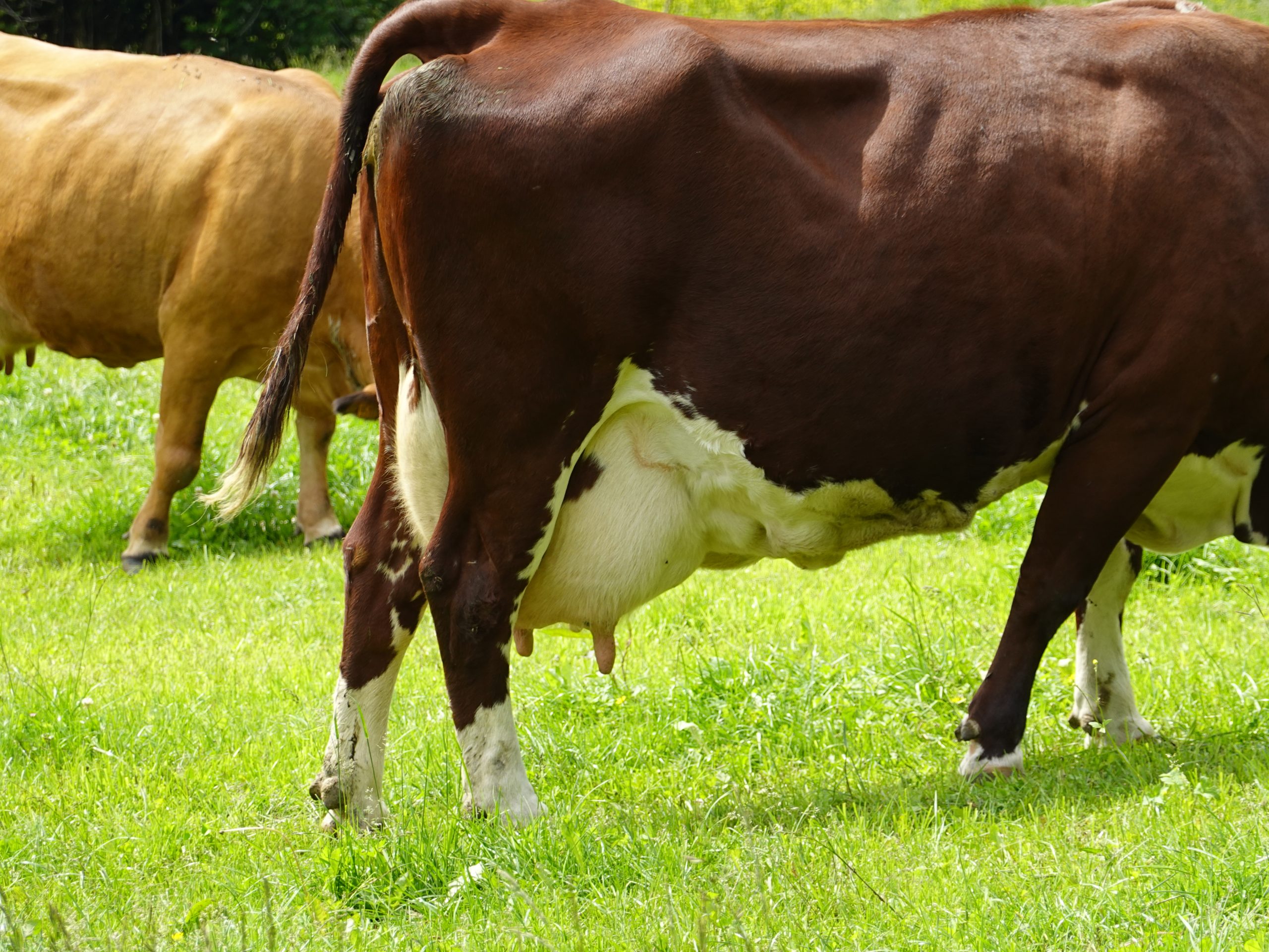
A cow’s udder is four glands in one bag. Each quarter makes its own milk and drains through its own teat, the four shut off from one another inside the one bag. The scan reads each quarter on its own, the probe laid on the skin of the udder to read the gland beneath. The four quarters hang in two halves, a left and a right, each half its front and its hind quarter. The scan works around the udder quarter by quarter, every gland read on its own and set against the others.
Each quarter has the same parts to read. The gland tissue makes the milk, a fine even tissue that fills the bulk of the quarter. The milk gathers in a cistern below the gland, a space that drains down into the teat. The teat carries the milk out through a narrow canal at its end. The scan reads each of those parts for the marks of mastitis. A quarter holds a surprising lot of milk, the cistern of a high-yielding cow a wide dark space the scan reads with ease. The gland, the cistern, the teat: each has its own normal look a vet carries in the eye.
Mastitis is the trouble the udder scan is run to read. An infection in a quarter inflames the gland, fills the milk with clots, walls off pus in abscesses, and in the worst of it kills the tissue outright. The scan reads how far the damage has gone, the picture that says what a quarter has left. The udder is a thing a hand has read for as long as there have been cows, the quarter felt for heat and hardness and the milk drawn off and looked at. The scan adds the one thing the hand never had, a look inside the gland itself.
A hard quarter the hand cannot read
A hand on the udder feels the heat and the swelling of a sick quarter, and no more. It tells the vet a quarter is in trouble without telling what the trouble is. The hand stops at the surface. The scan reads what lies inside the quarter. The milk read in a strip cup tells of the infection, the curd of a mastitic quarter plain in the cup. The scan adds the state of the gland the infection has reached.
The scan reads which trouble has hardened the quarter. A quarter hard with an abscess asks for one thing, a quarter hard with scar another, a quarter hard with the gas of gangrene another again. The hand feels all three as one hard quarter. The scan tells them apart inside, the picture naming the trouble the hand could only feel. Three hard quarters can need three different hands, one drained, one dried off, one treated and milked on, the scan the only thing that sorts them from the outside.
The inflamed gland
An infected gland thickens and coarsens on the scan, its fine grain lost under the swelling, the tissue gone dark on the screen. The vet reads that change to find the quarter the trouble sits in. The change reads first as a haze through the gland, the fine speckled grain of the tissue blurring under the fluid of inflammation that moves into it. A vet learns to read that early haze, a quarter caught before it has built an abscess.
The swelling reads the heat of the infection. A gland packed tight with the fluid of inflammation reads swollen, the tissue dark with the body’s fight against the infection. The vet reads how angry the gland looks, the swelling a measure of how hard the infection has hit. The gland of a quarter in the grip of a hot infection swells against its skin within hours, the gland read swollen tight the same day the cow goes off her milk. The scan reads the swelling for what it is, the gland’s own fight against the bug inside it.
The scan reads the gland for fluid gone where it should not be. Pockets of fluid through an inflamed gland mark the infection breaking the tissue down, the start of the abscesses to come. The vet reads those pockets early, the picture catching a gland on its way to worse.
The inflamed gland may yet come back. A quarter caught early, inflamed and not yet broken down, can heal once the infection is cleared, the gland reading its tissue back toward normal on a later scan. The scan that reads a gland still whole tells the vet a quarter is one to treat. A gland read whole under its swelling is a quarter the drugs can reach, the tissue there to come back once the infection is beaten. The scan that finds the gland still made of gland, with abscess and scar still kept off, tells the vet the cow is one to keep milking.
The abscess walled in pus
An abscess reads as a pocket of pus walled off in the gland. The infection gathers in a spot and the body walls it round, a pocket the scan reads as a dark or speckled fluid space with a bright firm wall around it. The pus inside reads as a cloudy fluid, flecked with the debris of the fight, sometimes swirling under the probe when the quarter is pressed. The wall reads as a bright rim, the body’s line drawn around the infection to keep it in. The vet reads the size of the abscess and where it sits in the quarter, the picture a map of the trouble for the treatment to come. An abscess near the skin can be drained, the vet guiding a needle or a knife to it on the scan, the pus let out and the quarter saved. An abscess set deep in the gland is a harder case, the pus out of a needle’s reach, the quarter often broken past coming back. The scan reads the one from the other, the count and the place of the abscesses the call between a quarter drained and a quarter lost.
The abscess is the finding that can be acted on. A single walled abscess in a quarter otherwise sound is a thing a vet can drain, the cow milking from that quarter again once it heals. The scan finds the abscess, sizes it, and places it, the read that turns a hard quarter into a job with a plan. A vet handed an abscess on the scan knows the next move, the spot to drain and the depth to reach it marked before a blade is laid on. The picture turns a blind cut into a placed one, the abscess opened where it sits.
The pus reads its own story in the abscess. A thin watery pus reads near-black, a thick cheesy one bright with speckle, an old walled one ringed with a heavy rind of scar. The vet reads the look of the pus and the wall to judge the age of the abscess, the read that tells a fresh pocket from a long-settled one. The age of an abscess shows in the gland around it, the dark angry tissue of a fresh one settling quiet once the body has walled the pocket off. The scan reads the gland around the abscess as much as the pus inside it.
The abscess reads for whether it has burst. An abscess that breaks open spills its pus through the gland, the walled pocket gone and the infection loose in the tissue. The scan reads a burst abscess by the pus spread where the wall once held it, a turn for the worse the vet reads at once. A burst abscess spreads the infection wide and turns a walled trouble into a loose one, the quarter harder to save once the pus is through the gland. The scan reads the spread and tells the vet a quarter has slipped from one a knife could fix toward one that may be lost.
The many-abscess quarter is read for what it has left. A gland riddled with pockets of pus is a quarter the infection has taken, the tissue more abscess than gland. The scan reads the spread of them, the count and the size that tell the vet the quarter is past saving. A quarter more pus than gland gives no good milk and stays a seat of infection, a danger to the udder around it. The scan reads it riddled and tells the farm to let the quarter go before it ruins the rest.
Treat or cull
The scan’s one great call is the treat-or-cull. A quarter saved or a quarter lost, read on the screen.
The clots in the milk
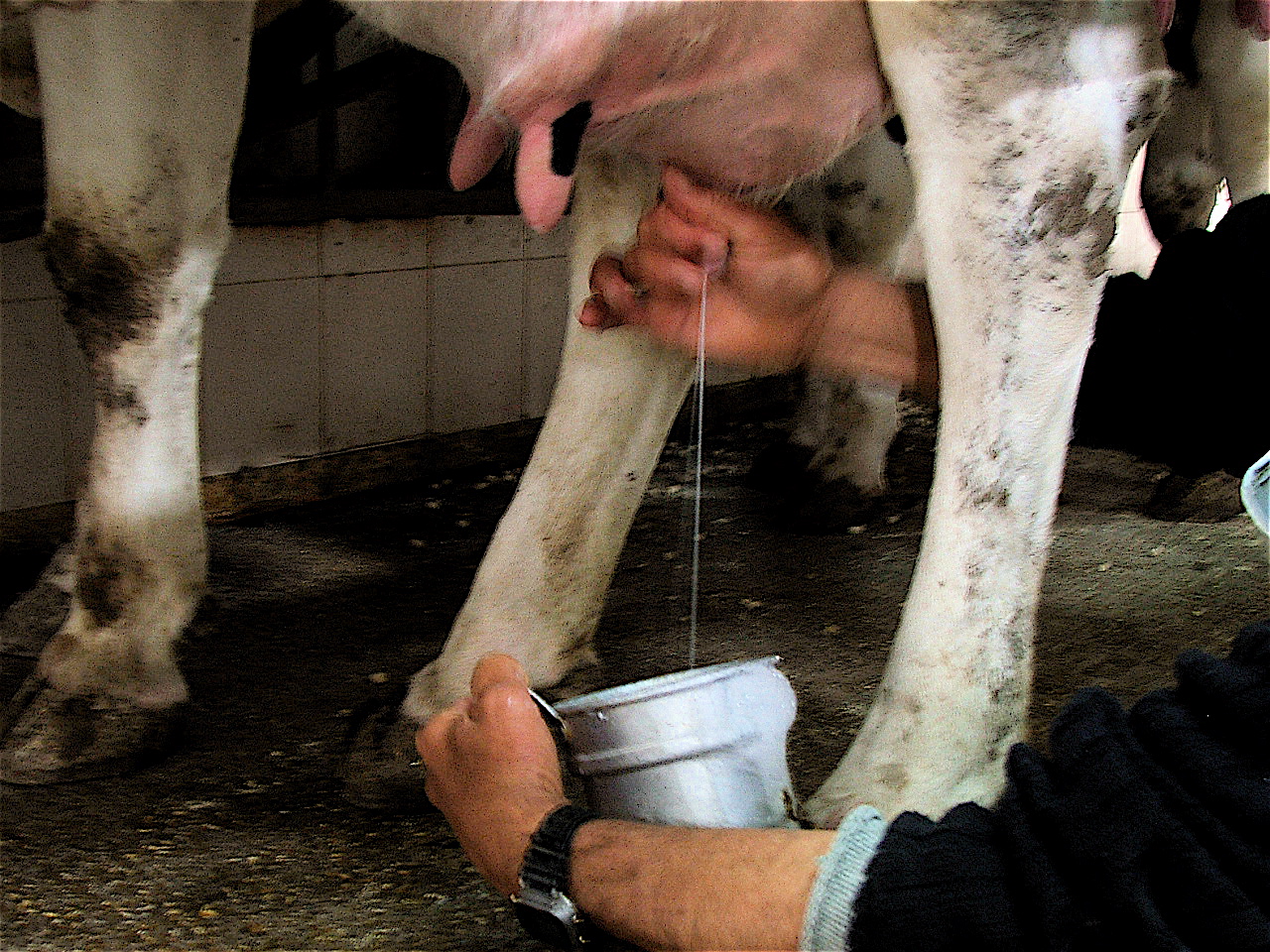
The milk in the cistern reads for the marks of mastitis. Clots and flecks of fibrin floating in the cistern read the infection in the milk itself, bright bits scattered through the dark where the milk should sit clean. The vet reads the cistern for the debris a healthy quarter never carries. The clots read as bright clumps drifting in the dark of the cistern, the fibrin the body throws into the milk to wall the infection off. A vet reads the load of them, a cistern thick with clots a quarter deep in mastitis.
The cistern reads how the milk runs out as well. A cistern full of clots and clumps reads a quarter that milks thick, the debris clogging the flow down to the teat. The scan reads the milk the cow gives before it is ever drawn, the trouble in the cistern read on the screen. A quarter that gives a thin clotted milk reads a cistern full of debris, the look of the milk and the look of the cistern telling the same tale. The scan reads on the screen what the milker reads in the pail.
The teat and its block
The teat carries its own troubles. The milk leaves a quarter through the teat, down a cistern in the teat and out a narrow canal at its end. A teat hurt or blocked stops the milk of a sound quarter, a cow that will not milk out from a gland with nothing wrong above. The scan reads the teat for what holds the milk back. A hard-milking cow loses time at every milking and risks the quarter going bad behind a teat that will not flow. The trouble is often the teat itself, the gland above it sound, the scan sent to read the teat when a quarter milks slow with no sign of mastitis.
The teat is read in a bath of water. A teat is a small thing right at the skin, read poorly by a probe laid straight on it. The vet stands the teat in a cup of warm water or lays a water-filled standoff against it, the water carrying the sound into the teat and drawing its small parts clear.
The block in a teat reads on the scan. A teat that milks hard carries something in its way, a flap of scar, a stone of hardened milk, a growth on the wall, a narrowing of the canal. The scan reads the block for what it is and where it sits, the picture that says whether a knife can open the teat or not. A flap of scar across the canal cuts the milk to a trickle, a hard pearl of clotted milk plugs it, a fleshy growth on the wall narrows the way. The scan names which it is, the block read for whether it sits where a knife can reach.
The teat wall reads for its own hurts. A teat trodden or cut heals with scar that narrows the canal, the milk slowed through a teat the cow has damaged. The scan reads the thickness of the teat wall and the state of the canal, the injury read where a hand feels only a hard teat.
The teat scan saves a quarter a sound gland would otherwise lose. A gland making good milk above a blocked teat is a quarter the vet will open, the cow milking from it again once the block is cleared. The scan reads the gland sound and the teat blocked, the call to open the teat made on the picture of both. A teat opened on a good gland gives the cow her quarter back, a year of milk saved off a small cut the scan placed. The teat read whole the vet leaves alone, the milk slow for some other cause the scan sends him to find.
The quarter that is lost
Some quarters the scan reads past saving. A gland scarred hard through with the fibrosis of an old mastitis will not make milk again, the tissue turned to a dense pale scar the scan reads in place of the gland. The vet reads a fibrosed quarter and calls it lost, the cow milked on her other three. A cow does not miss a single dry quarter, her three sound ones milking on for her. The vet dries off the lost quarter and leaves the cow her udder, the dead gland sealed away from the milking.
The fibrosed quarter reads dense on the scan. A gland gone to scar reads as a solid pale tissue with none of the soft pattern of a working gland, a quarter that feels hard to the hand and reads hard on the screen. The scan tells a fibrosed quarter from a swollen one by the dense scar that fills it, a quarter read past saving where the gland has gone to scar. A fibrosed quarter gives a little milk or none, the scar tissue making nothing where the gland once did. The scan reads how much of the quarter has gone to scar, the share of working gland that is left setting whether the cow is milked on from that quarter or it is dried off for good.
Gangrene reads the worst trouble of all. A quarter the infection has killed fills with gas, the dead tissue reading bright with the gas pockets through it, a black quarter going cold the scan reads as gas and ruin inside. A gangrenous quarter is a danger to the cow herself, the trouble read at once and the cow treated hard or the quarter taken off. Gangrene comes on fast, a quarter turned cold, its skin going blue within a day of a hot infection turning bad, the skin sloughing where the tissue has died. The scan reads the gas in the gland early, a vet given the hours that count to treat the cow or take the quarter before the rot reaches her blood.
The lost quarter is read so the cow is not. A cow milked on against a ruined quarter loses money and risks her health, the dead tissue a drain and a danger. The scan reads a quarter lost and tells the farm to dry it off or to cull the cow, the call that keeps a bad quarter from costing the whole animal.
Reading each quarter against the rest
A quarter reads best against its neighbours. The vet scans all four quarters of an udder, the sick one read against the three that are sound. A quarter swollen or scarred stands out plain beside its healthy mates, the comparison putting the trouble in relief. A vet who scans the sound quarters first carries their look in the eye for the sick one, the normal grain fresh in mind when the trouble comes up. The healthy three are the yardstick the fourth is read against.
The four quarters tell the spread of the trouble. The vet counts the quarters the mastitis has taken, the spread from a single sick gland to a whole udder inflamed telling how hard the cow has been hit. A local trouble in one gland is a different case from a mastitis run through all four. A whole udder inflamed marks a cow hit hard, sicker in herself than one with a single sore quarter, the vet reading her for more than the udder alone.
The healthy quarters set the read for the sick one. A vet learns the normal look of a gland from the three sound quarters on the same cow, the sick one read against them in the same scan. The comparison is the surest read the udder scan gives, the trouble named against the cow’s own healthy tissue. No two cows read the same, the grain of a gland a little different from one animal to the next. The three sound quarters give the vet that cow’s own normal, the sick one judged against the cow’s own healthy glands.
The probe and the water bath
The work runs on a high-frequency linear probe. The udder sits close under the skin. A high frequency draws the fine grain of the gland and the clots in the cistern in the detail the scan needs. The vet clips the hair, wets the skin, and lays the probe on the quarter, the gland read close under it. The udder skin scrubs up clean for the scan, the gel laid on and the probe worked over each quarter in turn. A vet reads a whole udder in a few minutes, four quarters and their teats covered on the one cow.
The teat asks for the water bath. The teat is a small thing right at the skin, read poorly by a probe laid straight on it, the water carrying the sound across a gap to draw its small parts clear. A cup of warm water or a water-filled standoff is the trick that brings a teat into focus.
What the udder scan settles
The udder scan reads a sick quarter from the inside, where a hand reads only its heat. It finds the gland inflamed, the abscess walled in pus, the clots in the milk, the teat blocked, the quarter scarred or gangrenous, each a different trouble behind the same hard quarter. The scan names which one the cow carries, the read a hand could never make. A hard quarter is a hard quarter to the hand, the same swelling whatever lies behind it. The scan opens the quarter to the eye, the abscess and the scar and the gangrene each read for what it is.
The call the scan makes is the treat-or-cull. A quarter with an abscess to drain, a teat to open, or a gland still whole is one to treat. The scarred, the gangrenous, and the abscess-riddled are the ones to lose, dried off or culled. The picture decides which way a hard quarter goes.
The udder scan keeps a cow’s quarters and the cow herself in good order, each quarter read from the outside and handled for what the scan finds inside it. The savable ones are treated and milking again, the lost ones dried off before they cost the cow her health. A dairy that scans its hard quarters keeps the cows it can keep and lets go the quarters it cannot, the udders read true, no quarter lost or kept on a guess. The scan is the tool that reads the inside of an udder a hand can only feel.
Common questions about udder and mastitis scanning
What does an udder ultrasound show in a cow with mastitis?
The damage inside the udder that a hand can only feel as a hard quarter. The scan reads the gland inflamed, the abscesses walled in pus, the clots filling the milk cistern, the teat blocked against the flow, and the gas of a gangrenous quarter. It reads how far the infection has gone and what a quarter has left.
Can ultrasound tell whether to treat or cull a cow with mastitis?
It is the main reason the udder is scanned. An abscess to drain, a blocked teat to open, or a gland still whole reads as a quarter to save. A gland scarred through, a quarter gone gangrenous, or an udder riddled with abscesses reads as one lost. The scan gives the vet the picture the treat-or-cull call is made on.
How is a cow’s teat scanned?
In a bath of water. The teat sits right at the skin, read poorly by a probe laid straight on it, so the vet stands the teat in a cup of warm water or lays a water-filled standoff against it. The water carries the sound into the teat and draws its small parts clear, the canal and the cistern read for a block that holds the milk back.
How does the vet know which quarter is infected?
By reading all four quarters against each other. The vet scans every quarter and picks out the one gone coarse and thickened, its grain lost and its cistern flecked with clots, standing out against the cow’s three sound glands. The comparison is the surest read the udder scan gives.
Why scan the udder when you can feel a hard quarter?
Because the hand stops at the surface. A hard quarter feels the same to the hand whether it holds an abscess, a scar, or the gas of gangrene, three troubles that need three different answers. The scan reads inside the quarter and tells them apart, the picture naming the trouble the hand could only feel.

About the Author
Julien Mercier
Senior R&D Engineer · Medical Ultrasound Transducer Development
Senior R&D Engineer with an M.S. in Applied Physics and over 15 years of experience in medical ultrasound transducer development, specializing in the design verification and performance testing of high-frequency imaging transducers. Currently leading the development and verification of the company’s next-generation high-frequency linear-array transducer, responsible for imaging performance evaluation and reliability analysis in preclinical testing. Brings extensive hands-on experience in piezoelectric element tuning, beamforming parameter optimization, and system-level performance testing.