





































Equine Tendon And Ligament Ultrasound Handheld Diagnosis
An equine tendon and ligament ultrasound reads the soft cords down the back of a horse’s lower leg for the damage of a strain, the probe drawing the flexor tendons and the suspensory ligament in fine detail. A pulled or torn tendon shows on the scan as a dark hole in the bright pattern of healthy fibres, its size measured and its healing followed over the months it takes to mend. The scan is the one tool that reads a tendon injury from the outside, telling a trainer how bad the damage is and when the leg can carry work again.
The tendons down the back of the cannon
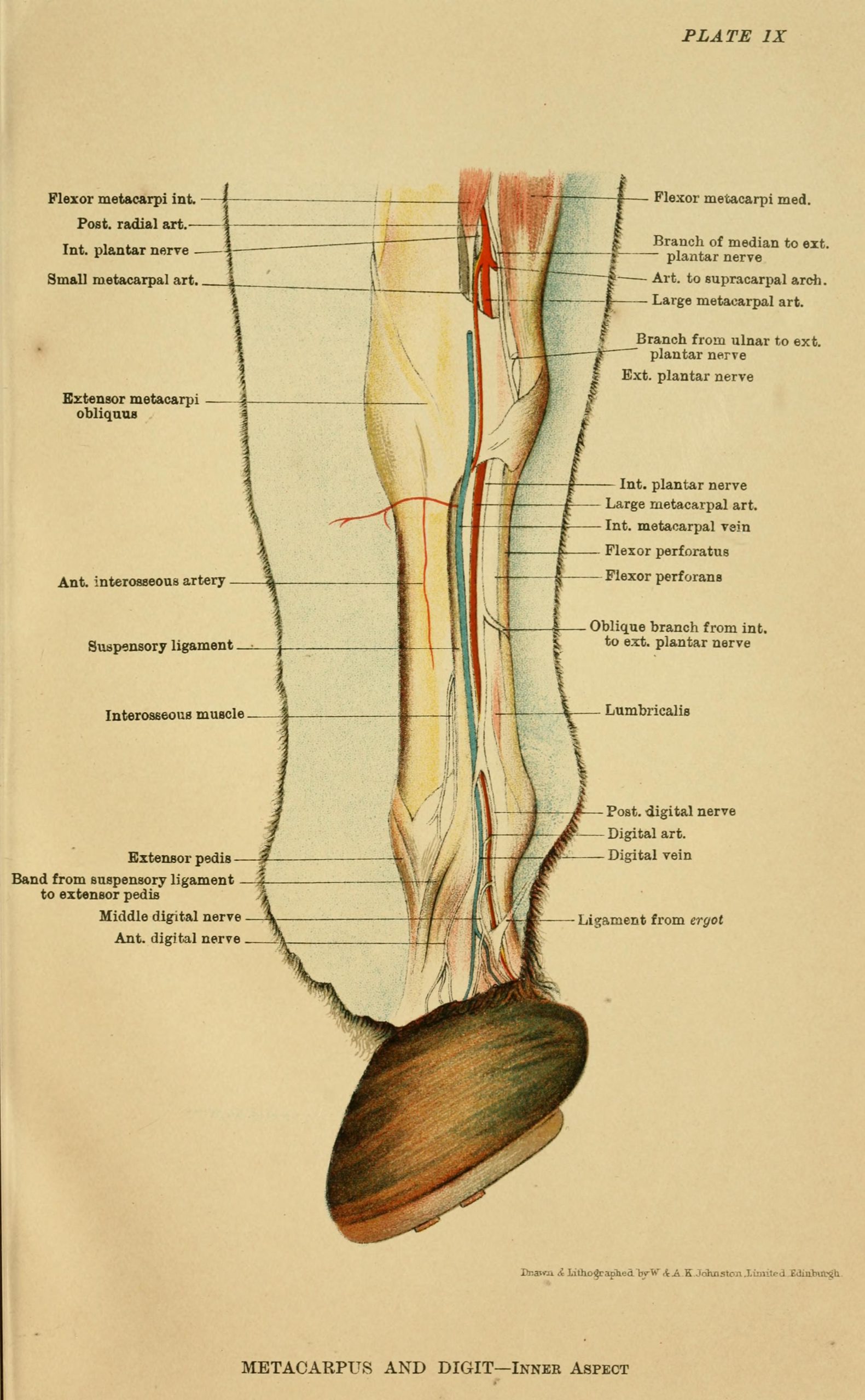
The cords the scan reads run down the back of the cannon bone, between the knee or hock and the fetlock. The superficial digital flexor tendon lies closest to the skin, the deep digital flexor tendon just beneath it, the suspensory ligament deeper still against the bone. A small check ligament joins the deep flexor partway down. The four sit in a stack the probe reads from the surface down. The cannon is the long bone between the knee and the fetlock on the foreleg, the hock and the fetlock behind. The cords run its whole length, the probe reading them from top to bottom in a single sweep down the leg.
Each cord carries the load of the gallop. The flexor tendons run from the muscles above to the bones of the foot, taking the pull of the leg against the ground. The suspensory ligament holds the fetlock up against the drop of the horse’s weight. A cord that tears under that load is the injury the scan is run to find. The cords are rope more than muscle, tight bands of fibre with little blood of their own, slow to heal for the want of it. A tendon takes far longer to mend than the muscle above it, the scan the way to watch a slow recovery.
The injury sits where the strain falls hardest. The superficial flexor tendon bears the brunt and bows the worst, a swollen tendon at the back of the cannon the classic sign of a strain. The suspensory and its branches go next, the deep flexor and the check ligament less often. The scan reads each in turn to find which cord has gone. A bowed tendon is the name a horseman gives the swelling, the back of the cannon curving out under the strained tendon. The bow tells a horseman a tendon has gone, the scan adding which cord and how badly.
The dark hole of a strain
A strain reads on the scan as a dark hole punched through the bright fibres of a tendon. Healthy tendon shows in cross-section as a tight pattern of bright dots, the cut ends of fibres packed close in an even field. A core lesion appears in the middle of that pattern as a black or grey hole, the fibres torn and the gap filled with blood and fluid in the first days. The vet measures how wide the hole runs across the tendon and how far it runs up and down the leg, the size of it the measure of the injury. In the days after a fresh strain the hole shows at its worst, the blood and the swelling at their peak. The scan run in the first week catches the injury at its fullest, the picture a trainer reads to know a tendon has gone before the leg has even fully swelled. A dark core in a tendon means months out of work, the worst lesions ending a galloping career.
The longitudinal view reads the fibres along their length. A probe turned to run up and down the leg shows the fibres of a healthy tendon as bright parallel lines, packed like the grain of wood. A lesion breaks those lines, the fibres in the damaged stretch lost in a dark gap. The break marks the length of the tear, the vet reading where the sound fibres stop, where the gap runs, and where they take up again past it. The length of that gap on the long view feeds into the grade.
The two views read together. The cross-section shows how much of the tendon’s width a lesion takes. The long view shows how far up and down the leg it runs. A vet turns the probe between the two on every scan, the lesion measured in both directions before it is graded.
The fresh lesion changes by the day. The dark hole of the first week fills slowly in the weeks after, the blood clearing and the body laying down new tissue, the gap turning from black toward grey. A scan repeated through the healing reads that change, the tendon followed from the tear to the mend. The first scan is often run a few days after the injury, once the worst of the swelling has settled enough to read through. A leg scanned in the first hot hours reads poorly, the picture clearer once the swelling has come down.
The look of the lesion grades its severity. The darkest, widest holes are full tears of the fibres, a horse facing the longest rest. The vet reads the darkness and the spread of the lesion to put a grade on it, the grade setting the rest a horse will need. A lesion graded at the first scan is the baseline every later scan reads against. The grade travels with the horse, the next vet reading the recovery from the number the first one set down.
Measuring the damage
The size of the lesion is put into a number. The vet measures the cross-sectional area of the tendon and of the dark core within it, the figures the hard measure of how much of the tendon has torn. A core that takes a quarter of the tendon’s width reads worse than one that takes a tenth. The cross-sectional area is read in square millimetres, a figure the vet writes down to compare scan against scan. The percentage of the tendon a lesion fills sorts a mild strain from a near-complete tear, the number doing the work an eye alone cannot.
The numbers are read against the sound leg. The vet scans the matching spot on the other leg and measures it the same way, the healthy tendon the yardstick for the hurt one. A tendon swollen to twice the area of its partner has taken a real injury, the comparison putting a figure on the swelling. The matching scan on the sound leg is run at the same level and the same angle, the only fair test of how much the hurt tendon has grown. A horse with both legs strained gives no clean comparison, the vet falling back on the known normal range for the breed.
The share of the tendon involved sets the grade. The more of the cross-section a lesion takes, the graver the injury, a lesion across half the tendon among the worst a leg can carry. The vet reads the percentage of the tendon lost to the lesion, the figure that decides how long the horse must rest. The grade runs from a mild strain healed in a few months to a full tear that takes the better part of a year. A vet sets the rest and the work-back from the grade, the plan built on the number the scan gives.
The figures turn a look into a record. A grade and a set of measurements written down at the first scan give the vet a fixed point to read the healing against. A later scan measured the same way shows whether the lesion has shrunk, the numbers telling the recovery a glance alone would miss.
The trap of the angle
A tendon read off-angle goes falsely dark. The bright fibres of a tendon show their brightness only when the probe sits square to them, the sound bouncing straight back. A probe tipped even a little lets the sound glance away, the tendon turning dark on the screen as though it held a lesion. The effect is strongest where a tendon curves, the fibres turning under the probe and catching the sound at a changing angle. A vet meets it often at the back of the fetlock, where the tendons bend around the joint.
That false darkness fools the unwary. A vet new to the work reads an off-angle shadow as a tear and calls an injury that the tendon does not carry. The skilled vet keeps the probe square, rocks it to bring the fibres to full brightness, and reads a dark patch as a lesion only once the angle is right. The artifact is the first thing a tendon scanner learns to beat.
The sound leg tells the truth
The vet scans the good leg the same way as the hurt one. A tendon means little read on its own.
Following the healing
A torn tendon is scanned again and again as it mends. The vet repeats the scan every couple of months, measuring the lesion each time against the figures from the last. The healing reads as the dark hole filling in, the new fibres laying down where the tear had been. The healing runs slowly, a tendon taking six months to a year to mend enough for full work. The scans are spaced across that time, one every couple of months reading the lesion smaller than the last.
The new fibres lay down out of line at first. A healing tendon fills its hole with tissue that reads bright in a tangle, the fibres crossed and disordered in the early weeks. The scan reads that tangle, the early mend filling the gap long before it can carry a gallop.
The fibres line up over months of careful work. Light, controlled exercise pulls the new fibres into line along the tendon, the scan reading them turn from a bright tangle to straight parallel lines. A tendon brought back too fast fills with disordered scar, the fibres never lining up to take the gallop. The work is dosed against the scan, a little more asked of the leg each time the picture shows the fibres holding. A program run by the scan brings a tendon back over months, the exercise raised step by step on the evidence of the fibres.
The scan calls the day a tendon can work again. A lesion filled in, the fibres lined up, the cross-section back near the size of the sound leg: those signs together tell the vet the tendon has mended enough to carry training. A horse put back to work on a scan that still shows an unfilled lesion tears the tendon again, the picture the guard against a second injury. The cross-section back to within a tenth of the sound leg, the fibres straight, the dark gap gone: the vet reads all three before the leg is cleared. A tendon cleared on a careful scan goes back to work with the odds of a second tear cut down.
The suspensory ligament
The suspensory ligament asks for its own read. It runs deep against the back of the cannon bone, from its origin just below the knee or hock, down the length of the bone, and into two branches that wrap to the fetlock. The scan follows it the length of the leg, the origin, the body, and the branches each read in turn.
The ligament reads a little differently from a tendon. Its normal tissue carries more grey and a coarser pattern, the fibres mixed with muscle in places. A vet learns the normal look of the suspensory to keep from calling its ordinary grey a lesion. The lesion shows as it does in a tendon, a dark hole and a swelling in the cross-section. The origin high under the knee is a common spot for the suspensory to strain, a proximal lesion that reads dark against the bone. The scan reaches it with the probe pressed high at the top of the cannon, the origin awkward to read low behind the knee.
The branches are the spot the suspensory tears more than any other. A branch torn where it wraps to the fetlock swells and reads dark the same as a tendon core. The vet scans both branches on a lame horse, the injury often hidden low where the ligament splits. A suspensory branch lesion is a long road back, the ligament slow to heal.
The probe and the prep
The work runs on a high-frequency linear probe. The tendons sit close under the skin. A high frequency draws their fibres in the fine detail the scan needs. A flat linear probe lays a wide window down the back of the cannon, the picture sharp on structures a centimetre or two deep. A standoff pad of soft gel sits between the probe and the skin, lifting the lens off a tendon that lies right at the surface. The nearest fibres lie so close to the probe they read poorly without it, the standoff bringing them into focus.
The prep makes or breaks the picture. The hair is clipped, the skin scrubbed and wetted, and a standoff pad or a thick layer of gel laid down to lift the probe off the tendon that sits right under the skin. The picture is only as good as the prep, a clipped and scrubbed leg the one a tendon reads sharp on.
Reading the leg in zones
The leg is read in a fixed set of zones. The back of the cannon is divided into levels down its length, the probe stopped at each to read the tendons in cross-section. The vet works down the leg zone by zone, a picture saved at each, the whole length covered in order.
The zones turn a scan into a map. A lesion named by its zone tells the next vet exactly where to look, the injury pinned to a level on the leg. A scan filed by zones reads against an earlier one spot for spot, the healing followed at the exact place the tendon tore. The zones are numbered down the leg, each a level the vet returns to on every scan of that horse. A tendon read by zones on the first visit is read by the same zones on the next, the levels lining up scan to scan.
What the scan cannot settle
The scan reads the soft cords. The bone behind them lies beyond its reach. A chip or a crack in the bone beneath a sound-looking tendon stays hidden from the soft-tissue scan. A horse lame past what the tendon shows is sent for an x-ray or a scan of the joint, the soft-tissue picture only one part of the workup.
An old injury reads much like a fresh one at a glance. A tendon that tore a year ago and healed with scar carries a thickened look of old damage that an untrained eye takes for a new lesion. The history of the leg reads alongside the picture, the scan told apart from a fresh tear by the story of the horse. A horse with an old healed bow carries a thicker tendon for life, the scan reading that thickening as the mark of the past injury. A fresh tear on top of an old one reads against the thickened baseline, the new damage told from the old scar by its darkness.
The scan reads the damage a tendon has taken. How much that damage hurts is a separate question. A lesion’s look on the scan does not always match the lameness it causes, a small tear sometimes the more painful of two. The vet reads the picture alongside the lameness, the scan one voice in the call, weighed with the rest of the exam.
The deeper structures test the scan. The deep flexor tendon low in the pastern, the suspensory where it runs under the splint bones: these sit deep or tuck behind bone where the probe reads them poorly. A lesion in those spots may need a scan from a special angle or a scan in the magnet to read at all.
The read comes with the eye for it. A vet who scans tendons through a racing yard reads the normal and the torn at a glance, the anisotropy beaten by habit and the lesions known by their look. A scan read by an eye new to tendons misses a fine lesion or calls an artifact a tear. The skill is built up over the legs passed under the probe, the same as any scan. The first lesion a vet calls is checked by a second pair of eyes or a repeat scan a week on, the early reads taken slowly and checked twice. By the time a vet has scanned a season of legs, the normal and the torn separate at a glance.
What the tendon scan gives
The tendon scan reads an injury that a hand can only guess at. A swollen leg tells a trainer something is wrong. The scan tells which cord has torn, how badly, and how long the road back will run. The picture turns a vague heat and swelling into a named injury with a grade and a plan.
The greater gift is the read on the healing. A tendon followed month by month on the scan shows the trainer when the fibres have knit and lined up, the one honest sign that a leg can carry the gallop again. A horse rested on the picture goes back sound, the scan guarding it from the second tear that ends so many careers. The trainer reads the scan as the leave to step the work up, the gallop added back only on a picture that says the fibres will hold. A scan that holds a horse back a month longer saves a career a rushed return would break.
The scan reads the tendon from the tear to the mend. It finds the dark hole of a fresh strain, measures it against the sound leg, grades it, and follows it through the months of healing to the day the leg works again. The cords down the back of the cannon are read end to end, the one tool that turns a tendon injury from a guess into a managed recovery.
Common questions about tendon and ligament scanning
What tendons and ligaments does the scan read?
The soft cords down the back of the lower leg. The superficial digital flexor tendon nearest the skin, the deep digital flexor tendon beneath it, the suspensory ligament deep against the bone, and the check ligament that joins the deep flexor. The scan reads each from the surface down for the dark hole of a strain.
What does a tendon injury look like on ultrasound?
A dark hole in the bright pattern of healthy fibres. In cross-section a torn tendon shows a black or grey core where the fibres have pulled apart, the tendon swollen around it. The long view shows the straight bright fibres broken by a dark gap. The vet measures the hole and grades the tear by its size and its darkness.
Why does the vet scan the other leg too?
To read the hurt tendon against a healthy one. The vet scans the matching spot on the sound leg and measures it the same way, the good tendon the yardstick for the injured one. A tendon swollen well past the size of its partner has taken a real injury, the comparison putting a figure on the damage.
Why does a tendon look dark when it is not injured?
From the angle of the probe. A tendon read off-square lets the sound glance away and turns falsely dark on the screen, an artifact called anisotropy. The vet keeps the probe square to the tendon and rocks it to bring the fibres to full brightness, reading a dark patch as a lesion only once the angle is right.
How is tendon healing followed?
By scanning the leg again every couple of months. The vet measures the lesion against the figures from the last scan, reading the dark hole fill in and the fibres line up over the months. A tendon back near the size of the sound leg with its fibres in line is one that can carry work again.

About the Author
Julien Mercier
Senior R&D Engineer · Medical Ultrasound Transducer Development
Senior R&D Engineer with an M.S. in Applied Physics and over 15 years of experience in medical ultrasound transducer development, specializing in the design verification and performance testing of high-frequency imaging transducers. Currently leading the development and verification of the company’s next-generation high-frequency linear-array transducer, responsible for imaging performance evaluation and reliability analysis in preclinical testing. Brings extensive hands-on experience in piezoelectric element tuning, beamforming parameter optimization, and system-level performance testing.