An infrared vein finder is a device that lights the skin with near-infrared light and shows the veins underneath as dark lines, so a clinician can see where to place a needle before touching the arm. It belongs to the KT-10 line of vein-finding tools. The device reads the surface veins by light alone, the ones a drip or a blood draw goes into, and lays a clear map of them on the skin or a screen. It carries no ultrasound and no radiation; the whole job is done with a band of light the eye cannot see.
Seeing the vein before the needle
Many veins are hard to find. A vein that lies deep, one hidden under a layer of fat, or a small rolling vein in a child can take several tries to hit, and each missed try means another jab for the patient and more time lost for the clinician. A vein finder sets out to take the guesswork out of that moment. It shows the veins before the needle goes in, so the person placing it can choose a good vein and aim with a clear picture in front of them. The whole point is to turn a stick that depends on feel and luck into one that depends on a picture of where the vein actually runs.
That is the whole idea of an infrared vein finder. It reads the veins through the skin with near-infrared light and draws them as a map the clinician can use. How an infrared vein finder sees the veins through the skin is the place to start, since everything else follows from that one trick of light. The device finds no veins the body does not have, and it places no needles. It shows what is there, clearly, before the work begins. It draws the veins on the patient’s own skin, in the spot the needle is going.
Everything else about the device follows from the light it uses. Near-infrared light, just past the red the eye can see, passes a few millimetres into the skin and is soaked up by the blood in the veins. The veins come back darker than the tissue around them. The device reads that difference and draws it as a line. No dye is injected, and no contact is made with the skin. The picture is live, updating many times a second. Because the light is invisible and harmless, the device can hover over an arm, a hand or a foot for as long as the search takes. A reading costs the patient nothing. It can be repeated as often as a search needs.
A vein finder reads the surface of the arm, the layer just under the skin where the useful veins run. That shallow reach is all the job needs, since a drip or a blood draw goes into exactly those surface veins. The device shows the clinician the veins a needle can reach, drawn on the very arm the needle is going into. Looking deeper into the body, at the deep veins and the organs, is a job for other tools. A vein finder does the surface job. It needs no cart, no probe and no drop of gel.
The light and the blood
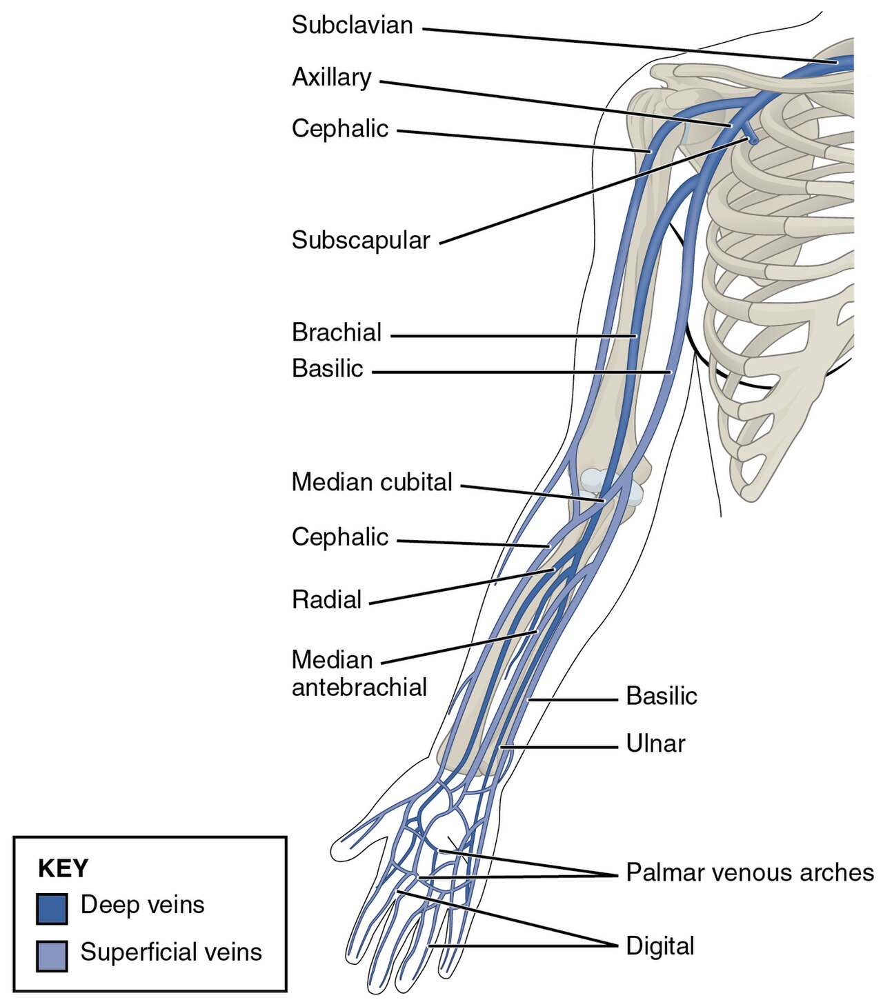
The veins of the arm and hand. The superficial veins near the skin, among them the cephalic, basilic and median cubital, are the ones a vein finder maps and a needle uses. The key tells the superficial veins from the deep ones.
The veins show up because of what blood does to this one band of light. Venous blood carries haemoglobin, the pigment that has just dropped off its oxygen. That pigment soaks up near-infrared light far more than the skin and fat around it. Where a vein runs, less light bounces back. The device paints that shortfall as a dark line. The blood-poor skin around the vein stays pale, so the veins stand out as a network of dark lines on the picture. The veins that carry the most blood, the broad surface ones a needle wants, give the boldest lines of all.
The light reaches only so far. It fades the deeper it travels, so a vein finder reads the veins near the surface best. Veins that run deep fall outside its reach. For the everyday veins on the back of the hand, the wrist and the forearm, that reach is enough, since those veins sit only a few millimetres down. A heavier arm, with more fat over the veins, gives a softer picture. The device leans on its processing to lift the lines. Most surface veins a needle uses sit within about three millimetres of the skin, which is where a plain reflection device reads best. The better projecting systems push that reach to around ten millimetres. The reach of the light matches the reach of the needle: both work on the surface veins, which is why the limit rarely gets in the way of the everyday job.
This is the fact the whole device is built around. The blood in a vein drinks a colour of light the eye cannot see. A camera tuned to that colour reads what the eye cannot. From there the device adds a source to shine the light, a processor to lift the faint lines, and a way to show the map back to the clinician. Everything else, the depth it reaches, the skin tones it handles, the two ways it shows the picture, follows from that one starting point. A reader who grasps the single idea behind it can make sense of every model on the market.
Where a vein finder earns its place
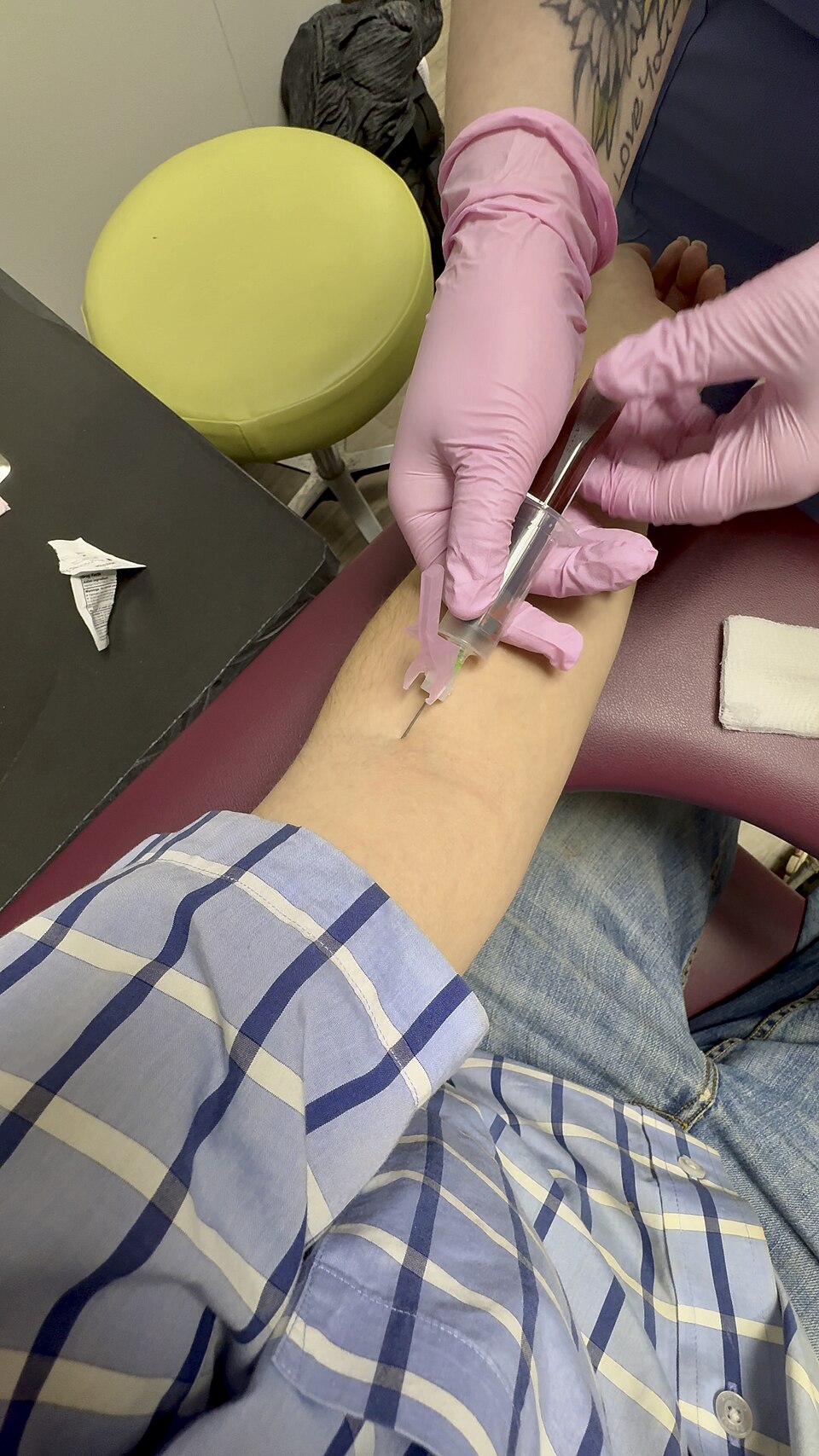
Drawing blood from a vein at the bend of the elbow. An infrared vein finder makes this everyday task easier on the arms where a vein is hard to see or feel.
A vein finder helps most where a vein is genuinely hard to find. On an easy arm a skilled nurse needs no help, and the device adds little. It shows its worth on the difficult stick: the patient who has been jabbed twice already, the arm where nothing can be seen or felt, the vein that hides under skin or fat. The case for it is strongest there, with the difficult-access patient that reviews of these devices look at most closely. Difficult venous access is common, and a failed first stick is an everyday event on a busy ward. A handful of groups run into that difficulty far more than the rest, and each has a page of its own in this group.
Children are the clearest case. A small child has small veins. A frightened child pulls away. A clean first stick is harder to win here, since a child’s small veins turn every attempt into a struggle for the child and the parents alike. A vein finder lets the clinician pick a tiny vein and aim for it. The long hunt of repeated tries is gone. A vein finder for paediatric IV access looks at how the tool is used on the smallest patients, where the veins are fine and the stakes for a calm, quick stick are high.
Patients on chemotherapy are another. Months of cytotoxic drugs harden and scar the veins. The easy veins get used up. What remains has to be protected, since the same patient will need access for many more rounds of treatment. Saving a vein, and hitting it on the first try, guards what is left. A vein finder for the chemotherapy patient covers the veins that long treatment leaves behind, and how seeing them first helps spare them.
A vein in a heavier arm brings its own problem. It can lie out of sight under a layer of fat, deep enough that the eye misses it and a finger only guesses. Here the device helps by finding the veins that lie within its reach and showing their exact line. The clinician can aim true on the first pass. A vein finder for venipuncture in the obese patient looks honestly at the depth limit and how to work within it, since the deepest veins lie beyond the light.
Across all of these, the gain is the same: fewer tries, less pain, and a vein found before the needle moves. The patients who need it most are the ones whose veins hide, from the smallest child to the patient left with few good veins after months of treatment. A vein finder does not make a hard stick certain. What it does is turn a blind search into a guided one, often the difference between one try and five on a difficult arm. The device sells itself on those arms, the ones a clinician used to dread.
When speed decides it
In an emergency the clock changes everything. A line has to go in fast, to give the fluids and drugs that turn a patient around. A patient in shock has veins that have gone flat, hard to see and hard to feel. The seconds spent hunting for a vein are seconds a sick patient cannot spare. A vein finder can show a usable vein quickly, on an arm where the usual landmarks have gone. That head start buys time a sick patient needs.
Speed is where a hands-free map pays off most. A device that throws the vein map onto the skin lets the clinician find a vein and place the line in one motion, with eyes on the arm the whole time. There is no glancing away to a screen and back. A vein finder for emergency IV access looks at rapid cannulation when a line cannot wait, and at how the tool fits a resuscitation where every second is spoken for.
Light that is safe to shine
Because a vein finder works by shining light on people many times a day, the safety of that light is set by a standard. The near-infrared source runs at low power and does not ionise tissue, nothing like an x-ray. The relevant rule is a photobiological-safety standard written for lamps and LED sources, and a vein finder is built and tested to meet it. It sets limits that keep the light safe for the eyes and skin of patients and staff alike, even with repeated use through a long day. The standard sorts a light source into risk groups by how much it emits and how close it is used, from an exempt group that poses no hazard up through the low-risk groups. A vein finder belongs in those lower groups. The IEC 62471 photobiological-safety standard for an infrared vein finder sets out what that rating means and how the source is classed for a device shone on people all day.
Across a nursing team
A vein finder is often bought for a whole department to share. A ward, an oncology unit or a paediatric clinic keeps one or two devices for the team to reach for when a difficult stick comes up. The value then is measured across many sticks a day, in first-attempt success and in the time a busy team saves. Across a year of difficult arms, the saved re-sticks add up to real time, less wasted kit, and many patients spared a second jab.
Used across a team, the device needs a few things a single owner can skip. It needs to be simple enough that every nurse can pick it up and use it the same way, with no manual at the bedside. It needs to be quick to clean, quick to charge, and ready at the couch the moment a hard vein turns up. A shared device that is awkward to use, or always flat, gets left in a drawer and the money wasted. A shared tool is judged on its practical side as much as on the picture it draws.
There is a training side to it as well. A vein finder gives a new nurse a way to see the veins an experienced colleague has learned to feel. The climb to sticking a vein with confidence gets shorter. The same picture, shown to a nervous patient, can settle the nerves of someone who dreads the needle by showing that the vein has been found. An infrared vein finder in nursing practice covers how a unit writes the device into a protocol, decides who uses it and when, and trains a team to get the most from it.
The gain to a unit is real even when it is hard to put a single number on. The studies show the benefit most clearly on the hardest sticks. That is why a unit aims the device at difficult access, where it does the most good. Fewer repeat sticks mean less wasted kit, fewer upset patients, and less time lost on the rounds. A team that finds veins faster runs a smoother clinic and sends fewer hard cases up the line for someone else to attempt. The device pays its way through all the small sticks that now go right the first time, on arms that used to take several.
Choosing a vein finder
Two kinds of vein finder are on the market. One kind throws the vein map straight onto the skin in visible light, so the veins appear drawn on the arm, and the clinician works with both hands free. This projecting kind suits a bedside and an emergency, where the clinician wants to watch the patient. The screen kind shows the map on a monitor or a phone, runs smaller and cheaper, and can store the image for teaching or a record. The choice between them shapes how the device fits a given ward, and it is the first thing a buyer settles.
Beyond the display, the things that separate one device from another are the depth it reads, how well it handles different skin tones, the size and weight in the hand, and the battery that has to last a shift. An infrared vein finder compared with the AccuVein AV500 sets a portable device against a well-known one on exactly these points, so a buyer can see where a lighter, cheaper unit holds up and where it gives ground.
Buying for a ward adds its own checks. A device passed from nurse to nurse has to clean quickly and survive daily handling. It has to charge fast and hold that charge through a busy list. A wipeable surface helps, since the device meets many patients in a shift and carries no cover of its own. The picture has to be clear enough that everyone trusts it, and simple enough that no one needs a manual at the couch. These practical points decide whether a device gets used every day or shelved after a week, and they weigh as heavily as the depth or the price.
An infrared vein finder, in the end, does one useful thing well: it shows the surface veins by light, before any needle moves. Nothing touches the patient. It does not replace the hand or the judgement of the person holding the needle. The hand still does the work. The device gives the clinician a clear view of the vein to work from. On the hardest arms, the small child, the scarred vein, the patient in shock, that early sight of the vein is the whole value of the tool.
An infrared vein finder at a glance
Light used
near-infrared, about 740 to 940 nm
Imaging depth
surface veins, a few mm, up to about 10 mm
Radiation
none; non-ionising light
Skin contact
none; the device works from above
Display
projected on the skin, or on a screen
Eye safety
built to the IEC 62471 standard
Common questions
What is an infrared vein finder?
It is a device that shines near-infrared light on the skin and shows the veins underneath as dark lines, so a clinician can see where to place a needle. It reads only the surface veins, the ones a drip or a blood draw uses, and shows them on the skin or a screen. It works by light, so it carries no ultrasound and no radiation. The KT-10 is one such device.
How does a vein finder show the veins?
The blood in the veins absorbs near-infrared light far more than the skin and fat around it, so less light comes back from over a vein. The device reads that shortfall and draws it as a dark line that traces the vein. The pattern of dark lines is the vein map, which the device shows back on the skin or on a screen.
Where does a vein finder help most?
On a difficult stick, where a vein is hard to see or feel. That includes small children, patients on chemotherapy whose veins are scarred, heavier arms where veins lie under fat, and emergencies where shock has flattened the veins. The device shows its worth on these arms, where repeated tries would otherwise be needed.
Is a vein finder the same as ultrasound?
No. A vein finder uses near-infrared light to show the surface veins, drawn on the skin or a screen. It does not use sound and does not look deep inside the body. For the deep veins and the structures below them, ultrasound is the tool. A vein finder handles the everyday job of finding a surface vein for a needle, with no gel and no contact.
Is the infrared light safe?
Yes. The light runs at low power and does not ionise tissue, nothing like the radiation of an x-ray. It adds no dose, needs no shielding, and touches nobody, since the device works from above the skin. A reading can be repeated as often as a search needs. The eye safety of the source is set by a published photobiological-safety standard the devices are built to meet.
Does it work on every skin tone?
It works on all skin tones, with some difference in how clearly the picture comes out. Heavier pigment absorbs a little of the light, so a very dark skin can give a fainter image, which the device answers with stronger processing and a choice of wavelength. Warming the arm and dimming a harsh light help on any skin. The veins still show; the operator may lean a little more on the device’s adjustments to read them.
About the Author
Julien Mercier
Senior R&D Engineer · Medical Ultrasound Transducer Development
Senior R&D Engineer with an M.S. in Applied Physics and over 15 years of experience in medical ultrasound transducer development, specializing in the design verification and performance testing of high-frequency imaging transducers. Currently leading the development and verification of the company’s next-generation high-frequency linear-array transducer, responsible for imaging performance evaluation and reliability analysis in preclinical testing. Brings extensive hands-on experience in piezoelectric element tuning, beamforming parameter optimization, and system-level performance testing.