How Infrared Vein Finder Sees Blood Vessels Through Skin
An infrared vein finder is a small device that shines near-infrared light on the skin and shows the veins below it as dark lines, so the person about to place a needle can see where the vein runs. It works by light alone. The same band of light a television remote uses passes a short way into the skin. The blood in the veins soaks it up. No needle, no dye and no radiation plays any part. The picture appears the instant the light is on. It updates many times a second.
The light that passes through skin
The light an infrared vein finder uses sits just past the red end of what the eye can see. It is near-infrared light, with a wavelength of roughly 740 to 940 nanometres. The eye reads nothing there, so the beam itself stays invisible on the skin. A camera built for that band reads it well. This is the same kind of light a remote control sends to a television, and the same band a night-vision camera works in. The device floods a patch of skin with it and watches what comes back.
Skin lets this band of light through better than most. Between about 700 and 900 nanometres lies a gap that researchers call the optical window, or the biological window. In that gap the main things the body is made of soak up very little light. Water makes up most of the body. It stays fairly clear to light in this window. The pigment and the tissue of the skin let much of it pass too. A beam in this window sinks a few millimetres into the arm before it fades. That is far enough to reach the veins that run just under the surface.
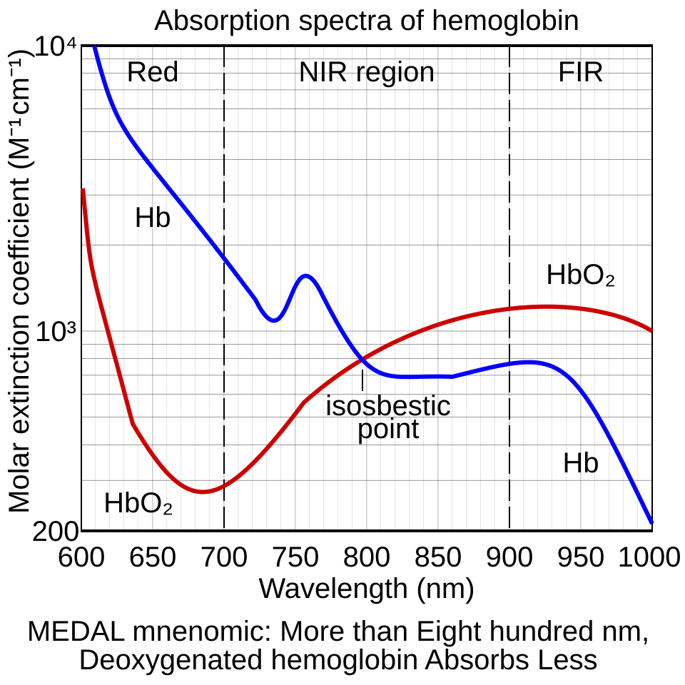
Below 700 nanometres the picture breaks down. There the blood pigment, haemoglobin, soaks up the light so strongly that the beam cannot reach far. Above 900 nanometres water begins to take over, soaking up the light before it can return a clear signal. The window between these two limits is the only band that both reaches the vein and comes back out. A vein finder is built to work inside it. The exact wavelength a device picks sits in this range. It is the wavelength that gives the cleanest contrast on the veins.
Within that window, a few wavelengths work better than the rest. Deoxygenated blood, the kind that fills the veins, has a marked absorption peak near 760 nanometres, where it takes up the light keenly. Some devices work close to that figure. Longer waves of 850 or 940 nanometres are common too, since they travel a little deeper into the arm, where the blood in a vein still absorbs far more light than the bloodless tissue around it. Each device settles on the point that suits the veins it is made to find. The figure is a deliberate choice.
None of this light is felt. Near-infrared light at the levels a vein finder uses carries no warmth the skin can notice. It does no harm to the tissue. It is simply light, a shade the eye cannot register, shone on the surface and read back. It adds no radiation dose, since infrared light is not the ionising kind. A person under a vein finder feels nothing. The arm can rest under it for as long as the search takes.
Why blood shows up dark
The near-infrared absorption of haemoglobin. The blue curve is deoxygenated haemoglobin (Hb), the form carried in venous blood; it has a peak near 760 nm. The red curve is oxygenated haemoglobin (HbO₂). The curves cross at the isosbestic point near 800 nm. A vein finder works across this region, where the blood in a vein absorbs the light and reads darker than the skin around it.
The veins look dark for one reason. The blood inside them drinks up the infrared light. Venous blood carries haemoglobin that has given up its oxygen, the deoxygenated form. That form soaks up near-infrared light far more strongly than the skin, fat and muscle around it. Less light comes back from the line of a vein than from the skin beside it. The device reads that shortfall of returning light as a dark stripe. The stripe traces the vein.
The size of that difference is what makes the method work. The blood in a vein absorbs far more near-infrared light than the bloodless tissue beside it, a contrast a clinical study of a near-infrared vein finder relied on to map veins through the skin. That gap in absorption becomes a gap in brightness on the sensor. A wider gap gives a clearer line. The veins that carry the most blood, the ones most useful for a needle, give the boldest lines of all.
Arteries show up far less, which is why a vein finder draws mostly veins. The arteries run deeper in the limb and carry thick muscular walls, so little of their blood sits within the light’s short reach. The veins a needle wants run just under the skin, holding a broad pool of blood. That pool soaks up the light and casts the dark line. So the boldest marks on the picture are the surface veins, the very ones a nurse uses for a drip or a blood draw.
The picture, then, is a map of where blood pools just under the skin. Wherever a surface vein runs, less light comes back, and the device paints a dark line along it. Everywhere else the picture stays pale. The pattern of dark lines is the vein map a clinician reads before placing a needle. It is built from the way blood treats one narrow band of light.
How an infrared vein finder reads a vein
Property
Figure
Light used
near-infrared, about 740 to 940 nm
Deoxygenated-blood peak
near 760 nm, in the venous blood
Optical window
about 700 to 900 nm, where skin lets light through
Depth read
a few mm, up to about 10 mm in better units
Radiation
none; non-ionising infrared light
Image shown
projected on the skin, or on a screen
Reading the light that bounces back
Catching that faint returning light is the device’s main job. A ring or a grid of small near-infrared sources, usually light-emitting diodes, lines the head of the device and floods the skin evenly. A camera sensor in the middle, the kind that is sensitive to near-infrared, reads the light that bounces back from the arm. Because the sources are spread around the sensor, the skin is lit flatly, with no single bright spot to wash out the picture.
What the sensor catches is faint, so the device works on the raw image before anyone sees it. A small processor lifts the contrast, so the dark veins stand out from the slightly-less-dark tissue. It smooths the speckle that fine tissue scatters into the light. It traces the edges of the vein lines to make them crisp. All of this happens many times a second, fast enough that the picture keeps up with a moving arm. The clean image a clinician sees is the one the processor has built.
This way of working, lighting the skin and reading the reflection, lets a vein finder do its whole job from above the arm. The device never touches the limb. Nothing has to be pressed against the skin, and nothing has to wrap around it. The device hovers a set distance above, shines its light down, and reads what returns. That hands-off design is part of why the picture appears at once and why the same device serves an arm, a hand or a foot.
Drawing the map onto the skin
The boldest kind of vein finder does something striking with the picture: it throws it straight back onto the skin. A device of this kind reads the veins with infrared light, builds the map inside, and then shines that map down onto the arm in visible light, so the dark lines fall exactly over the real veins beneath them. The clinician looks down at the patient’s own skin and sees the veins drawn on it in light.
The trick of it is registration: the projected lines have to land precisely over the veins they stand for. The device measures where each vein sits in its view and aims the projector so the bright line falls on that same spot. Done well, the line of light sits right on the vein, to within a fraction of a millimetre. A clinician can mark the skin along the projected line, or simply follow it with the needle. The map and the arm become the one surface, read together.
Because the map is painted on the skin, the clinician keeps both hands free and both eyes on the patient. There is no screen to glance away to. The vein is shown in place, on the limb, where the needle is going. The light tracks the arm in real time, so a small shift of the limb does not throw the map off. The picture stays locked to the skin it is drawn on.
The projected image can show more than the resting veins. Some devices follow the faint movement of blood and pick out which lines are flowing, helping tell a flowing vein from one that has clotted off. Some let the operator zoom the projected map, or freeze it, or change the colour of the lines for the skin it shows against. The projection is a live drawing the operator can adjust.
This painted-on-skin approach is the one most people picture when they think of a vein finder. It suits a bedside well, where a clinician wants to look at the patient and work with both hands. The cost of it is a device with a projector and the optics to aim it, which makes the unit larger and dearer than a plain viewer. For many wards the hands-free map on the skin earns that cost.
Showing it on a screen
The other kind of vein finder shows the map on a screen. It reads the veins the same way, with near-infrared light, then displays the processed picture on a small monitor, a tablet or a phone. The clinician sees the pattern of veins on the screen, then turns to the arm to place the needle.
A screen device is usually smaller, simpler and cheaper than a projecting one, since it needs no projector and no fine aiming. It can also store and share the image, which helps for teaching or for a record. The trade is that the eyes travel between the screen and the arm, since the map and the needle no longer share one surface. For many uses that is a small price, and the screen type is common in clinics and on mobile carts.
How deep the veins can be
A vein finder reaches only so far into the arm. Knowing that reach is part of using it well. The infrared light fades the deeper it goes. Every millimetre of tissue scatters and absorbs a little more of it. By a few millimetres down, most of the light is gone. So the device reads the veins that lie near the surface, the shallow ones a needle reaches anyway. Deeper veins fade from view, because too little light returns from them.
For a plain reflection device, the useful depth is around three millimetres. That is enough for most of the veins a nurse uses on the back of the hand, the wrist or the forearm, which sit just under the skin. The better projecting systems reach further. Some are reported to show veins as deep as ten millimetres, and the faint motion of blood deeper still. The exact figure depends on the device, the wavelength and the person’s skin.
Depth is also why a heavy arm gives a fainter picture than a slim wrist. Where a thick layer of fat lies over the veins, the light has more tissue to cross before it reaches them and comes back, so the lines read softer. The device still helps there, showing the shallower veins and the general run of the deeper ones, even when it cannot draw them sharply. Knowing the reach keeps an operator from trusting a blank patch as proof that no vein is there.
None of this makes the depth limit a flaw. The veins a needle can actually reach are the shallow ones. Those same surface veins are the ones the light reads best. A drip or a blood draw goes into a vein near the skin, the kind a vein finder draws most clearly. The reach of the light and the reach of the needle line up. The tool shows the clinician what the clinician can use.
Light is all that touches the skin
One feature stands out in daily use: nothing has to touch the patient. The device works from above, by light alone, so there is no probe to clean between patients, no gel to wipe on, no pad to press on a sore arm. A clinician can hold it over a frightened child, a burn, or a fragile elderly hand. Nothing rests on the skin. The reading is instant and bears repeating as often as needed, since a harmless light costs the patient nothing.
What sharpens or blurs the picture
Several things decide how clear the vein map comes out. Skin tone is one. Heavier pigment in the skin absorbs some of the light on the way in and out, so a very dark skin returns a fainter picture. The device leans on its processing to lift the lines there. Body fat is another factor, since a deeper layer over the veins softens them. Temperature changes the picture too. A warm arm has well-filled veins that read boldly, the reason a warm compress is a common trick before a difficult draw.
Hair, sweat and surface marks can each scatter the light and add noise to the picture. Movement blurs it, the same way a moving subject blurs a photograph. The live processing keeps up with a slow shift well enough. Strong room light with its own infrared content, bright sunlight or a heat lamp, can wash the picture out, so the better devices filter for their own wavelength to hold the contrast. None of these stops the device working; each just asks the operator to read the picture with them in mind.
Good technique answers most of these. Warming the arm, dimming a harsh light, holding the device at the distance it is built for, and giving the processing a moment to settle all sharpen the map. The picture a vein finder gives is a real reading of the arm in front of it. A little care in how it is taken pays off in how clearly the veins appear. An operator who knows what helps gets a cleaner picture from the same device.
Where the picture helps
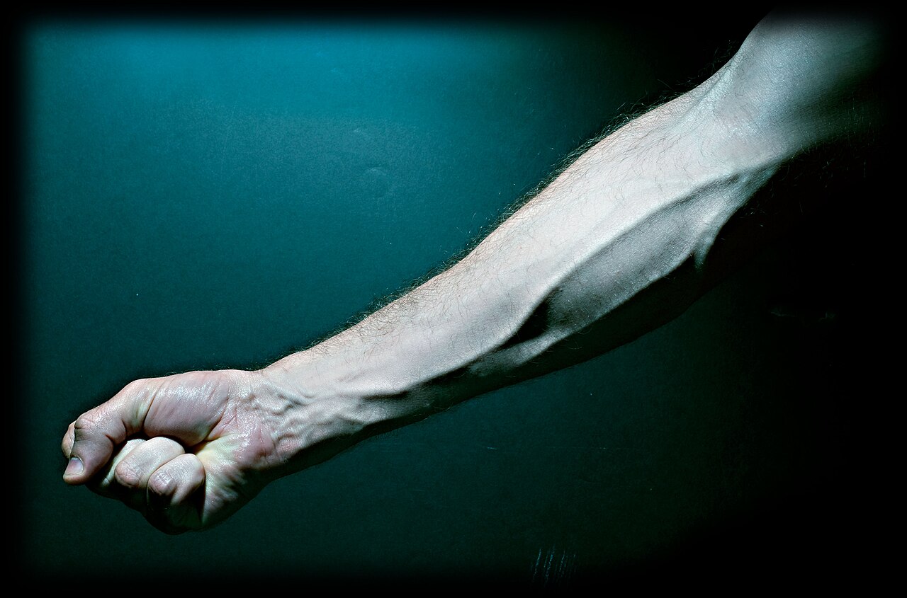
The surface veins of the forearm and hand, the kind a vein finder maps before a drip or a blood draw. These run just under the skin, within the few millimetres of depth the near-infrared light reads best.
Behind all its optics, a vein finder does one plain job: it shows where the veins run before a needle goes in. A clinician about to place a drip or draw blood can see the map of veins on the arm, pick the one that looks fullest and straightest, and judge its path and depth before touching the skin. The guesswork of feeling for a vein that cannot be seen is what the device sets out to remove.
Seeing the vein first changes how the stick is planned. The operator can choose a vein clear of a valve or a fork, follow its line up the arm, and place the needle along its length. A vein that tends to roll can be held and watched while the needle enters. A vein that runs shallow can be taken at a low angle. The map turns a by-feel task into one done in plain sight.
The device does not place the needle, and it does not replace the skill of the person holding it. It shows the vein; the clinician still reads the map, picks the spot, sets the angle and feels the give as the needle enters. A vein finder in skilled hands makes a good operator faster and surer. It also helps a learner see what an experienced finger has come to feel. Either way the hand still does the work.
A vein finder, in the end, is a clever use of one fact: the blood in a vein drinks a particular colour of light the eye cannot see. From that single fact the whole device is built, the near-infrared source, the camera tuned to catch the reflection, the processor that lifts the faint lines, and the projector or screen that hands the map back to the clinician. It reaches the surface veins a needle uses, draws them in real time, and lays the picture where the work is done. A frightened patient, a hard-to-find vein, a busy ward: in each the device does the same quiet thing, turning veins the eye cannot see into a map anyone can read. Light goes in, a map comes back, and the needle goes where the eye can now follow.
Common questions
What kind of light does an infrared vein finder use?
It uses near-infrared light, just past the red end of what the eye can see, with a wavelength of roughly 740 to 940 nanometres. That band passes a few millimetres into the skin before it fades, far enough to reach the surface veins. The light is invisible and harmless, the same kind a television remote sends. Many devices work near 760 nanometres, where the deoxygenated blood in the veins absorbs strongly, or at 850 to 940 nanometres, which reach a little deeper.
Why do the veins show up as dark lines?
Because the blood in the veins absorbs near-infrared light far more strongly than the bloodless skin and fat around it. The haemoglobin that venous blood carries soaks up this band of light, so less of it bounces back from over a vein than from the plain skin beside it. The device reads that shortfall of returning light and draws it as a dark line that traces the vein.
How deep can a vein finder see?
A plain reflection device reads veins about three millimetres down, enough for most of the veins on the back of the hand, the wrist and the forearm. The better projecting systems reach further, with some reported to show veins as deep as about ten millimetres. The reach depends on the device, the wavelength and the person, since a thick layer of fat over the veins softens the picture. The veins a needle actually uses are shallow, which is the depth the light reads best.
Is the infrared light safe for the patient?
Yes. The near-infrared light a vein finder uses runs at low power and does not ionise tissue, nothing like the radiation of an x-ray. It adds no dose to the patient and needs no shielding. The light is shone gently on the surface and read back, and nothing touches the skin. A reading bears repeating as often as a search needs. The eye safety of the infrared source is set by a published standard that these devices are built to meet.
Does a vein finder work on dark skin?
It works on all skin tones, with some change in how clear the picture comes out. Heavier pigment absorbs a little of the light on its way in and out, so a very dark skin can return a fainter image. The devices answer this with stronger processing and a choice of wavelength that holds the contrast. The veins still show; the operator may lean a little more on the device’s adjustments to read them. Warming the arm and dimming harsh light help on any skin.
About the Author
Julien Mercier
Senior R&D Engineer · Medical Ultrasound Transducer Development
Senior R&D Engineer with an M.S. in Applied Physics and over 15 years of experience in medical ultrasound transducer development, specializing in the design verification and performance testing of high-frequency imaging transducers. Currently leading the development and verification of the company’s next-generation high-frequency linear-array transducer, responsible for imaging performance evaluation and reliability analysis in preclinical testing. Brings extensive hands-on experience in piezoelectric element tuning, beamforming parameter optimization, and system-level performance testing.