Transthoracic Echocardiography TTE Handheld Ultrasound For Cardiac POCUS
A clinician with a handheld probe can look straight at a failing heart and read its squeeze, its filling, and the fluid around it, often before a cardiologist could reach the bedside. That focused cardiac scan asks a few large questions in a short, fixed sequence of views, far from the hour-long study of an echo lab. On a wireless handheld, it travels to the resuscitation bay, the ward, and the clinic with no machine to wheel in.
The questions a focused scan answers
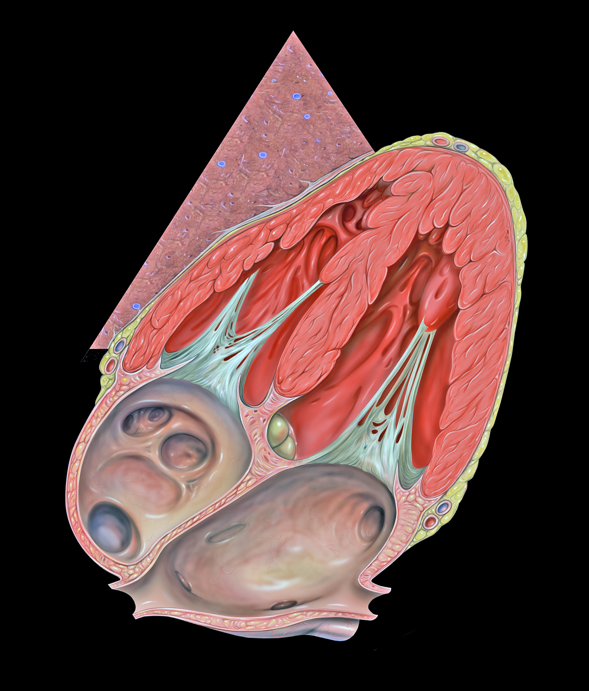
An anatomical illustration of the heart turned as the subcostal window frames it, the small triangle marking the probe’s beam. The four chambers, the valves between them, and the sac wrapping the whole are what each echo view lays out in gray. Illustration: Patrick J. Lynch, CC BY 2.5.
The focused scan does not try to be a full echo. It asks a short list of questions, each one big enough to change what happens to the patient in the next few minutes. Is the heart squeezing well or barely at all. Is there fluid around it, pressing it shut. Is the patient empty and short of blood to pump, or already full to the brim. Is the right side of the heart strained by a clot lodged in the lungs. Each question has an answer the probe can read at a glance. None of these is a subtle finding. A heart that has all but stopped, a sac swollen tight with blood, a vein flat and empty, a right chamber ballooned by a clot: these are signs big enough for a trained eye to catch in a single loop. The focused scan gives up the fine detail of a full study for speed and certainty on the questions that decide whether a patient lives the next hour. That trade is the whole point of it.
Answering them takes a handful of standard views and a few simple measurements, learned as a fixed routine. A clinician runs the same sequence every time, so that under the pressure of a crashing patient nothing is skipped. The whole scan takes a few minutes, repeated whenever the patient changes, something a formal study booked for next week cannot offer. That repeatability is its own kind of power. The same probe that found a poor squeeze at midnight can check an hour later whether the drug is working. The changing picture guides the treatment in a way a single snapshot never could. The scan becomes a monitor at the bedside, not only a test run once and filed. In a busy department that one fact, that the scan costs nothing to repeat, changes how a sick patient is watched through a long night.
The windows on the heart
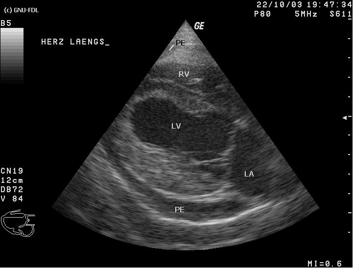
A real parasternal long-axis scan, the view a reader starts on. The right ventricle, left ventricle, and left atrium are labelled (RV, LV, LA); the black rim marked PE is a pericardial effusion, fluid in the sac around the heart. The “GE” and “5 MHz” note the machine and its probe. Image: Kalumet, CC BY-SA 3.0.
The heart hides behind the ribs and the lung, so it is read through a few narrow gaps where it comes close to the chest wall. A focused study works through a fixed set of five windows each time, each a different angle on the same beating organ, and together they leave no chamber unseen. Each window is a knack of its own, a spot on the chest and an angle of the probe the hand learns only with practice. A patient’s body fights the reader at every turn, the lung sliding across the heart, the ribs blocking the beam, the belly full of gas that scatters the sound. Learning to find all five cleanly, on a fat patient and a thin one, on a calm chest and a heaving one, is the real work of the focused scan.
The first window sits a little left of the breastbone. Looking at the heart along its length from there lays out the left ventricle, the two valves on its left side, and the sac around it in one clean picture. It is the view a reader starts on, the one that shows the squeeze and the pericardium at a glance. The picture should lay the left ventricle across the screen, the mitral valve flapping at its inflow, the aortic valve guarding its outflow, and a thin bright line of pericardium wrapping the back wall. A dark stripe behind that line is fluid in the sac. The size of the chamber, the thickness of its walls, and the vigour of the squeeze are all read here before the probe turns.
Turn the probe a quarter circle and the same window gives a short-axis ring through the heart. The left ventricle appears as a circle that should thicken in evenly from every side. A ring flattened on one edge points to a right heart under strain. The slice read low, at the muscles that anchor the valve, grades the squeeze wall by wall. At that level the chamber should close in evenly from every side. A segment that fails to thicken stands out plainly against its neighbours, the bedside sign of a blocked artery feeding that patch of muscle.
Move down to the apex, where the heartbeat taps the chest, and the probe looks up through all four chambers at once. This view sets the right side beside the left and reads their sizes against each other, the clearest window for a strained right ventricle. The valves swing in plain view here too. The squeeze of the whole left chamber reads cleanly from below. The trick is to find the true apex and not cut the chamber short, a common slip that makes a weak heart look better than it is. Done well, the view weighs the two ventricles side by side, watches all four chambers fill and empty in time, and shows each valve opening and closing in real time. It is the workhorse of the focused study, the view a reader returns to for the size of the right heart and the strength of the left.
The last windows come from below the breastbone. With the probe tucked under the ribs and aimed up through the liver, the subcostal view catches the heart and the sac around it, the view that often shows a tamponade best and the one a team falls back on during a resuscitation when the chest is busy with compressions. Flattening the probe and tilting it up under the breastbone brings that picture into focus when the parasternal windows fail. This is the view a code team reaches for, taken from the belly with the chest left clear for compressions, the heart and its sac caught in the pause of a pulse check. A still heart with no squeeze, a sac tight with blood, a fine quiver of fibrillation: the subcostal window reads them in the few seconds a rhythm check allows, which is why it earns its place at the head of a resuscitation. Nowhere else does the focused scan change an outcome so directly, the difference between calling a death and finding the one beat that says keep going.
Reading the squeeze
The first thing a focused scan wants is the strength of the squeeze. A healthy left ventricle clears more than half its blood with each beat. A reader who watches the walls drive inward learns to call the squeeze strong, fair, or barely there by eye alone, a judgment that guides the next drug or drip within seconds. The eye is quicker than any caliper here. A heart whose walls leap in and nearly meet is squeezing well, a call the reader makes in a single beat, long before a measurement could be set up. That first impression sets the early treatment. The numbers follow to confirm it. This is why a focused reader trusts the gestalt, the whole moving picture taken in at once, more than any single frozen frame or figure pulled from it.
The walls also tell their own story one by one. A segment that lags or falls still where its neighbours drive in marks the territory of a blocked artery, the bedside trace of a heart attack in progress. A focused reader learns to watch the whole ring, not only the overall squeeze, since a heart can pump a fair average with one wall already dead. The rule is to read each wall against the ones beside it, since the eye judges a lagging segment best by contrast with its working neighbours. The focused scan is content to flag the patch and send the patient on for the formal study that maps which artery has closed. That single skill, telling a wall that has died from one merely stunned, takes more practice than anything else in the scan.
Where a hard number is needed, putting a figure on the squeeze turns that impression into a percentage, the share of blood the ventricle ejects with each beat. The eyeball estimate and the measured figure usually agree. The trend over hours, the squeeze improving or fading on the same probe, matters more than any single reading. The figure guides how hard to push the drugs that help a weak heart squeeze. Whether that figure rises or falls over the next hour says whether the treatment is working. It means little alone, read always alongside the size of the chamber and the patient in the bed. Tracked this way, the figure becomes a dial the team turns the treatment against, the clearest proof that what they are doing is helping the heart or hurting it.
The same views give the chamber’s size. Measuring the ventricle’s width at the end of filling and again at the end of the squeeze shows how thick the muscle has grown and how far the chamber shrinks with each beat, a second read on a heart that may be failing or merely small. The width at rest separates a wall thickened and stiffened by years of high pressure from a thin one stretched wide, two different hearts, each managed its own way. These are the measurements that turn a glance into a record, the same numbers another reader can check tomorrow and watch for change. A stiff little heart fills poorly even when its squeeze looks strong, and the dimensions catch that failure to fill.
How much blood the heart moves
A strong squeeze does not always move much blood, so a focused scan can measure the flow itself at the mouth of the outflow valve. The distance the blood travels with each beat, read there as a velocity-time integral, gives the length of one stroke, and multiplied by the width of the outflow it gives the volume pushed out per beat. The beauty of the measure is that it needs no guess about the heart’s shape, only the speed of the blood leaving it. A stroke distance that drops when a patient bleeds, or rises when fluid is given, tracks the output beat to beat, the closest a focused scan comes to a flow meter held against the heart. On a handheld it reads in under a minute, the kind of number that once meant a trip to the echo lab and now waits on no one.
From that stroke and the heart rate comes the output, the litres a minute the heart delivers to the body. Whether more fluid will help a struggling patient turns on watching that stroke rise or stay flat when the legs are lifted or a small bolus is given, one of the genuinely useful things a focused scan can settle at the bedside. The scan watches the stroke after a small bolus or a passive raise of the legs. If the stroke jumps, the heart has room for more, the green light to keep the fluid running. If it stays flat, the heart is already on the steep part of its curve, the cue to stop before the lungs flood, and reading that line saves a patient from litres they cannot use.
The measurement asks for care. The reader lines the gate up in the outflow of the left ventricle, a finger’s width below the aortic valve, and traces the bright envelope each beat draws. The width of that outflow, measured once in the long-axis view, holds steady for a given patient, so once it is set the stroke can be followed from the trace alone, beat after beat, as fast as the heart changes. It is a number a handheld takes in under a minute, on a ward where a formal study is hours away.
The volume the heart is working with
The great vein returning to the heart reads the pressure filling it. The width of the inferior vena cava and the way it narrows with each breath tells a reader whether the heart can take more fluid or has reached its limit. A vein that stays fat through every breath points to a heart already full, the cue to hold the fluid and look elsewhere for the problem. The reading has its traps. A patient on a ventilator breathes backwards, so the vein moves the opposite way. A calm, well-trained athlete can carry a fat vein that barely moves on a perfectly healthy heart. The width is a clue read together with the rest of the scan, never a verdict standing on its own. Read in series, the same vein shows whether a treatment is filling the patient or draining them, and it answers, faster than any other view, the question that haunts the care of a shocked patient: give fluid, or hold it.
The right side of the heart
The right ventricle usually sits quietly in the background of a focused scan, thin-walled, smaller than the left chamber beside it. When it swells to match or outsize the left, the strain showing on the right heart becomes the finding that matters, the sign of a large clot lodged in the lungs or a long-standing pressure load on the chamber. A strained right ventricle rounds out from its usual thin crescent and pushes back against the left, stealing room from the chamber beside it. The ratio of the two, read at a glance in the apical view, is the clearest single sign of a heart fighting a pressure in the lungs it cannot overcome. A reader new to the scan learns this ratio early, since it needs no measurement and no calculation, only the eye to see one chamber crowding the other.
A right ventricle larger than the left, a wall that bows across into the left chamber, a base that barely lifts: each points the same way, toward a right heart fighting a pressure it cannot overcome. In a breathless patient, that picture turns a guess into a working diagnosis the moment the probe finds the apex. The classic look is a right chamber as wide as the left or wider, a septum shoved across into a flat-sided D, and a base that barely lifts with each beat. Set against a breathless patient with a swollen leg, that picture points hard at a clot in the lungs and starts the clock on the treatment that breaks it up. A clot caught early is treatable. The look at the right heart is what catches it, which is why a breathless patient earns that view before any other.
Why a handheld carries the cardiac scan
The focused echo began on a cart and now lives wherever the patient is. A probe the size of a thick pen, run from a phone, brings every one of these views and measurements to the bedside, the ambulance, and the clinic far from any echo lab. The same phone stores the clips, runs the measurements on the screen, and sends a loop to a cardiologist for a second read in seconds. A scan that once meant moving a sick patient to a machine now arrives in a coat pocket. For a small hospital with one echo machine and a waiting list, a few handhelds move the urgent scans out of the queue and onto the wards. For a clinic with none, the handheld is the difference between seeing the heart and guessing at it from the outside. The wireless link does more than cut a cable; it lets the clip a junior captures travel across the city to a senior for a read, turning a lone bedside guess into a shared decision in minutes.
What the focused scan leaves out
The focused scan answers big questions fast. It reads the squeeze, the fluid, the volume, and the right heart, and it leaves the subtle valve, the small hole, and the fine clinic numbers to the full study. A normal focused scan rules out the emergencies it was built to catch, no more than that. It will not catch a small hole between the chambers, a growth on a valve, or a narrowing that needs careful Doppler to grade. Those belong to the full study, run by a sonographer with time and a high-end machine. The focused scan knows its own edges and stops cleanly at them. That honesty is part of its strength, since a tool trusted to know its limits is trusted everywhere else. A clinician who knows exactly what a scan can and cannot say carries real authority with it. It is the rare tool that earns trust by naming what it cannot do. A clinician who carries that honesty is believed when the picture is hard to read.
Every one of these views depends on the hand that finds it. A heart read from a poor angle, a chamber cut short, an effusion mistaken for the fat pad beside it: each is a trap for the untrained, and the focused scan rewards practice as much as any skill in medicine. The number is only as good as the picture it came from. A reader who logs a hundred scans next to a formal study learns where their eye is right and where it drifts, and that calibration is what makes a focused number safe to act on. Skill, more than the probe, is what the focused scan asks for. A year of scanning beside a mentor teaches what no manual can.
The focused scan opens a question as often as it closes one. A poor squeeze seen at the bedside still goes to a formal study for its cause. A strained right heart still sends the team looking at the lungs for a clot. The picture points the next step, fast, and the full workup follows behind it. Used as the opening read of a case, the focused scan speeds every step that comes after it, and leaves the final word to the full study behind it.
The heart, read at the bedside
Five windows, a few measurements, and a probe in a pocket: enough to read a failing heart where it lies and act before the cart arrives.
Common questions about handheld cardiac ultrasound
What is a focused cardiac ultrasound?
A short, set sequence of heart views aimed at a few urgent questions: is the heart squeezing, is there fluid around it, is the patient full or empty, is the right heart strained. It is run at the bedside by the treating clinician and answers in minutes, where the full echo-lab study takes an hour and a booking.
What are the five standard cardiac views?
The parasternal long axis and short axis, taken from beside the breastbone; the apical four-chamber, from the point where the beat taps the chest; and the subcostal heart and inferior vena cava views, taken from under the ribs. Together they show every chamber, the valves, the sac around the heart, and the great vein that fills it.
Can a handheld measure ejection fraction?
Yes, by eye and by calculation both. A trained reader estimates the squeeze from the way the walls move. The device measures it from the chamber’s dimensions or the blood it ejects. The handheld figure tracks a patient well from hour to hour. The finest single measurement still belongs to a formal echo lab.
What does the inferior vena cava show on ultrasound?
The volume the heart is working with. Its width, and the way it collapses as the patient breathes, reads the pressure filling the heart. A vein that stays wide through every breath points to a heart that cannot take more fluid. It is a quick read on whether fluid will help or harm.
Does a focused scan replace a full echocardiogram?
No. It answers the urgent questions a sick patient turns on, fast and at the bedside, and it repeats as often as the patient changes. The full echocardiogram grades the valves, measures the fine numbers, and names the cause behind a finding the focused scan only flags. The two do different jobs.
About the Author
Julien Mercier
Senior R&D Engineer · Medical Ultrasound Transducer Development
Senior R&D Engineer with an M.S. in Applied Physics and over 15 years of experience in medical ultrasound transducer development, specializing in the design verification and performance testing of high-frequency imaging transducers. Currently leading the development and verification of the company’s next-generation high-frequency linear-array transducer, responsible for imaging performance evaluation and reliability analysis in preclinical testing. Brings extensive hands-on experience in piezoelectric element tuning, beamforming parameter optimization, and system-level performance testing.