





































Subxiphoid window
Subxiphoid Tilt Technique Comparison Wireless Ultrasound Probes
In the subxiphoid view the probe hardly moves on the skin; the angle does all the work. It reads the heart from below the breastbone, aiming up through the left lobe of the liver, where a degree or two of tilt can turn a clean four-chamber heart into a slab of liver. Each setting of the angle brings up its own structure, and each needs its own steady hold.
Why a couple of degrees change everything in this window
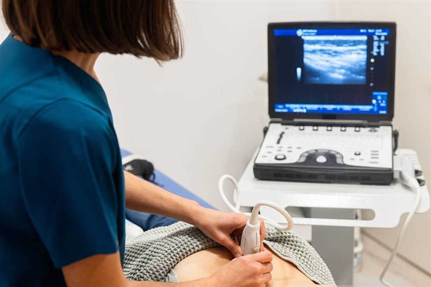
The subxiphoid window works through a long acoustic path. That path is the whole reason the angle rules this view. The probe lies almost flat on the upper belly, just below the xiphoid process, with the beam aimed up toward the left shoulder. The sound crosses the left lobe of the liver first. The liver serves as an acoustic window that carries the beam on to the heart, which sits well behind it. Because the beam travels so far before it reaches the chambers, a small change in tilt at the skin swings the far end of the beam through a wide arc by the time it arrives. The one-degree nudge that barely shifts a parasternal image moves the subxiphoid plane across a whole chamber. The geometry turns a gentle wrist motion into a large change deep in the field. So a skilled operator reads the heart by rocking the tail of the probe in tiny increments, eyes on the screen the whole way. Depth compounds it. The heart sits far from the footprint in this view, often fifteen centimeters or more into an adult, so the probe runs at a low frequency to reach that depth. A low frequency widens the beam and softens the picture, so the tilt has to lay the thin sharp core of the beam right on the four-chamber plane. Miss the angle by a little and the chamber walls dissolve into the speckle at depth. The payoff for getting it right is a view nothing else delivers from a single window. The subxiphoid plane shows all four chambers, the pericardium wrapped around them, and the place where the inferior vena cava meets the right atrium, in one sweep of the wrist. A pericardial effusion often shows against the right heart first in this window, so the angle that brings up the right ventricle free wall is the angle that catches tamponade early. None of that arrives by luck. It arrives by reading the screen as the angle changes a degree at a time. The pace of the search matters at this depth too. The machine builds a deep image more slowly than a shallow one, since each pulse has a long round trip and the frame rate falls as the field lengthens. A wrist that sweeps the whole arc in one quick pass outruns the screen. The plane of interest flashes past between frames and the operator never sees it. The practised move is a rock of two or three degrees, then a beat of stillness for the image to settle, then the next step. The angle search runs at the pace the frame rate allows.
The grip sets up the whole motion. The operator holds the probe overhand, fingers draped over the top of the housing, heel of the hand resting on the belly. That hold keeps the hand clear of the beam and frees the wrist to tilt without sliding the footprint. A fist-style grip fights the small movements this view needs.
The screen guides the angle better than any landmark on the body. The operator sets a deep field first, fifteen or twenty centimeters, so the far heart sits in view at all. The gain then comes up enough to part the dark blood from the grey wall. Only after that does the tilt search begin, since a hunt run at the wrong depth or gain chases a target the machine cannot show. A probe with a presets menu starts closer to right, since the cardiac preset loads the depth and the gain this window expects before the first sweep.
Breathing shifts the angle the way the wrist does. The heart swings under the diaphragm through each cycle, so a plane that reads clean at the end of a breath drifts off at full inspiration. A reader either freezes the clip at the same point each time or holds the breath to stop the drift. The tilt and the breath act as a single control here.
What each tilt setting brings up
A flat probe, pressed gently and aimed at the left shoulder, lands the subcostal four-chamber plane. The right-sided chambers sit nearest the probe, so they appear at the top of the screen, the mirror of the apical view that puts them at the bottom. A reader new to the window has to hold that flip in mind to call the chambers right. A correct plane shows the crux of the heart, where the atrioventricular valves meet the septum, with the septum running away from the probe toward the apex. The right ventricle reads as the chamber hugging the near field under the liver edge. When the crux is missing the plane has cut the heart obliquely. The fix is a small rock of the tail, watched on the screen, until the crux forms.
Tilting the tail of the probe toward the patient’s feet, then rotating it upright, brings the inferior vena cava into a long-axis view as it runs into the right atrium. This is the angle for a volume read, since the vessel diameter and its swing with each breath track the filling pressure. A small rotation peels the IVC away from the aorta lying next to it, so the two are not confused. The angle here is steeper and more sagittal than the one that found the four chambers, which is why the operator treats it as a separate move rather than a tweak. The vessel also collapses with a sniff in a patient who is volume down, so a reader watches the diameter through a quick inspiration for the swing that reports the pressure. The sagittal angle has to stay locked through that breath, since a tilt drifting mid-sniff smears the diameter and ruins the number. A steady angle is what turns the IVC sweep into a measurement a team can act on.
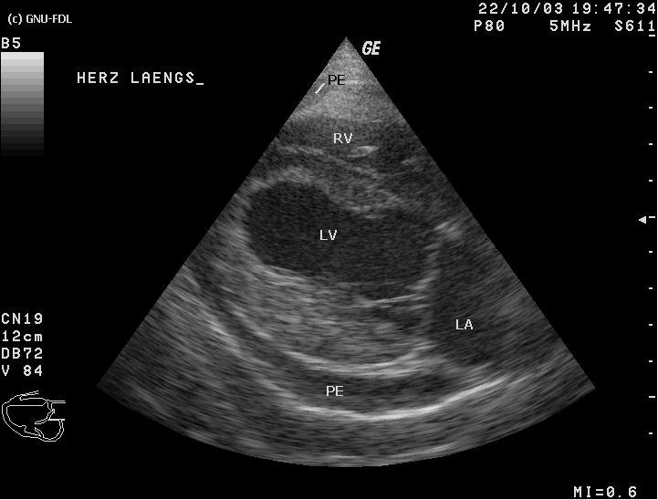
Firmer pressure with a tilt back toward the head opens the pericardial space around the apex and the right heart. An effusion reads as a dark stripe that stays with the heart through the cycle, set apart from the bright pericardium. The angle that lays the right ventricle flat across the screen is the one that catches early fluid, since the free wall gives way there first when pressure climbs. The reading is timed as well as angled. Fluid is judged at its widest in diastole, frozen and measured there. A thin rim of a few millimeters behind the posterior wall can be a normal finding. A stripe that wraps the right ventricle and deepens through the exam is the one that changes the plan. The same tilt that found the fluid holds steady as the clip records.
The breath that rescues the view
A held breath drops the diaphragm and pulls the heart toward the probe. That single move often saves the view.
Where the subxiphoid wins and the angle runs out
The subxiphoid window is the one left open when the views between the ribs are shut. A patient on a ventilator with the lungs pushed full of air closes them. So does a chest in the middle of compressions, or a barrel chest from long-standing COPD. In each of those bodies the soft belly stays open from below. The parasternal and apical planes have their own pages, so the point here is the narrow one, that the subxiphoid steps in from below when the chest wall blocks the rest.
During a resuscitation the view carries a value the others cannot. The probe sits low under the sternum, clear of the hands working compressions, so a rhythm check during the pulse-check pause costs a two-second look. The flat approach keeps the operator off to the side, out of the team’s way at the head and chest. A handheld probe sharpens that further, since there is no cart to wheel in against a crowded bed. The two-second look also leaves a record. The operator saves the clip during the pause, then reads it again between cycles without holding up compressions. The saved clip lets the team review the call once the pulse check ends.
The angle has limits no wrist can fix. A heavy panniculus thickens the path ahead of the liver, so the low-frequency beam loses what edge it had. Bowel gas trapped under the footprint scatters the sound into grey noise. A fresh abdominal incision or a guarding, tender belly rules out the steady pressure this view leans on. A distended belly after a large meal pushes gas up under the ribs, so even a textbook angle reads through a curtain of scatter. Post-operative dressings and drains across the upper abdomen block the footprint outright. The operator reads these bodies before the probe lands, since in some of them the window is gone before the first sweep. That time is better spent on another window.
Tilt technique buys back some of that ground. Sliding the footprint a little to the patient’s right brings more liver under the beam and less bowel. The held breath adds depth. Dropping the frequency one step trades a touch of resolution for the penetration to reach the far heart. Each move is small. Together they often turn a grey screen into a usable one.
A view that still will not resolve after those moves is the signal to change windows rather than grind the angle harder. A wireless probe makes the switch quick, since one footprint and a tap on the phone swaps the preset from cardiac to abdominal with no cart and no second machine to boot up. The operator loses a few seconds when the subxiphoid will not give. The exam moves to the next window with the patient barely touched.
A wrist skill before anything else
The subxiphoid view rewards a slow, deliberate wrist. The footprint lands in a second. The angle takes the minute that follows, rocked in small steps against the live screen until the heart forms.
On a handheld probe such as the C10CW the whole window rides in one hand at the bedside. From the four chambers to the pericardium, everything sits a wrist-tilt apart.
Common questions about the subxiphoid tilt
What is the subxiphoid cardiac window?
It is the view a probe reads from just below the xiphoid process, aimed up through the left lobe of the liver toward the heart. The liver carries the beam to all four chambers and the pericardium in one plane.
Why does a small tilt change the image so much?
The beam travels a long path through the liver before it reaches the heart, so a one-degree change at the skin swings the far end of the beam across a whole chamber. The view rewards rocking the probe tail in two- or three-degree steps.
How do you land the four-chamber plane?
Hold the probe almost flat, aimed at the left shoulder, and rock the tail until the crux of the heart forms, where the valves meet the septum. The right chambers sit nearest the probe, so they read at the top of the screen.
Which angle brings up the inferior vena cava?
Tilt the tail toward the feet, then rotate upright into a sagittal plane to catch the inferior vena cava running into the right atrium. A small rotation separates it from the aorta alongside.
When does the subxiphoid window fail?
A heavy panniculus, trapped bowel gas, a tender or post-operative belly, or a distended abdomen after a meal all scatter or block the beam. In those bodies the window is often gone before the first sweep.
Why does the window matter during resuscitation?
The probe sits low under the sternum, clear of the hands doing compressions, so a rhythm check costs a two-second look in the pulse-check pause, and the saved clip reads again between cycles.

About the Author
Julien Mercier
Senior R&D Engineer · Medical Ultrasound Transducer Development
Senior R&D Engineer with an M.S. in Applied Physics and over 15 years of experience in medical ultrasound transducer development, specializing in the design verification and performance testing of high-frequency imaging transducers. Currently leading the development and verification of the company’s next-generation high-frequency linear-array transducer, responsible for imaging performance evaluation and reliability analysis in preclinical testing. Brings extensive hands-on experience in piezoelectric element tuning, beamforming parameter optimization, and system-level performance testing.