The subcostal view comes at the heart from below, the probe tucked under the breastbone and aimed up through the liver, which serves as a clear acoustic window onto the chambers above it. It is the view that works when the others fail: it needs no rolling of the patient, survives the press of chest compressions, reaching a heart that a barrel chest hides from every parasternal angle. Two readings make it indispensable. It catches fluid in the sac from the side gravity pulls that fluid toward; it brings the great vein returning blood to the heart into clean view, the one window that reads the fullness of the circulation directly. For a crashing or arrested patient flat on a trolley, it is often the only window left.
Probe under the breastbone, laid flat, aimed up at the left shoulder.
That placement, and its order in the wider sweep, sits in the overview of the standard windows. The rest is the subcostal itself: why it survives a resuscitation, how it reads the great vein, and how to tell that vein from the artery beside it.
The window the others lack
The subcostal view earns its place by working in the conditions that defeat the rest. A patient in cardiac arrest cannot be rolled onto their side for an apical window; the hands doing compressions crowd the parasternal spots; the subcostal probe, laid flat on the upper belly, reaches the heart from a corner the resuscitation leaves free. In the brief pause of a pulse check it answers the questions that matter: is the heart moving at all, is there a great clot of blood in the sac stopping it, is there any organised beat beneath the chaos.
A heart standing still on the subcostal screen through a pulse check tells the team their efforts have not yet restarted it; a heart stirring weakly tells them a rhythm may be returning, a reading taken in the few seconds the protocol allows for the pause, the probe lifted again the moment compressions resume.
The liver is what makes the window work. Sound passes cleanly through the dense, fluid-rich liver where it scatters off air-filled lung, so aiming up through the liver from below opens a path to the heart that the lung would otherwise block. The fuller the breath the patient takes, the lower the diaphragm drops and the more heart slides into that hepatic window. A liver shrunken or pushed aside narrows that window; a stomach full of gas right under the probe can blot it out, the price the subcostal view pays for borrowing another organ to see through.
It works when nothing else will.
The inferior vena cava running through the liver into the right atrium. Wide and barely moving (left) reads a full, high-pressure tank; narrow and collapsing with the breath (right) reads a low one. Original illustration.
The vena cava and the tank
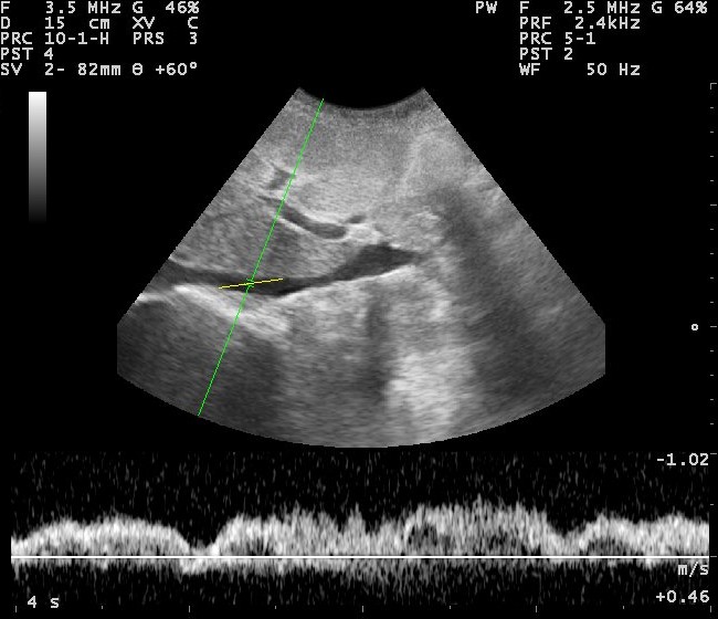
The subcostal window holds the one reading no other view gives cleanly, the state of the great vein that carries blood back to the heart; learning it is the deeper reason to reach under the ribs. Rock the probe from the four-chamber toward the patient’s spine and the inferior vena cava comes into long axis, a broad dark channel running through the liver into the right atrium, the hepatic veins joining it like tributaries near its mouth. Its size and the way it changes with the breath read the pressure filling the right heart, which is to say the fullness of the tank. A spontaneously breathing patient draws the diaphragm down on inspiration, the chest pressure falls, a healthy vena cava sucking narrower with each breath in; ask the patient to sniff sharply and a normal cava collapses by more than half its width, the sign of a low, comfortable filling pressure and a tank with room in it. A vena cava that stands wide and barely stirs with the breath, collapsing little or not at all, reads the opposite: a high filling pressure, a right heart backed up, a tank already full or overfull, the picture behind a failing heart or a strained right side. A narrow cava that collapses to nothing with each breath reads a tank run low, a patient who may take more fluid to fill it. The measure is taken at a fixed spot, a short distance from where the vein meets the atrium or just past the hepatic vein inflow, since the channel narrows and widens along its length. Two traps wait here for the unwary. The vein slides bodily through the imaging plane as the patient breathes, so a single line of M-mode can show a false collapse as the channel swings out of the beam instead of truly narrowing, a pseudo-collapse undone by reading the width in the moving picture or across the short axis instead. And a patient on a ventilator breathes backward to a natural one, the machine’s positive pressure pushing the cava wider on inspiration, so the familiar rules reverse and the reading must be taken with the breathing pattern firmly in mind. Read with those cautions, the vena cava turns a question of volume that once needed a needle in a great vein into a glance from a probe on the belly.
The single number has limits to keep in view. The vena cava reads the pressure filling the right heart, not the need of the whole body for fluid, so a wide cava in one patient and a flat one in another can both mislead taken alone. It is one pointer among several, the trend across a resuscitation often telling more than any single width, the vein watched as fluid runs in to see whether the tank fills or stays flat.
The inferior vena cava on ultrasound, running through the liver toward the right atrium, the channel whose width and collapse read the fullness of the circulation. Nevit Dilmen, Wikimedia Commons, CC BY-SA 3.0.
Wide and still means full; narrow and collapsing means dry.
Telling the vein from the artery
The vena cava has a near neighbour easy to mistake for it, the error flipping a reading on its head. The aorta runs alongside the cava through the upper belly; a clinician who measures the aorta thinking it the cava reads a volume that is not there. A few plain marks tell them apart at a glance.
The cava sits to the patient’s right and runs into the right atrium; follow it up and it joins the heart. It is thin-walled, changes its width with every breath, and gathers the hepatic veins near its mouth. The aorta sits to the left, runs straight down past the heart instead of into it, carries a thick muscular wall that pulses hard with each beat, throwing off branches the cava never has. A channel that pulses and ignores the breath is the artery; a channel that joins the heart and breathes with the patient is the vein. A moment spent following the channel up to where it ends settles the matter past any doubt: the one that empties into a beating chamber is the vein the reading wants, the one that marches on past the heart the artery to leave alone.
Into the atrium, it is the cava; pulsing past, it is the aorta.
The sac, caught from below
The subcostal window is the surest place to find fluid around the heart. A patient lying on their back lets a pericardial collection pool in the space nearest the probe coming from below, so a rim that a parasternal view might skim is laid open here, the dark band sitting between the liver-side wall of the heart and the bright sac around it. A small collection shows here as a thin dark stripe against the moving wall; a large one wraps the heart in a black halo it appears to swing within, the size and the swing together gauging how much the sac holds.
The view reads the squeeze of that fluid as well as its presence. When the collection presses hard enough to stop the heart filling, the thin-walled right chambers buckle inward in the phase between beats, a collapse the subcostal four-chamber catches plainly, the warning that a drain is needed before the heart is choked. The deeper reading of tamponade belongs to the dedicated study of the effusion; the subcostal view is where the focused exam first catches sight of it.
Holding the probe flat
The subcostal view rewards a flat hand more than a firm one. The probe is laid almost against the skin of the upper belly, the beam aimed up under the ribs toward the left shoulder at a shallow angle instead of jabbed down into the abdomen, the operator’s hand often draped over the top of the probe to flatten that angle. A jab straight down finds only liver and bowel; a long, low aim finds the heart. A lower-frequency probe, the kind built for the belly, reaches the heart from this depth better than the small high-frequency one laid on the lung, since the heart sits far from the skin when seen from below.
The breath is the other lever. Asking the patient to take a deep breath and hold it drops the diaphragm and slides the heart down into reach, turning a hidden heart into a clear one for the seconds the breath is held. The depth is set deep enough to lay the far wall of the heart and the sac behind it in view, since a sac missed off the bottom of the image is a sac not read. The view also takes a hand reaching across the patient from the right, the operator standing clear while another works the airway, a reach that keeps the subcostal probe out of the chest where the team is busy.
Lay it flat; aim it long.
Where it fails
The window has its own enemies. Gas in the bowel beneath the probe scatters the beam and swallows the heart, a belly full of air defeating the cleanest technique. A chest blown wide by old lung disease hyperinflates and pushes the heart up and away from the subcostal reach, the same patients whose parasternal windows are also poor. A belly tender from recent surgery or injury will not take the pressure the view sometimes needs. A recent large meal swells the stomach into the path; the same window read an hour later, or after a breath drops the heart lower, often opens where it was closed.
The reading is weighed with the patient and the rest of the scan. A vena cava that looks full is read against the heart that fills it and the lungs above; a sac seen from below is confirmed from a second angle when time allows. The subcostal view is a powerful first look and a poor last word, strongest as one window among the five rather than a verdict standing alone.
The view of last resort and first reach
The subcostal window is the one a clinician keeps for the moments the others are lost. When a patient cannot roll, when hands are busy on the chest, when a barrel chest closes every parasternal door, the probe slides under the ribs and finds the heart from below, the view of last resort in a crashing patient. It is also the first reach for a plain question of volume, the great vein read in seconds where the answer once waited on a line threaded into the neck.
The deeper value is reach without disturbance. The subcostal view asks nothing of the patient but a breath, takes the heart and the great vein from a single flat placement, giving back the squeeze, the sac and the fullness of the circulation while the resuscitation carries on around it. A window that began as the awkward last option has become, for the sickest patients of all, the one a trained hand reaches for first. The view that asks the least of a patient gives back the richest reading when the patient can give the least, the flat placement and a single held breath all it needs to read a heart, a sac and a vein at the bedside of someone too sick to be moved at all, the answers arriving in the seconds a resuscitation can spare, no roll and no wait asked of a patient who has neither to give.
About the Author
Julien Mercier
Senior R&D Engineer · Medical Ultrasound Transducer Development
Senior R&D Engineer with an M.S. in Applied Physics and over 15 years of experience in medical ultrasound transducer development, specializing in the design verification and performance testing of high-frequency imaging transducers. Currently leading the development and verification of the company’s next-generation high-frequency linear-array transducer, responsible for imaging performance evaluation and reliability analysis in preclinical testing. Brings extensive hands-on experience in piezoelectric element tuning, beamforming parameter optimization, and system-level performance testing.