Parasternal Short Axis PSAX View Technique Handheld Ultrasound
The short axis cuts the heart across instead of along, slicing it into a round cross-section the way one cuts a loaf. Where the long axis lays the chambers out in a line, the short axis stacks them into rings; that single change of angle opens readings no lengthwise slice can offer: the whole circumference of the left ventricle seen at once, the shape of the wall it shares with the right, the even or uneven squeeze of muscle all the way round a circle. It is the view that reads the right heart’s burden on the left, the one that lays every wall of the pump side by side for a fair comparison.
A quarter-turn from the long axis, marker to the left shoulder.
That turn, and where it falls in the wider sweep, is held in the overview of the standard windows. The rest is the short axis itself: the rings it stacks, the shapes it reads, and the one sign that makes it the view a strained right heart cannot hide from.
A stack of rings, base to apex
The short axis is not one picture but a series, each found by tilting the beam a little further down the heart. Aim it high, toward the base; the aortic valve appears in cross-section, ringed by the right-sided structures. Tilt the nose of the probe down toward the apex and the mitral valve comes into the ring, then the body of the left ventricle at the level of its papillary muscles, then the narrowing apex itself. Each tilt slides the slice along the heart, a stack of cross-sections from the valves at the top to the tip at the bottom. The base sits highest and nearest the left shoulder; the apex lies lowest and points toward the left hip, so the tilt that walks the slice from one to the other follows the heart’s own lean across the chest. A clinician who pictures that lean fans the right way the first time instead of searching blindly up and down, the small apex ring drifting out of plane with each beat while the papillary ring just above it carries the bulk of the exam’s work.
The skill is knowing which ring answers which question. The papillary level reads the pump and the walls; the valve levels read the valves; the base reads the right-sided chambers wrapped around the aorta. Fanning through the stack in order, top to bottom, is the short axis exam; skipping a level is skipping a question. The order is always the same, base to apex, so nothing is forgotten in a hurry.
Tilt down the heart; the rings change one by one.
At the papillary level the left ventricle is normally a round ring (left). A right ventricle under heavy load flattens the shared septum and pushes it across, turning the O into a D (right). Original illustration.
The D-sign, when the septum gives way
The short axis holds one finding no other quick view shows as plainly; learning to read it is the deeper reason to turn the probe across. A healthy left ventricle, cut across at the papillary level, is a near-perfect circle, a round ring of muscle with the right ventricle draped over one side as a thin crescent. The wall between them, the interventricular septum, curves gently away from the left ventricle, bowing toward the lower-pressure right side as it should. That circle is the baseline; its loss is the sign. When the right ventricle comes under a sudden, heavy load, its pressure climbs until it matches or passes the pressure on the left, and the septum, caught between the two, stops bowing toward the right and flattens, then buckles the other way, pushing into the left ventricle. The round O of the cross-section becomes a D, the straight back of the letter being the flattened septum shoved across by the overloaded right heart. That is the D-sign, naming a right ventricle in trouble at a glance. The timing of the flattening tells one kind of trouble from another, a reading to take with care. A septum that flattens hardest at the peak of the squeeze, in systole, points to a right ventricle fighting high pressure, the picture of a large clot in the lungs or a long-standing pulmonary hypertension. A septum that flattens in the relaxed filling phase, in diastole, rounding out again as the heart squeezes, points instead to a right ventricle merely overfilled with volume, the load of a leaking valve or a hole between the chambers. Pressure flattens the septum when the heart contracts; volume flattens it when the heart fills. The short axis lays that distinction open in a single cross-section, a round ring or a D, read in the time it takes to recognise a letter, the one window where the strained right heart writes its burden plainly on the shape of the left.
A round ring is calm. A D is a warning.
The doughnut and the walls
At the papillary level the left ventricle is a thick ring of muscle, the shape that earns the view its nickname of the doughnut. A healthy ring thickens evenly all the way round with each beat, every arc of muscle closing inward together. The eye reads the squeeze here as it does on the long axis, judging the pump strong or weak from the change in the cavity, the short axis adding the round, even, all-at-once view the lengthwise slice cannot give. Thickening counts for more than inward motion here: a wall dragged inward by its lively neighbours can fake a squeeze it never makes, so the eye watches each arc grow fatter through the beat before it trusts that arc is working.
The ring carries more than a global reading. Each arc of it belongs to the territory of one coronary artery: the front to one vessel, the side wall to another, the back and the septum to a third. A segment that lags, failing to thicken while its neighbours close in, marks a regional weakness, the print of an old or fresh injury to that one artery’s share of the muscle. The short axis is the one quick window that lays all the walls in a single circle, so a lazy segment shows itself against the lively ring around it rather than hiding in a slice that never included it. The arc that lags points back toward the vessel that feeds it, a clue the short axis hands over almost for free: a dead front wall speaks of one artery, a dead inferior wall of another.
The doughnut reads the whole wall at once.
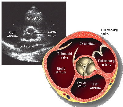
The short axis at the aortic valve, the three cusps meeting in the Mercedes sign, ringed by the right-sided structures. Patrick J. Lynch and C. Carl Jaffe, Wikimedia Commons, CC BY 2.5.
The aortic valve as a three-pointed star
Tilt up to the base and the aortic valve sits in the centre of the ring, its three cusps closing to a shape clinicians call the Mercedes sign, a three-pointed star where the leaflets meet. A valve that opens wide and closes to that clean star is normal; a valve that shuts on two cusps with a single line, in place of three, hints at the bicuspid valve a younger patient with a murmur may carry.
The same slice catches the right-sided structures wrapped around the aorta. The right ventricle’s outflow drapes over the top, the pulmonary valve sits at its end, the tricuspid valve opens to the side; the right atrium completes the ring. A single tilt to the base, then, reads the aortic valve and takes in the right-sided valves around it, a quick survey no other parasternal cut frames so completely. The base earns a stop in a breathless patient even when the left heart looks well, since a swollen right atrium or a sluggish right outflow can be the first hint of the strain the D-sign confirms a level below.
Three points, a healthy valve; two, suspect a bicuspid.
The fish-mouth mitral
Tilt a little down from the base and the mitral valve fills the ring, its two leaflets opening and closing in a shape likened to a fish’s mouth. A healthy mitral opens to a wide oval; a valve stiffened by old rheumatic disease opens to a narrow slit, its edges thickened and tethered, the fish-mouth pinched nearly shut.
The view can gauge the narrowing, not just name it.
Tracing the open mouth of a stenosed mitral valve at this level gives a rough measure of how tight the narrowing has grown, a planimetry the short axis offers that the lengthwise slice cannot. A handheld scan reads it coarsely, enough to flag a tight valve and send it on; the fine grading waits for the full study, as it does for every valve the focused exam only points at.
Keeping the ring round
The short axis lives or dies by a true cross-section; a true cross-section is a circle. The beam must cut the left ventricle square across its long axis; tilt it off square and the round ring stretches into an oval, an ellipse that can fake a flattened septum where none sits or hide a real one in a distorted shape. The fix is to set up from a clean long axis first, then rotate exactly a quarter-turn, keeping the two papillary muscles balanced at the sides of the ring as the mark that the cut is square. Centring the ventricle in the middle of the screen keeps the ring whole as the heart swings with each beat, since a chamber drifting to the edge slips out of the slice at the moment a reading is taken.
The level matters as much as the angle. Reading the pump at the valve level instead of the papillary level misjudges the squeeze, since the ring changes shape and meaning as it climbs and falls through the heart. A disciplined exam fixes the papillary muscles in view before judging function, fans deliberately through the stack, level by level, in place of drifting between them; it rounds the ring before trusting its shape.
An oval is an angle error until proven a true D.
Where it misleads
The commonest trap is the false D, an oblique cut that elongates the ring and mimics the septal flattening of a strained right heart. A clinician who calls a D without first rounding the ring can invent a right-sided emergency out of a tilted probe. The opposite error hides as easily: a real D read as an innocent oval when the operator assumes a poor angle rather than checking it.
Load and level lay further traps. An eyeball estimate of the squeeze swings with how full the heart is, a tank run dry faking a vigorous pump; a reading taken at the wrong ring misreads a normal heart. Each is undone by the same care that serves the whole view: round the ring, fix the level, and read the shape and the squeeze against the other windows before the verdict is set.
Why the short axis pairs with the long
The two parasternal cuts are made to be read together, one the answer to the other’s blind spot. The long axis lays the chambers in a line and reads the valves side-on; the short axis cuts across and reads the circumference, the septum’s shape and every wall at once. A finding hinted at lengthwise is confirmed across; the round ring or the telltale D adds a dimension the single long slice can never hold. Run one without the other and the parasternal study is half done.
The deeper value is the right heart made visible. A focused exam that watched only the left ventricle would miss the strained, swelling right side until it was far advanced; the short axis, with its septum and its D, puts the right heart’s burden in plain sight from the same parasternal spot, a turn of the wrist away from the long axis, among the strongest turns a handheld probe is ever asked to make. A breathless chest read from the long axis alone leaves the right heart half in shadow; the quarter-turn brings it into the light, the round ring or the leaning D telling in one glance what the lengthwise cut could only guess at.
About the Author
Julien Mercier
Senior R&D Engineer · Medical Ultrasound Transducer Development
Senior R&D Engineer with an M.S. in Applied Physics and over 15 years of experience in medical ultrasound transducer development, specializing in the design verification and performance testing of high-frequency imaging transducers. Currently leading the development and verification of the company’s next-generation high-frequency linear-array transducer, responsible for imaging performance evaluation and reliability analysis in preclinical testing. Brings extensive hands-on experience in piezoelectric element tuning, beamforming parameter optimization, and system-level performance testing.