18 to 24 MHz Ultra High Frequency Aesthetic Medicine Ultrasound
An 18 to 24 MHz probe is the ultra-high-frequency ultrasound an aesthetic clinic uses to see the skin and the vessels just beneath it. Its picture stops at about a centimeter down, the shallow layer where injectors do their work. That overlap, between what the probe reaches and where the work happens, is why it belongs in the clinic.
What the frequency buys
The number on the probe sets everything that follows. At 18 to 24 MHz the sound draws fine detail and fades fast in tissue, so the beam reaches only about a centimeter under the skin. Across that centimeter the resolution runs near 80 microns. Axial resolution is the one that matters here. It is the smallest gap the probe can still split into two. At 80 microns it tells the top of a filler pocket from its bottom. It shows the wall of a small artery as its own bright line. Lateral detail, across the beam, is coarser, so an injector sweeps a structure in two directions before trusting what the screen shows. That resolution separates the epidermis, the dermis and the fat beneath into distinct layers, with hair follicles and glands beginning to appear inside them. The same probe carries color Doppler. Color marks the larger vessels at a glance. Spectral Doppler reads the speed and direction of flow in a single one. Together they turn an invisible artery into a labeled line on the screen. Freeze the image and the depth of a vessel or a filler pocket reads straight off the scale at the side. An aesthetic clinic leans on those two abilities, reading the layers and seeing the flow, for the better part of its work. A filler deposit, a thread, and the facial arteries an injector must avoid all sit within that first centimeter. The needle shows as a bright point moving through the tissue in real time. The cost of the high frequency is depth. Anything deeper than a centimeter or so is gone, which is why the probe is made for the face and the skin. Inside its shallow range it sees more finely than any general probe a clinic owns.
That shallow window is where the work lives.
The probe is a flat linear array. Its picture updates in real time. An injector watches the needle move and the filler spread while it happens. The device is handheld and runs from a phone or a tablet, so the scan happens in the treatment room. That makes it practical to look before, during and after a procedure in the same visit.
Before a probe like this, an injector worked from anatomy and feel. A good one knew where the arteries usually run and stayed in the safer planes. That knowledge still matters. What the eye could not do was show the vessel in front of the needle in this particular face on this particular day. The probe adds that last piece, the one a textbook leaves out: the position, here and now.
One probe covers the whole range of this work. The same head that maps a vessel before filler also localizes a muscle for botox, reads the plane a thread sits in, measures the thickness of the skin and the fat, and checks how deep a lump reaches. The work changes from one use to the next. The probe and its shallow window stay the same. A clinic buys a single tool and finds a use for it in nearly every treatment it runs, which is a large part of why the probe spreads through a practice once it arrives.
Mapping the vessels before an injection
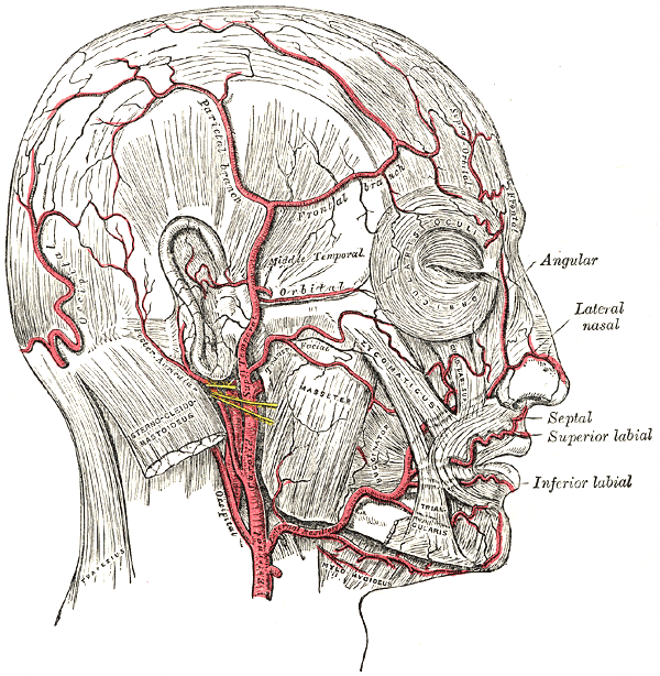
The facial arteries an injector maps before a needle: the angular and lateral nasal at the nose, the labial vessels at the lips, the facial artery running up the jaw. This is an anatomical engraving (Gray’s Anatomy), not an ultrasound image; the red vessels and the labels are part of the original.
The use that injectors talk about more than any other is finding the facial arteries before a filler needle goes near them. Filler pushed into an artery can block it. The skin that artery fed can die. Filler that travels backward toward the eye can take the sight with it. The danger is the artery. Filler forced into one travels where the blood would have gone, downstream into the skin or back toward the eye. These events are rare. They are also severe enough that avoiding them shapes how careful injectors work. The arteries do not run in the same place in every face, so a textbook map is only a starting point. An injector who has seen one occlusion rarely goes back to injecting these areas blind. The map is cheap insurance against the rare event that ends a career.
Color Doppler turns the search into something direct. The probe runs over the planned site. The arteries show up as lines of flow on the screen. The injector reads the actual path in this patient before choosing where the needle goes. A second sweep checks the depth the filler will sit at. Done well, the scan that maps the facial vessels before a filler injection shows the exact path the needle must stay clear of.
Some sites carry more risk than others. The glabella holds the supratrochlear and supraorbital arteries, the nose the dorsal nasal and angular, the mouth the facial and labial vessels. Several of these connect back to the artery behind the eye, which is the route by which a misplaced filler can reach the retina. Knowing which arteries run through each facial danger zone is the larger part of the safety in these areas. The probe is how an injector checks them in this particular face. A textbook shows only the average one.
The nose deserves its own mention. It carries a dense supply from the dorsal nasal and angular arteries. That supply varies from one nose to the next. It is also a frequent site for both filler and surgery. A scan before a nose procedure shows where those vessels run in this patient, which is the point of vascular ultrasound before a rhinoplasty. The same caution applies whether a needle or a blade follows.
Mapping shades into guiding. One described technique uses an 18 MHz probe in three steps: map the arteries, inject under live ultrasound, then confirm the blood is still flowing. A retrospective report followed this in around 480 patients as a way to lower vascular events in high-risk areas. The map and the live needle together give more control than landmarks alone.
When a vessel is blocked
Even with care, a vessel sometimes blocks. The signs come quickly: blanching, then a dusky color, then pain out of proportion to the procedure. Speed matters from the first minute, because the skin downstream is losing its supply. An occlusion near the eye can threaten sight within hours. Ultrasound shortens the time from suspicion to action, since it shows whether filler is sitting in or against a vessel. Color marks the trapped filler against the still tissue around it, so the clinician acts on a picture, not a hunch. The same scan tells an occlusion from a bruise or simple swelling, which look alike from the surface and call for different care.
The fix for a hyaluronic acid filler is the enzyme that dissolves it. Ultrasound steers that enzyme straight into the deposit, then watches for flow to return, which is the work of locating a filler embolism in an emergency. Seeing the deposit beats flooding the area blindly and hoping.
The speed of the response decides the outcome. A clinic ready for this keeps the dissolving enzyme on hand and a plan rehearsed before the day it is needed. The scan supports that plan. It does not replace the drug or the urgency. Finding the deposit fast is the part the probe adds.
The other injections: botox and threads
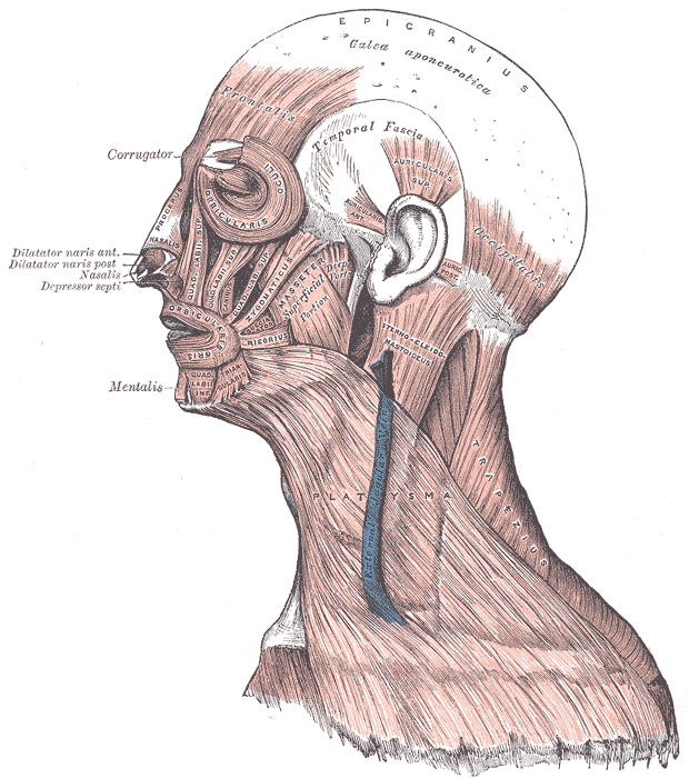
The facial muscles botulinum toxin targets and the planes a thread is anchored in: the masseter at the jaw, the corrugator and orbicularis around the eye, the platysma down the neck. An anatomical engraving (Gray’s Anatomy), not an ultrasound image; the labels are part of the original.
Filler is not the only thing the probe guides. Botulinum toxin works only if it reaches the right muscle. Some muscles are hard to hit by feel. A dose that drifts into the wrong one weakens a muscle the patient needs. Ultrasound shows the target muscle directly, so the dose lands where it should and stays off the nerve, gland or neighboring muscle it should not touch. The toxin spreads a short way from where it lands, so the depth of the placement decides which fibers it reaches and which it spares. A dose a few millimeters off can weaken the wrong muscle.
The masseter is the clear example. Treating it for a wide jaw or for clenching means placing the toxin in a muscle that varies in size from face to face. Watching the needle enter it is steadier than estimating from the surface. That reliability is the case for localizing muscle for a botox injection with ultrasound. Reviews report better precision than the blind, landmark-based method.
Thread lifts raise their own question of depth. A barbed thread holds only if it sits in the right plane, deep enough to grip and clear of the muscle. Placed too deep it can catch a nerve or the parotid gland. Ultrasound reads the layers and the SMAS, the sheet the thread anchors to, so the thread goes where it holds.
After the procedure the same probe finds a thread that migrated or bunched. Doppler flags a spot where a thread presses on flow. Checking placement and complications is the substance of assessing a thread lift with high-frequency ultrasound. Reported adverse events under ultrasound guidance stay low, below roughly one in twenty.
Reading the skin and the fat
Past injections, the probe reads the skin as a structure. The epidermis shows as a bright top line, the dermis as a band below it, and the fat as a darker layer beneath that. The dermis is thin, a millimeter or two over much of the face and thinner on the eyelid, so reading it at all takes the resolution this probe gives. Each has a thickness that shifts with age, with sun damage and with disease, which is what analysing the skin layers with ultrasound sets out to measure. The reading is quick and repeats the same way each visit, so it fits a routine appointment without adding much time. A number taken today sits beside one taken months ago, which turns a vague impression of thinning skin into a measured change.
Below the dermis the fat has a thickness of its own. Measuring it matters before body contouring, before some filler placements, and when a treatment claims to reduce it. The probe puts a number on what the eye and the pinch can only estimate, which is the point of assessing the subcutaneous fat layer. A baseline taken before a treatment and a second reading after it turn a claim of fat reduction into two numbers a patient can compare. The pinch and the eye cannot do that.
Because the measurement repeats, the probe also tracks change. A dermis thinning with age, a scar maturing, a filler slowly absorbing: each can be measured on one visit and compared on the next. That turns a vague sense of progress into a figure on the chart. A clinic that records the numbers can show a patient the change treatment by treatment and can spot the treatments that do little.
The same measuring power follows what a treatment does over time. After an energy device tightens the dermis, or a course of microneedling thickens it, the probe shows the change as a number on the next visit. A claim of a firmer dermis becomes a figure on the chart, set beside the last one.
Which use comes first
A clinic rarely adopts every use at once. The one that drives purchases is the vascular map before filler, because it guards against the complication that ends careers. A practice that injects the nose, the glabella or the nasolabial fold has the strongest reason to start there. The botox and thread uses follow as the clinic grows comfortable with the probe in hand.
The skin and fat measurements tend to come later, once the probe is a habit. They turn a vague before-and-after into a number a clinic can show. The lesion check sits apart, a safety net for the lump that turns out not to be cosmetic. Each use rests on the same one fact: the probe shows the layer the work happens in.
The order is not fixed. A clinic that does more threads than filler starts with the thread plane. One that screens lesions for a dermatology arm starts there. The probe earns its place at whatever the clinic does day to day, then spreads to the rest.
Cost rarely decides against the probe. A handheld unit runs from a phone or a tablet the clinic already owns and costs a fraction of a cart-based machine. It needs no dedicated room. The barrier to adoption is habit, not money. A clinic that builds the scan into its routine draws on it every day. One that buys it and leaves it in a drawer gains little for the spend.
Screening a skin lesion
Sometimes the lump in the skin is not cosmetic. A high-frequency probe estimates how deep a growth reaches. A melanoma’s depth tracks closely with the Breslow thickness that guides surgery, with reported correlations above 0.9. The probe does not diagnose the cancer; a biopsy does that. What it adds, in screening a skin cancer with high-frequency ultrasound, is a depth before the knife, more reliable the thicker the lesion.
Starting with the probe
A clinician new to the probe faces a short learning curve. The hand has to learn to hold the probe still, to read a gray picture, and to line the beam up with a needle. Knowing the few things that trip people up first is the whole point of what to know before using ultrasound the first time. Much of it comes with a few dozen scans. The clinic that makes the scan a fixed step for its high-risk injections draws far more from the probe than one that reaches for it only when a case already looks hard. The dangerous case is so often the one that seemed routine.
For the full picture of where the probe fits across a practice, what an 18 to 24 MHz probe does for an aesthetic clinic walks the range from the first consultation to the management of a complication. The short version is plain. The probe shows the layer the work happens in. That single fact sits behind every use a clinic finds for it.
What it does not do
The probe is shallow on purpose. That sets its limits. It does not reach much past a centimeter, so deeper structures need another tool. It does not diagnose what a biopsy diagnoses. It does not replace skill; a clear picture still has to be read by someone who knows what they are looking at.
Common questions about aesthetic ultrasound
What frequency probe is used for aesthetic ultrasound?
An ultra-high-frequency probe, in the 18 to 24 MHz range. It reaches about a centimeter under the skin and resolves detail near 80 microns. That is fine enough to separate the skin layers and to show the small facial vessels an injector needs to avoid.
Can ultrasound prevent filler complications like blindness?
It lowers the risk. Removing it entirely is beyond any tool. Mapping the facial arteries before an injection shows where not to place the needle. Guiding the injection live keeps the filler out of a vessel. If an occlusion happens anyway, ultrasound finds the blocked spot and steers the dissolving enzyme into it. The events are rare to begin with. These steps make them rarer.
Is ultrasound used before the filler injection or during it?
Both. Before, it maps where the arteries run in this face. During, it shows the needle and the filler spreading in real time. Afterward, it can find a deposit that ended up in the wrong place. The same probe covers all three points, changing only where the clinician looks.
Does ultrasound help with botox?
Yes, by showing the muscle the toxin is meant for. Some muscles, like the masseter, vary in size and sit under others, so hitting them by feel is uncertain. Watching the needle enter the target muscle places the dose where it works and keeps it off the nerves and glands nearby. Studies report better precision than blind, landmark-based injection.
Can the probe identify what filler is in the face?
Not exactly. Hyaluronic acid reads as a dark pocket that looks like fluid, which the probe locates and measures well. The probe shows where a filler sits and how much is there. Naming the brand is beyond it. Location alone is enough to guide the enzyme that dissolves a hyaluronic acid filler.
Does ultrasound replace a biopsy for a suspicious mole?
No. It estimates how deep a lesion reaches. In a melanoma that depth tracks with the Breslow thickness that guides surgery. That measurement helps plan an excision before it happens. The diagnosis still comes from a biopsy and the pathologist who reads it.
How is this different from a pregnancy or abdominal scan?
It uses a much higher frequency. The probe for pregnancy or the abdomen reaches deep, twenty centimeters or so, at a resolution near a millimeter. The aesthetic probe trades that reach away. It covers the first centimeter at a resolution near 80 microns, which the deep probe cannot approach. The work decides which probe to pick up. Facial work calls for the high-frequency one.
About the Author
Julien Mercier
Senior R&D Engineer · Medical Ultrasound Transducer Development
Senior R&D Engineer with an M.S. in Applied Physics and over 15 years of experience in medical ultrasound transducer development, specializing in the design verification and performance testing of high-frequency imaging transducers. Currently leading the development and verification of the company’s next-generation high-frequency linear-array transducer, responsible for imaging performance evaluation and reliability analysis in preclinical testing. Brings extensive hands-on experience in piezoelectric element tuning, beamforming parameter optimization, and system-level performance testing.