A preoperative vascular ultrasound for rhinoplasty is a scan of the nose’s blood supply taken before the surgeon makes an incision. A handheld high-frequency probe maps the arteries that feed the skin of the nose and finds any filler left from an earlier treatment. The skin of the nose lives on a fine network of vessels. A rhinoplasty lifts that skin off the framework beneath it. The scan shows what supplies the skin, in time to change the plan before the cut.
The blood supply a rhinoplasty depends on
The skin of the nose is fed from a few directions at once. The lateral nasal artery runs up each side from the facial artery and reaches the tip. The dorsal nasal artery comes down from above, off the ophthalmic artery behind the eye. Small columellar branches climb the central strut from the lip. These vessels meet in a network just under the skin, the subdermal plexus. That plexus is the layer that keeps the skin of the nose alive. The dorsal nasal artery carries a second meaning. It comes off the ophthalmic artery, which runs behind the eye. That link is the path by which a filler embolism can reach the back of the eye. The same connection means the dorsal nasal feeds part of the upper nose from above. A surgeon working high on the bridge stays aware of a vessel that ties the nose to the orbit.
A rhinoplasty works under that network. The surgeon lifts the skin off the bone and the cartilage to reshape the framework underneath. A clean dissection stays deep to the plexus, in a plane that leaves the feeding network on the underside of the skin. The skin then keeps its supply through the lift. The whole craft of the soft-tissue part of the operation is to raise the cover and keep its blood supply intact under it.
The supply has little to spare in some noses. Thick sebaceous skin sits on a richer network and tolerates more. Thin skin over a narrow tip runs closer to the edge of what the plexus can keep alive. A nose operated on before has given up some of its supply to scar. On each of these the surgeon wants the blood supply known before the skin is lifted. The alternative is learning it on the table, once the skin over the tip has gone dusky and the operation has become a rescue.
A rebuilt framework still needs its own skin to cover it. That skin has no replacement in kind.
The plane that keeps the skin alive
The safety of the skin comes down to the plane the surgeon lifts in. A clean rhinoplasty lifts in the plane deep to the SMAS, down against the framework. That plane leaves the subdermal plexus on the underside of the flap, so the skin keeps its supply through the lift. A dissection that drifts up toward the skin, above the SMAS, runs into that plexus and into the supply it carries. The deeper plane is the safe one for that reason. The flap carries its own blood supply up with it.
The scan reads the layers that define the plane. It shows the skin, the SMAS beneath it, and the framework under that. It measures the thickness of each one. A surgeon planning the lift sees how much SMAS there is to stay deep to, and how thin the cover runs over the tip. The thickness numbers steer the choice point by point. Over thick skin the plane has room to spare. Over the thin skin of the tip the surgeon stays exactly in the safe layer, since there is no margin to give back. The plane is easier to hold in a nose the surgeon has already seen in cross-section.
Filler or scar in that plane makes a clean lift harder. Filler can sit in the same layer the surgeon wants to stay deep to. Scar can glue the plexus down to the framework, so the clean plane is gone where it formed. The scan shows both before the lift, where they sit and how they cross the intended plane. The surgeon adjusts the dissection to what the picture holds.
Why the scan comes before this operation
The complication this scan guards against is the loss of the skin. A rhinoplasty that strips too much of the supply, or that operates on a nose already short of it, can leave the skin to break down. The tip is the part farthest out, the last reached by the vessels feeding in from the sides. Necrosis of the skin over the tip is rare. The repair that follows it is long and the scar it leaves is permanent. A surgeon plans the whole operation around keeping it from happening. The cost of the loss is high. The skin of the nose has no like-for-like replacement, so a flap that dies leaves a defect that grafts and further surgery only partly repair. A nose left scarred from a dead flap is a worse result than the one the patient came in to fix. The whole point of the scan is to keep that result off the table.
Some noses raise the stakes. A revision case carries scar from the first operation, on a supply that has been cut once already. Thin skin shows every irregularity and forgives little. A heavy smoker brings vessels that already struggle to feed the skin. A nose that has held filler is its own category, the one that has grown common in recent years. The scan reads each of these risks as a picture before the plan is fixed.
A surgeon judges the thickness of the skin by feel. The vessels beneath it need the probe to be seen.
The filler problem
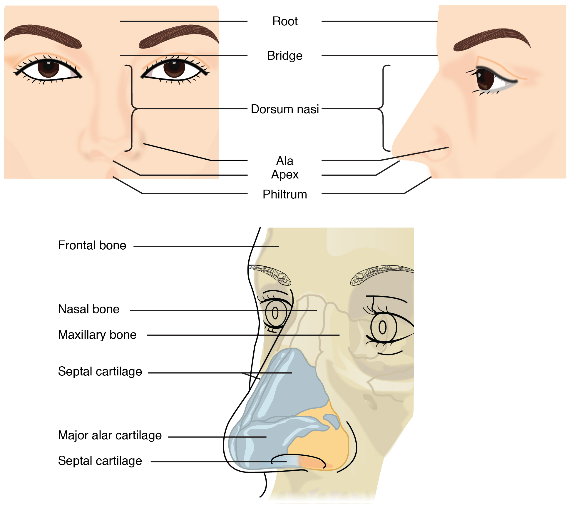
The external nose and the framework beneath it: the dorsum, the apex and the ala above, the nasal bone and the alar cartilage below. A rhinoplasty reshapes that framework and lays the skin back over it. This is an anatomical diagram, not an ultrasound image.
More patients arrive for rhinoplasty with filler already in the nose. A non-surgical nose job uses filler to smooth a bump or to lift a drooping tip. The result fades over months. Some of these patients later want the lasting change a surgery gives. The filler from the earlier treatment does not always clear on its own by then. It can sit in the tissue for years. The non-surgical nose job has spread fast, so the pool of patients carrying nasal filler has grown with it. A clinic that offers rhinoplasty now sees these patients often. The scan has become part of working that out safely, since the filler from another clinic, placed by another hand, is otherwise unknown to the surgeon.
Filler in a nose bound for surgery is a real hazard. Hyaluronic acid placed by an earlier injector can sit right against the arteries that feed the skin. It can wrap a vessel or push it out of its usual place. A surgeon cutting into that tissue works around vessels that have been moved from where the anatomy says they should be. The filler can also hide a vessel from the fingers that would otherwise feel a pulse through the skin. Operating on a nose full of old filler raises the chance of cutting or compressing a vessel the skin depends on. The danger climbs higher when the filler has caused quiet trouble already, a small past occlusion that scarred the supply without anyone naming it at the time. A scan before the operation finds the filler, shows where it sits against the vessels, and measures how much of it remains. The surgeon then knows whether the nose is ready for a knife or whether the filler needs to go first. None of this can be read from the surface. A nose that looks clear can hold a pocket of filler deep against an artery, left from an injection the patient half-remembers from years before.
This is the strongest reason the scan has caught on for rhinoplasty. A surgeon who could once only ask the patient about past injections can now look for the answer. A patient’s memory of what was placed, and when, is often vague. The filler shows on the screen whether the patient recalls it. A history taken in words becomes a picture taken in the room.
The finding changes the timing of the operation. A surgeon who sees filler against the vessels can dissolve it with hyaluronidase and wait for the tissue to settle before operating. The nose comes to surgery closer to its natural anatomy. The vessels sit back where they belong. A wait of a few weeks buys a safer operation. The scan is what tells the surgeon the wait is needed at all. Old filler does more than wait to be found. It can draw a slow reaction around itself, a firm nodule the body has built to wall it off. That nodule reads on the scan as a mass gathered around the filler. A surgeon who finds one expects firmer tissue at that spot under the knife. Hyaluronic acid also lasts longer in the nose than a patient tends to expect, since the tissue holds it still and little there breaks it down. A scan years after the injection can still find a pocket of it.
The patient sees the filler too. The screen settles a question that words alone leave open.
What the probe shows before the incision
The scan reads four things on a nose before surgery. The arteries come up on color Doppler as lines of flow along the sides and the dorsum. The probe follows each one and notes the depth it runs at. A surgeon planning the plane of the dissection wants the vessels mapped against that plane. The map shows where a cut would pass close to a feeding artery. The arteries of the nose run at known depths, the lateral nasal in the soft tissue of the side wall, the dorsal vessels higher on the bridge. The probe confirms where each one runs in this nose. A vessel sitting shallower than usual, pushed up by old filler or by scar, is exactly the one a routine dissection would catch.
Old filler shows as dark pockets in the tissue. Hyaluronic acid reads as a space that looks like fluid against the gray of the skin and the fat. The probe measures how deep each pocket sits and how close it lies to a mapped vessel. A pocket pressed against an artery is the finding that changes a plan more than any other. The closer a pocket lies to a vessel, the more the surgeon plans around it. A pocket sitting clear of the arteries weighs less on the lift. The scan gives the distance, so the surgeon ranks the pockets by the risk each one carries.
Scar shows as bright tissue where the clean planes of an untouched nose have broken up. A revision nose is full of it. The scan shows how deep the scar runs and what it has done to the supply beneath. The skin and the SMAS layer below it each carry a thickness the probe can read. That reading tells the surgeon how much cover there is to work with over the framework.
Four readings come off the one scan: the arteries, the old filler, the scar, the thickness of the cover. Each one feeds the plan.
Reading a nose that has been operated on
A nose operated on before is the hardest to read by hand. A scan helps furthest there. The first operation moved the anatomy. It left scar that tethers the skin and reroutes the vessels from their textbook course. A second operation lifts a skin flap that has already given up part of its supply once. The margin for error is narrower the second time around. The first operation rearranged the supply, so the revision surgeon cannot lean on the textbook layout. The scan is how that new layout gets learned before a plane is chosen. Without it, the surgeon lifts a flap on assumptions the first operation already broke.
The scan reads what the first surgeon left behind. It shows the scar planes the new dissection has to cross. It shows the vessels that survived the first operation and the places a supply was lost. A revision planned against that picture lifts the flap where the supply is best preserved. The surgeon then cuts with a map of a nose that is no longer the standard one in the atlas. The scan also flags where the first operation thinned the cover too far. A patch of skin left thin by the earlier surgery, with a supply already poor, shows on the scan as a danger spot for the second. The surgeon plans the new lift to spare it. A revision done blind to that history risks finishing what the first operation started.
What the scan changes about the plan
The scan changes the operation in a few concrete ways. A nose with filler lying against a vessel gets the filler dissolved first, with the surgery put back a few weeks to let the tissue settle. A nose with thin skin over a marginal supply gets a dissection plane chosen to keep the plexus on the flap. A revision nose gets its approach picked around the scar the scan has mapped. Each of these is a decision made before the first cut, from the picture the scan gives. The plan can also go the other way. A scan that shows a healthy supply and a clean plane tells the surgeon the nose is ready as it stands. That reading saves a delay the surgeon might otherwise add out of caution. The picture gives a reason to wait or a reason to proceed, each backed by what the nose shows.
It also changes what the surgeon tells the patient. A patient with old filler hears why the operation has to wait. A patient with thin skin over a poor supply hears the real risk to the skin before agreeing to anything. The consent rests on a picture of that patient’s own nose. A risk read off the screen is easier to explain than one quoted from a textbook average.
The cut comes after the picture. The picture is what makes the cut safer.
What the scan does not do
The scan does not perform the operation. It maps the supply, finds the filler, reads the scar, measures the cover. The skill of lifting a flap without harming the vessels on its underside stays in the surgeon’s hands. A clear scan in an unsteady hand changes nothing about the result. The probe gives the surgeon a picture of the nose before the first cut. What is done with that picture is the operation itself. The scan is one input among several. It sits beside the photographs, the examination and the patient’s goals. A surgeon weighs the picture of the supply along with everything else that shapes the plan. The probe adds a layer the eye and the hand cannot reach, and stops there.
Common questions about preoperative nasal ultrasound
Why scan the nose before a rhinoplasty?
To see the blood supply of the skin and to find any old filler. A rhinoplasty lifts the skin off the framework. The skin survives on a network of small vessels just beneath it. The scan maps those vessels and measures the skin, so the surgeon plans a dissection that keeps the supply on the flap. It also finds filler from a past treatment that could sit against a vessel.
How does the scan help with old filler in the nose?
Hyaluronic acid shows on the scan as a dark pocket that looks like fluid. The probe measures how deep it sits and how close it lies to an artery. A surgeon who finds filler against a vessel can dissolve it with hyaluronidase and let the nose settle before operating. The picture replaces a vague history of past injections with something the surgeon can see.
Can a rhinoplasty be done on a nose that has had filler?
Often, yes, with care and timing. The risk is that old filler sits against the vessels the skin depends on. A scan shows where the filler is and how much remains. Many surgeons dissolve the filler and wait a few weeks, so the nose comes to surgery closer to its natural anatomy. The scan is what tells the surgeon whether that wait is needed.
What complication is the scan guarding against?
Loss of the skin over the nose, above all over the tip. A rhinoplasty that strips too much of the supply, or that operates on a nose already short of it, can leave the skin to break down. Necrosis of the tip skin is rare. The repair is long and the scar is permanent. The scan reads the supply before the operation so the plan can protect it.
Is this the same as mapping vessels before a filler injection?
No. Both look at the nasal vessels, for different reasons. Mapping before an injection keeps a needle clear of an artery. This scan protects the blood supply of a skin flap during surgery and finds filler that surgery would disturb. The procedure, the risk and the plan that follows are all different. The shared part is the high-frequency probe.
Does a revision rhinoplasty need the scan more?
It gains more from it than any other nose. A revision nose carries scar from the first operation, on a supply that has been cut once already. The anatomy has moved from its textbook course. The scan shows the scar planes and the vessels that survived, so the second operation lifts the flap where the supply is best kept. The margin for error is narrower the second time.
Does the scan replace the surgeon’s judgment?
No. It maps the supply, finds the filler and reads the scar. The surgeon does the rest. Lifting a flap without harming the vessels under it is a skill of the hands. A clear scan in an unsteady hand changes nothing. The picture informs the plan and the consent. The operation stays the surgeon’s work.
About the Author
Julien Mercier
Senior R&D Engineer · Medical Ultrasound Transducer Development
Senior R&D Engineer with an M.S. in Applied Physics and over 15 years of experience in medical ultrasound transducer development, specializing in the design verification and performance testing of high-frequency imaging transducers. Currently leading the development and verification of the company’s next-generation high-frequency linear-array transducer, responsible for imaging performance evaluation and reliability analysis in preclinical testing. Brings extensive hands-on experience in piezoelectric element tuning, beamforming parameter optimization, and system-level performance testing.