What Plastic Surgeons Need to Know Before Using Ultrasound First Time
Handheld ultrasound, for a plastic surgeon trying it the first time, is a small high-frequency probe placed on the skin to show the layers beneath on a phone or a tablet. What a surgeon needs before leaning on it is a short list: what the picture shows, what its gray shades mean, and how the eye for reading it is built. The learning is quick. The payoff lands in the cases where a look under the skin changes the plan.
The learning curve is shorter than it looks
The hardest part sounds harder than it is. A surgeon already carries a deep map of the anatomy in three dimensions, built over years of operating. Ultrasound asks that surgeon to match a gray picture to that map. The matching comes within a few dozen scans for a new user. The hand that already knows where the facial artery runs learns to find it on the screen fast. A structured first session helps the match along. An hour with the probe on a model or a willing colleague, naming each layer as it comes up, ties the picture to the anatomy faster than scanning alone does. Many surgeons pick up the basics in a short hands-on course, then build speed in their own clinic over the weeks that follow. The groundwork takes an afternoon. The fluency builds over the season of use that follows.
The first sessions go best on a known target. A surgeon new to the probe can start on a wrist or a forearm, where the artery pulses plainly and the layers stack in a clean order. The same moves carry over to the face. Scanning a part of the body the surgeon knows cold builds the link between the picture and the anatomy, before that link gets tested on a delicate area. The forearm teaches the moves that matter. Sliding the probe along a vessel, turning it from a long view to a cross-section, holding it still on a target: these are the same hands a surgeon will use on the face. The technique carries straight from the forearm to the face. Only the site is new.
The skill splits into two. One half is holding the probe and working the machine, which a few dozen scans settle. The other half is reading what comes up, which keeps growing for as long as a surgeon scans. A beginner reaches useful competence quickly. Mastery of the harder pictures comes over years, the way it does with any tool a surgeon picks up. The plateau a beginner reaches is already useful. A surgeon who can find a vessel, read the planes and guide a needle has enough to work safely in the common cases. The growth past that point sharpens the reading of the unusual picture, the early complication or the odd anatomy. Each stage of skill pays for itself before the next one arrives.
Reading the gray picture
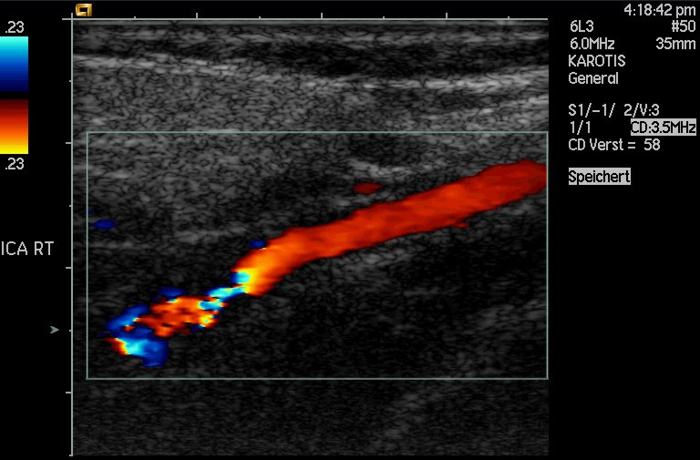
Color Doppler fills a flowing artery with color over the gray picture, here a carotid in the neck. This is the mode a surgeon switches to for finding a facial vessel before a needle. The labels and the green box are the machine’s own.
The screen shows a slice through the tissue. The probe sends sound straight down and draws what lies along that single plane, from the skin at the top of the screen to the deeper tissue at the bottom. A surgeon used to seeing the whole field at once learns to think in slices, sweeping the probe to build the third dimension in the mind. The picture is a cross-section the surgeon moves through the tissue. Building the third dimension is the mental move that takes practice. The surgeon sweeps the probe across a structure and stacks the slices into a shape in the mind, the way a CT reader builds an organ from its sections. A vessel seen in cross-section as a black dot becomes a long black tube when the probe turns ninety degrees along it. Reading both views of the same structure confirms what it is.
Brightness carries the meaning. Tissue that throws the sound back reads bright, so a dense structure like a tendon or a scar lights up. Tissue that lets the sound pass reads dark, so fluid and the fat-poor parts of the skin read black. Bone and the surface of metal reflect almost everything and read as a bright line with a shadow below. A few hours of scanning teach the eye which shade goes with which tissue. A handful of landmarks anchor the reading. Fat reads as a dark field crossed by bright strands. Muscle reads as darker bundles wrapped in bright lines. A nerve reads as a cluster of dark dots in a bright frame, the honeycomb a surgeon learns to recognize. Once these patterns are fixed in the eye, the gray blur reads as a map.
Depth reads from top to bottom. The skin sits at the top of the screen, the entry line where the sound meets the surface. The dermis follows as a bright band, the fat below it darker, the muscle and the bone deeper still. A surgeon reads the depth scale at the side of the screen to know how far down a structure sits. A vessel a centimeter down on the screen is a centimeter down in the tissue. The machine measures for the surgeon. Freezing the picture and dropping two calipers on a structure reads its size or its depth in millimeters. A surgeon checking how deep a plane sits, or how thick a layer runs, gets a number off the frozen frame. That number goes into the note and into the plan, a measured fact the next visit can be set against.
Color marks the blood. Switched into color Doppler, the machine paints moving blood over the gray picture, so an artery fills with color and stands out from the still tissue around it. This is the mode a surgeon reaches for to find a vessel before a cut or an injection. The color shows where the blood runs and how deep it sits, the two facts that keep a needle or a blade clear of it. Color Doppler needs its own tuning for the face. The small facial vessels carry slow blood, so the scale comes down low to catch it. The box that marks where the machine looks for color stays small and sits over the spot in question, which keeps the reading quick and sensitive. A surgeon learns to read the color as flow, with the gray picture underneath showing the depth and the surroundings.
The whole picture updates live. The probe shows the tissue as the surgeon moves it, the needle as it advances, the filler as it spreads. That live feedback is the heart of what ultrasound adds to the surgeon’s hands.
The mistakes a beginner makes
The first mistake is anisotropy. A structure like a tendon or a nerve reads bright only when the beam meets it square. A few degrees of tilt make the same structure go dark, looking for a moment like a tear or a gap. A beginner who does not know the effect reads a false finding off it. The fix is to rock the probe until the structure flares bright, which confirms it is there and intact. Every new user meets this trap. Learning to rock the probe past it is one of the first real skills. The demonstration sticks once a surgeon sees it. Resting the probe on a tendon and tilting it slowly turns the bright fibers dark, then back to bright across a few degrees of swing. After watching that once, a surgeon reads a dark patch with the angle in mind, rocking the probe before calling anything a gap. The brightness is a property of the angle as much as of the tissue.
The second mistake is too much gain. Turned up high, the gain floods the screen with bright speckle that buries the real structures. Turned too low, the picture goes dark and a vessel hides in it. A beginner learns to set the gain so the fluid in a vessel reads black and the tissue around it reads an even gray. The right setting comes from a few dozen tries, the eye learning the look of a balanced picture. Some machines split the gain by depth. A near and a far control, or a row of sliders down the screen, let a surgeon even out a picture that reads bright up top and dark below. A beginner can leave these on the preset at first, then reach for them once the eye knows what an even field looks like. The aim throughout is a picture where the same tissue reads the same shade at every depth.
The third mistake is pressure. A vein collapses flat under the weight of the probe, vanishing from a picture it should fill. A beginner pressing hard to get a clear image squeezes the soft structures out of view. The fix is a light touch, with a thick layer of gel carrying the sound so the probe barely dents the skin. Reading a vein at all depends on letting it stay round. The same light touch serves the artery. Heavy pressure flattens the soft tissue and pushes a vessel deeper than it sits, throwing off the depth a surgeon reads. A thick layer of gel does the coupling so the probe floats on the surface. The picture a surgeon trusts comes from a probe resting on the skin, barely denting it.
The fourth mistake is losing the orientation. A probe has a mark on one side that matches a mark on the screen, so the surgeon knows which edge of the picture is which side of the patient. A beginner who ignores the mark maps a finding to the wrong side. Setting the mark the same way for every scan, toward the patient’s head or the surgeon’s left, builds a habit that keeps the picture honest. The convention matters more than which way it points. A surgeon who always sets the mark toward the head, or always toward one side, reads every picture the same way from then on. A finding on the left of the screen lands on the same side of the patient every time. The habit removes a whole class of error before it starts.
The settings to learn first
Four controls cover the early work. Frequency sets the trade between detail and depth, high for the skin and the face, lower for anything deep. Depth sets how far down the screen reaches, kept shallow for a facial scan so the skin fills the picture. Gain sets the overall brightness, balanced so the tissue reads an even gray. The color Doppler box marks the patch where the machine looks for blood, kept small and placed over the spot in question so the color stays sensitive. Two more controls reward a little attention. The focus marker sets the depth the beam is sharpest at, placed level with the structure in question for the cleanest picture there. Time-gain sliders even out the brightness from top to bottom. A beginner can leave both on the preset and still get a clean facial scan, then learn them as the eye grows more particular.
A surgeon learns these four by changing one at a time and watching the picture answer. The presets a machine ships with handle a first scan well enough, so a beginner can start there and adjust by feel. The settings become second nature within the first dozen sessions, the hand reaching for the right control without a pause. The presets carry a beginner further than expected. A machine built for the skin ships with a setting tuned for it, so a first scan needs little adjusting. The learning is in knowing which control to nudge when a picture comes up wrong, the gain for a dark field, the depth for a structure off the screen, the focus for a blurred target. A surgeon builds that reflex one scan at a time.
What first use realistically gives
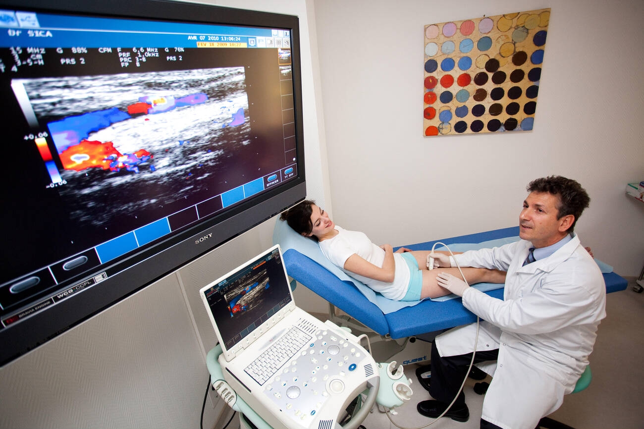
A handheld ultrasound probe, wiped down between patients, run from a phone or a tablet. A probe like this is the whole machine a surgeon learns to hold steady. The one shown is a convex probe; aesthetic work uses a higher-frequency linear head.
A surgeon new to the probe gains the clearest benefit where the stakes are highest. Finding the facial vessels before filler, checking a plane before a thread, reading old filler before a rhinoplasty: these are the first uses that earn the learning, because each guards against a complication a surgeon dreads. The probe rewards the cautious case first. These first uses share a shape. Each one puts a question the eye cannot answer, where a vessel runs, how deep a plane sits, what a past injection left behind, in front of a surgeon about to commit a needle or a blade. The scan answers the question before the commitment. A surgeon who scans the high-risk case first builds confidence on the cases that call for it.
The picture informs the plan. It does not replace the judgment that reads it. A surgeon still decides where to cut, how much to lift, where to place the filler. The scan adds one layer to that decision, a look under the skin the eye and the hand cannot reach on their own. A clear scan in the hands of a surgeon who cannot yet read it changes nothing. The reading is the skill. It grows with use. The scan also leaves a record. A saved image of the vessel mapped before an injection, or the plane checked before a cut, documents the care taken. That record sits in the file alongside the consent, a picture of what the surgeon saw before acting. The habit of saving the scan builds a trail of careful practice, case by case.
The honest expectation for a first month is modest and real. A surgeon learns to find a vessel, to read the main layers, to spot old filler, to guide a needle under live view. The harder pictures, the subtle scar or the early complication, come with more scanning. A surgeon who expects to read everything on day one is let down. A surgeon who expects to read the common, high-value findings is rewarded from the first week. The week-by-week of it is steady. The first scans find the obvious vessel and the main layers. By a month, the eye picks out old filler and follows a needle live. By a season, the subtler readings come within reach. A surgeon who measures progress against that arc stays encouraged, each week adding a reading the last one lacked. The pace suits a busy surgeon. A scan adds a minute or two to a case. The skill builds inside the normal run of work, a few patients at a time.
Building it into the practice
The probe earns its keep once it becomes a routine step. A surgeon who scans only when worried scans too rarely to stay fluent. A surgeon who builds the scan into a fixed point of certain procedures keeps the skill sharp and meets the rare hard case ready. The first use is the start of a habit. The habit is where the value lives. A short audit keeps the habit honest. Looking back over a month of saved scans shows a surgeon where the probe changed a plan and where it confirmed one. That review turns scattered scans into a measure of what the tool does in the surgeon’s own hands. By the end of a few months the probe has earned a fixed place in how the careful cases are run.
Common questions about a surgeon’s first use of ultrasound
How long does it take a surgeon to get useful with handheld ultrasound?
The hands-on part settles within a few dozen scans, holding the probe still and working the controls. Reading the picture for the common, high-value findings, a facial vessel or old filler, comes within the first weeks. The harder pictures keep developing over months and years. A surgeon’s existing map of the anatomy makes the start faster than it is for someone learning the anatomy at the same time.
Where should a surgeon practice first?
On a part of the body the surgeon knows cold, like a wrist or a forearm, where the artery pulses plainly and the layers stack in a clean order. The moves carry straight over to the face. Practicing on familiar anatomy builds the link between the gray picture and the three-dimensional map already in the surgeon’s head, before that link is tested on a delicate area.
What is the first trap a beginner runs into?
Anisotropy. A tendon or a nerve reads bright only when the beam meets it square, . A few degrees of tilt make it go dark, looking for a moment like a tear. The fix is to rock the probe until the structure flares bright. Learning that the brightness depends on the angle, and reading past it, is one of the earliest real skills.
Which settings matter at the start?
Four. Frequency for the trade between detail and depth, depth for how far the screen reaches, gain for the overall brightness, and the color Doppler box for finding blood. A surgeon learns them by changing one at a time and watching the picture answer. The machine’s presets handle a first scan, so a beginner adjusts from there by feel.
Does the scan replace a surgeon’s judgment?
No. It adds a look under the skin to a decision the surgeon still makes. The scan shows where a vessel runs or how deep a plane sits. The surgeon decides what to do with that. A clear picture in the hands of someone who cannot yet read it changes nothing, which is why the reading is the skill that matters.
What can a surgeon realistically read in the first month?
The common, high-value findings: a facial vessel on color Doppler, the main skin layers, a pocket of old filler, a needle under live view. The subtle pictures, an early complication or a faint scar plane, come with more scanning. A first month gives a surgeon the readings that guard the riskiest cases, which is where the probe pays off first.
About the Author
Julien Mercier
Senior R&D Engineer · Medical Ultrasound Transducer Development
Senior R&D Engineer with an M.S. in Applied Physics and over 15 years of experience in medical ultrasound transducer development, specializing in the design verification and performance testing of high-frequency imaging transducers. Currently leading the development and verification of the company’s next-generation high-frequency linear-array transducer, responsible for imaging performance evaluation and reliability analysis in preclinical testing. Brings extensive hands-on experience in piezoelectric element tuning, beamforming parameter optimization, and system-level performance testing.