What 18 to 24 MHz High Frequency Probe Does for Aesthetic Medicine Clinics
An 18 to 24 MHz probe is the safety equipment an aesthetic clinic uses to see under a needle. The probe changes the injection itself. It also changes the consultation that comes before the needle and the records kept after it.
Why a clinic brings one in
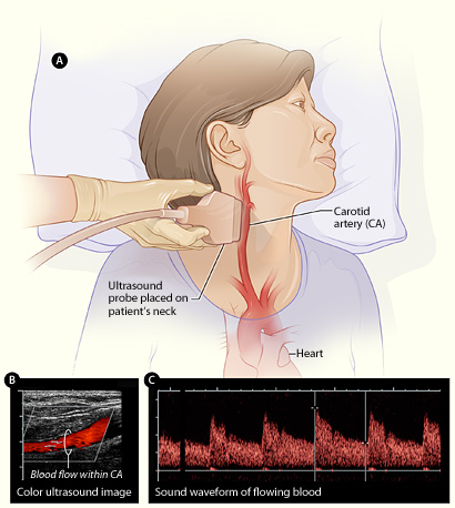
Color Doppler fills a vessel with the color of its flow, shown here on a carotid. The power to find an artery this way, in one patient, is the safety the clinic is paying for. This is a labeled illustration with an ultrasound inset. The panels and labels are part of the original.
A clinic buys this probe to lower one risk. Filler can enter a facial artery. A blocked artery starves the skin it feeds. Through links that run back toward the eye, a misplaced bolus can reach the retina and take sight. These events stay rare. A clinic can run for years without one. A single case still closes practices and draws lawsuits. The probe lowers the odds of that case. It shows the arteries in the patient on the table. The injector plans the needle around them. The technical work of finding the vessels belongs to the probe itself. The value to the clinic is simpler. Every injection now rests on this patient’s own anatomy. Many aesthetic and ultrasound bodies teach a pre-injection scan of the high-risk areas. Boards and insurers look for that scan after an adverse event. Patients ask whether a clinic scans at all. A clinic that injects high-risk areas with no scan is the one left to explain the choice.
The case for it grows each year. A pre-injection scan started as a mark of a few careful injectors. It is becoming routine in the higher-risk parts of the face. The glabella between the brows, the nose, and the folds beside it carry arteries that link to the eye. A scan of those areas now reads as ordinary practice. The clinic that adopts it early stays ahead of where the standard is heading.
The money behind the decision is lopsided. A handheld probe costs a small fraction of one vascular complication. One complication carries emergency care, corrective work, a refund, lost time, and legal exposure. The probe pays for itself the first time it heads off such an event. The clinic can never know which event that was. A clinic buys the probe the way it buys any safety measure. It pays once and hopes never to draw on it.
The everyday gains are quieter than the rare disaster. A clinic that scans before it injects places less product in the wrong plane. It leaves fewer lumps to dissolve. It calls fewer patients back for a correction. The patient feels this as work that holds up over the months that follow. A reputation builds on results that needed no reworking. The whole purchase is a decision about risk. The picture on the screen is only how that decision gets made.
The case that turns a clinic
Many clinics adopt the probe the hard way. A near-miss tends to change a practice faster than any article does. A vessel blanches and recovers. A full occlusion takes an afternoon to rescue. After one such day, the scan stops being optional. The clinic that adopts before that day skips the lesson. The probe lets a clinic learn the lesson at a far lower price.
The same path runs across the whole field. The early adopters were often the injectors who had seen a complication up close. Their stories spread through training, through conferences, and through the cases that reach the news. The rest of the field follows behind them. A clinic weighing the purchase today is rarely asking whether scanning works. It is asking how long it can keep injecting high-risk areas blind.
What it does in the consultation
The probe changes the consultation before any needle appears. A scan during the consult shows patients their own face on the screen. They see the vessel that runs close to a planned site. They see how thick the skin is in one area. They see the filler an earlier clinic left behind. Many patients have never seen any of this. The conversation moves from a list of risks read aloud to a picture the patient can follow. A patient who follows the reasoning makes a sounder decision. That decision is the kind least likely to end in regret.
Consent built on a picture holds firmer than consent built on a warning. A patient who has seen the risky vessel understands why the plan is cautious there. The same patient understands why the clinician declines a request outright. The view explains a conservative result on its own terms. The clinic that says no to a risky request keeps the trust of the person it turned down. A picture carries that explanation where words alone struggle.
The probe also settles questions the patient cannot answer. Someone who had filler elsewhere often does not know what was used, how much, or where it sits. The scan finds the old product. A new plan can then account for what is already in the face. A patient who sees the old filler explained avoids a worse surprise later. The clinic avoids being blamed for what a previous one left behind.
There is a commercial side to this. A clinic that scans looks careful because it is being careful. Patients notice the difference. They tell other people about it. Anyone can buy filler and a syringe, so a clinic competes on the safety it can show on a screen. The patient who felt looked after comes back. That patient brings others who want the same care.
The screen also softens a hard conversation. A patient pushed toward a risky amount of filler can hear a flat refusal as a sales line. The same patient, shown the artery running under the spot they want filled, reads the refusal as care. The picture carries the reason where words alone sound like an excuse. A clinician who points at the vessel keeps the trust of the person turned down. That person often returns for the treatment that is safe.
During the work, and on the record afterward
What the probe shows can be saved. That record outlasts the visit. A clinic can store an image of where the filler went, at what depth, in which plane. A patient returns months later for a touch-up or with a complaint. The record shows what the clinic did. A field that ran on memory and a few handwritten notes now has something close to evidence.
The record earns its place when something is disputed. A complication can turn into a question of whether the clinic acted reasonably. A stored scan answers part of that question. It shows what was placed, where it went, and whether a vessel was checked first. A saved image lets the clinic show its work. The picture that guided the injection becomes the document that defends it. The probe goes on protecting the clinic long after the syringe is gone.
The record serves the ordinary case too. A patient moves between clinicians inside a practice, or returns after a year away. The next person is not starting blind. They can see what was placed and build the next treatment on a known picture. Written notes alone rarely carry that much. The probe lets a clinic treat a returning patient as the same patient over years. An image says in a glance what a page of notes struggles to put in words: where the product sits, in which plane, how much of it went in. The clinician who opens the file a year later reads the face as it was, not a guess at what an earlier hand meant by a line of shorthand.
The record also speaks to the people who judge a clinic. An insurer pricing a policy, a board reviewing a complaint, a lawyer weighing a claim, each asks whether the clinic acted with care. A file that shows a vessel was checked before the needle answers that in the clinic’s favor. A clinic that scans and saves builds, case by case, the evidence that it works to a standard. That evidence is hard to assemble after the fact.
The capability this rests on
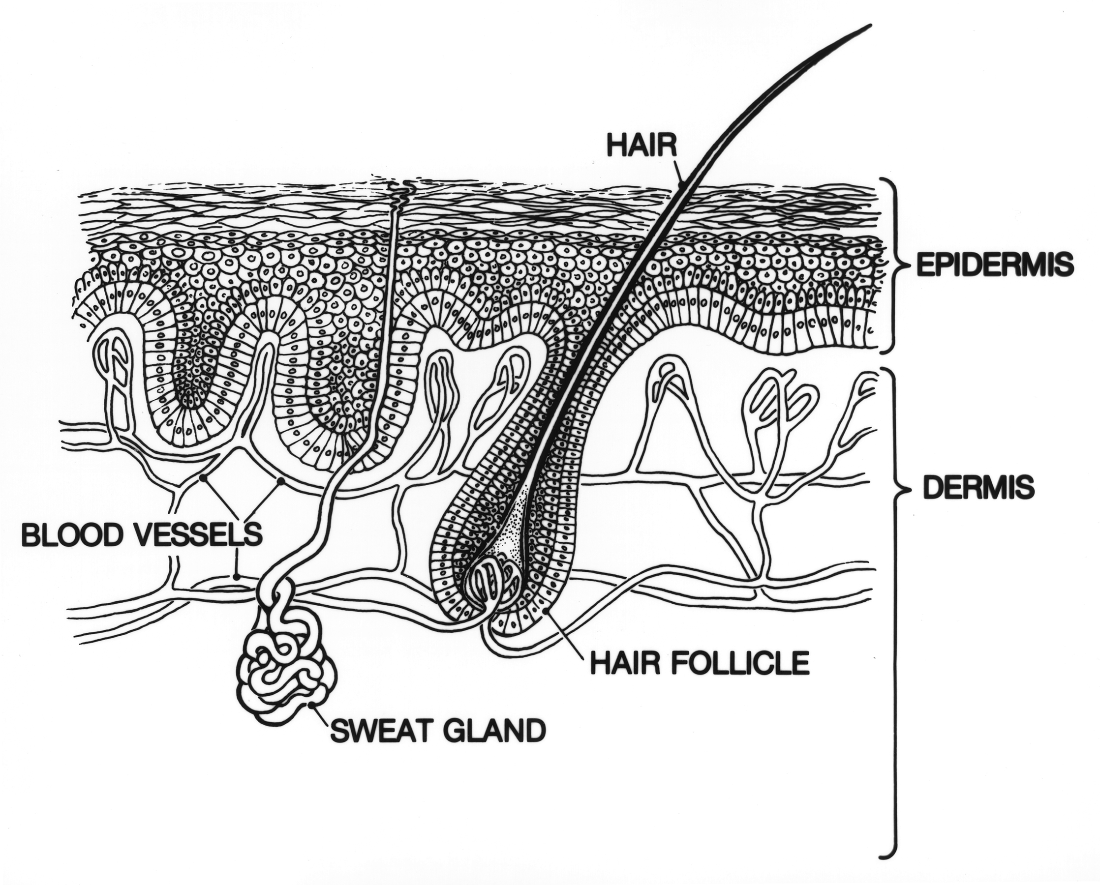
What the probe resolves in the first centimeter: the epidermis, the dermis below it, the small vessels and follicles within. This is an anatomical line diagram. It is not an ultrasound image. The labels are part of the original. A clinic’s value from the probe rests on seeing this layer clearly.
All of the value above rests on one technical fact. At 18 to 24 MHz the probe sees about a centimeter under the skin in fine detail. It reads the skin as separate layers. With color Doppler it shows the facial vessels by their flow. That shallow, high-resolution window is the layer aesthetic work lives in. The exact figures for resolution, depth, and Doppler are the probe’s own subject. Everything a clinic gains from the probe is built on that one capability. Lose the resolution or the depth and the rest of the value goes with it. No clinic buys the probe for the physics. It buys what the physics makes possible: a clear look at the one layer where the whole of aesthetic work happens. The filler, the toxin, the threads, the arteries an injector must miss all sit inside that first centimeter. A probe that reads it cleanly is the foundation under every use a clinic finds.
What it costs and what it returns
The cost of entry has dropped to where the decision is easy. A handheld probe runs from a phone or a tablet the clinic already owns. It costs a fraction of a cart-based machine. It needs no dedicated room. A clinic buys the one head its work calls for and adds others later. A single practitioner or a small clinic pays a price close to a piece of office equipment. That low cost is a large part of why the tool has spread the way it has.
One avoided complication pays for the probe many times over.
The return is harder to put on an invoice. Much of it lives in events that never happened. The clearest line is the complication avoided, with its emergency care, its refunds, and its legal cost. Below that sit the steadier gains: fewer corrections, patients who come back, a reputation for care, a record that holds up. None of these arrives as a single payment. Together they make the probe one of the cheaper forms of insurance a clinic can carry. The avoided complication never appears on a ledger, so the return hides in plain sight. A clinic pays for the probe once. It never tallies the emergencies that did not happen, the refunds it did not write, the bad reviews it never earned.
The probe also opens revenue a clinic might turn away. A practice nervous about the nose or the glabella can take those cases on once it can see the vessels first. Work that felt too risky to offer becomes work the clinic can do with a margin of safety. The tool adds to what a clinic can sell, beyond protecting what it already does.
The probe also helps a clinic charge for what it does. A clinic known for scanning holds its prices against cheaper competitors. It sells the safety around the filler. Patients who want the careful version will pay for it. Those patients tend to be the ones a clinic wants: the people who come back, follow advice, and refer others like them. The probe lets a clinic compete on care.
Fitting it into how a clinic works
The probe does nothing sitting in a drawer. The change that matters is making the scan a fixed step. It comes out for every high-risk injection, every time. A clinic that scans only when worried captures little. The dangerous case is so often the one that seemed routine going in. Turning the look into a habit is the work.
Learning to use the probe is the smaller part of that. The hand picks up the basics in a few dozen scans. The harder change is cultural. The habit has to spread across everyone who injects. The clinic has to agree on which procedures always get a scan. The records have to stay consistent from one clinician to the next. That is a matter of clinic policy. A clinic that treats the probe as a shared standard draws far more from it than one where it stays one enthusiast’s hobby.
A clinic that keeps its scans can also learn from them. Gathered across many patients, the records show patterns no single case reveals. They show where corrections clustered, where a plane was missed, how a filler behaved over the months after it went in. A clinic can study its own work and tighten its practice over time. Reviewed in a batch, the saved scans show a clinic its own patterns: the plane that caused trouble, the area where corrections clustered, the filler that behaved oddly over the months after it went in. A practice that studies its own work tightens it season by season, in a way no single case could teach.
Which clinics gain first
The probe earns its place fastest in the practices that inject the riskiest areas. A clinic built around the nose and the midface works over the arteries that link to the eye. For that clinic the scan guards against the worst events it faces. A clinic centered on lip work runs a lower vascular risk, so its case for the probe leans more on the consultation and the record than on the rare disaster. Even that clinic meets the occasional patient who wants the one risky treatment. The probe lets it take that case on with a margin of safety it would not have had blind.
Volume tips the math too. A high-throughput clinic injecting many patients a week meets the rare complication sooner, by the numbers alone. The probe spreads its cost across many treatments and shortens the odds of the day no clinic wants. A low-volume practice buys it more as cheap insurance against a single career-ending event.
The newest injectors gain in a different way. An experienced hand has a feel for the planes built over years. A clinician earlier in that journey leans on the picture while the feel develops. The probe shortens the gap between a new injector and a safe one, which matters to a clinic training its own people.
The clinic without a clear high-risk caseload still has a reason. Even a practice that largely avoids the danger zones meets the patient who wants the one risky treatment. The probe lets that clinic say yes safely, once, rather than turn the patient away or work blind. A tool that sits ready for the rare hard case pays off the day that case walks in.
What it does not do
The probe is a tool. It is not a guarantee. It does not make a hurried or careless injector safe. It gives a careful one a clearer view. It does not diagnose disease. It does not replace a clinician’s training. It does not remove the need for judgment. It reaches only the first centimeter, so anything deeper is a job for another probe. A clinic that expects the device to carry the safety on its own has misread what it is for. The judgment of where and how deep to place the filler stays with the person holding the syringe. The probe informs that decision. It does not make it.
Common questions
Why would an aesthetic clinic invest in ultrasound?
The main reason is risk. Filler injected into a facial artery can cause serious harm. Those rare events bring lawsuits and reputational damage that can close a clinic. Seeing the vessels before the needle lowers the odds of one. A clinic buys the probe the way it buys other safety equipment, against a cost it hopes never to meet.
Does scanning protect a clinic legally?
It helps. A stored scan records what was placed, where it went, and whether a vessel was checked. A later dispute can then rest on evidence. The image that guided the injection can document it afterward. A clear record stands up far better than recollection.
How does it change the patient consultation?
It lets the clinician show patients their own anatomy on the screen: the vessel near a planned site, old filler from another clinic, the plane a new injection will fill. Consent built on a picture the patient can see holds firmer than consent built on a spoken warning. Many patients also find that watching the care taken builds trust.
Is a handheld probe affordable for a small clinic?
Yes, by current standards. It runs from a phone or a tablet the clinic already has. It costs a fraction of a cart machine. It needs no special room. A clinic buys the one probe its work needs. A small practice pays a price close to office equipment.
Does every injection need a scan?
Not every one. The case for scanning is strongest in the high-risk areas: the nose, the glabella, the regions over named arteries. The harder discipline is deciding which procedures always get a look and then doing it every time. The dangerous case is often the one that seemed routine.
Does ultrasound replace injector skill?
No. It gives a skilled, careful injector a clearer view and a record. It does not make a hurried one safe. It does not diagnose disease. It does not replace training and judgment. A clinic that treats it as a substitute for skill has misread what it does.
Will patients come to expect a clinic to scan?
More and more, yes. Ultrasound is spreading in the higher-risk parts of aesthetics. Patients read about it and ask whether a clinic uses it. A clinic that scans can say that it does and show the patient why it matters. A clinic that does not may find itself explaining the choice.
About the Author
Julien Mercier
Senior R&D Engineer · Medical Ultrasound Transducer Development
Senior R&D Engineer with an M.S. in Applied Physics and over 15 years of experience in medical ultrasound transducer development, specializing in the design verification and performance testing of high-frequency imaging transducers. Currently leading the development and verification of the company’s next-generation high-frequency linear-array transducer, responsible for imaging performance evaluation and reliability analysis in preclinical testing. Brings extensive hands-on experience in piezoelectric element tuning, beamforming parameter optimization, and system-level performance testing.