Melanoma Skin Cancer Ultrasound Screening Handheld High Frequency
High-frequency ultrasound for melanoma is the use of a skin probe to read what lies below a known tumor: how deep it reaches, whether the lymph nodes it drains to are involved, and whether it has come back after treatment. The diagnosis itself is made on the surface, by the eye, the dermoscope and the biopsy. Ultrasound takes over from there, at the bedside, adding the depth and the spread to the picture the surface exam begins.
Where the diagnosis comes from
A melanoma is found and named on the surface. A clinician spots a mole that has changed, a dermoscope reads the pattern of pigment and vessels in its surface, and a biopsy sends the tissue to a pathologist who confirms the cancer under the microscope. That chain does the diagnosis. The reading that calls a spot a melanoma happens in the skin’s top layers and in the lab. That sequence is well worked out. A changing mole draws the eye, a closer look with the dermoscope sorts the harmless from the worrying, and a biopsy settles the rest under the microscope. Each step reads the surface and the cells. None of it needs to see how deep the tumor has grown, because depth is a separate question the surface tools were never built to answer.
Ultrasound looks at a different question. It sends sound into the skin and shows the tumor as a structure with a depth, a shape and a base. A melanoma reads on the scan as a dark, fairly well-defined mass set into the brighter skin around it, often carrying its own blood flow on color Doppler. The probe says nothing about the pigment pattern a dermoscope reads. It speaks to how far the tumor has grown down and how far it has spread. The image is a structure with a size. A melanoma on the scan has edges, a base and a height the probe can measure, set into the layered skin around it. The blood it has recruited shows on color Doppler as flow running into and through the mass. These are the marks of a tumor with depth and a supply, the things that matter once the diagnosis is settled and the treatment is being planned.
This division decides where ultrasound belongs. The surface tools answer whether a spot is a melanoma. The probe answers how thick it is and where it has gone. A clinic uses each tool for the question it suits. The probe joins the work once a melanoma is on the table as a diagnosis or a strong suspicion. The order holds in practice. A suspicious spot goes to the dermatologist for the surface read and the biopsy. A confirmed melanoma comes to the probe for the depth and the nodes. The two readings stack into one plan, answering what the lesion is and how far it reaches. A clinic that runs both has the fuller picture before the first cut.
Measuring the depth before the surgery
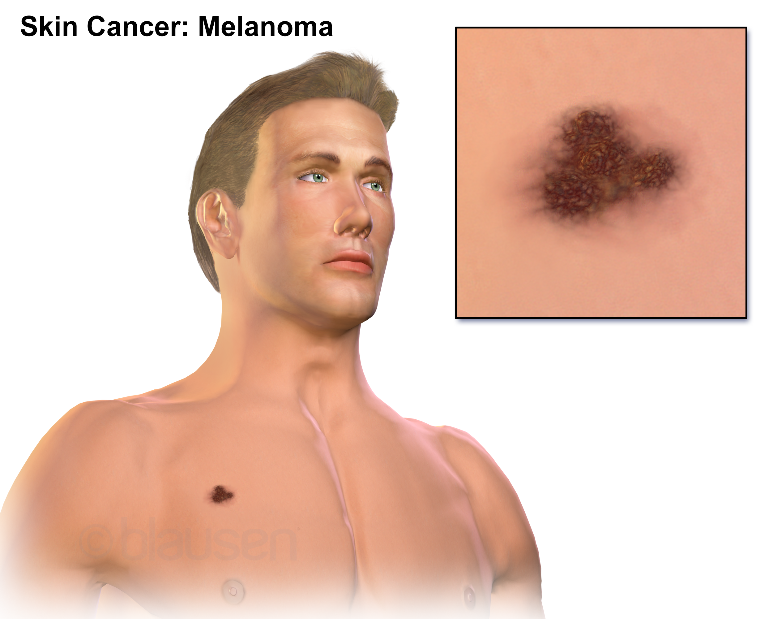
A melanoma on the skin, with the changing spot shown close up. The eye and the dermoscope read a surface like this to make the diagnosis. The probe then measures how deep the lesion reaches before the surgery. This is a medical illustration.
Depth is the number that drives the treatment of a melanoma. The thickness of the tumor, measured from the surface down to its deepest cell, sets how wide the surgeon cuts around it and whether the nearby lymph nodes need sampling. The pathologist measures that thickness on the excised tissue as the Breslow depth, the figure the whole plan turns on. Ultrasound offers an estimate of it before the knife. The thresholds are sharp. The thicker the tumor, the wider the margin the surgeon cuts, and the stronger the case for sampling the sentinel node. The line sits around a millimeter of thickness, below which a melanoma usually takes a narrow excision alone. The depth decides which path a patient is on, so an early estimate of it shapes the whole operation from the start.
A high-frequency probe reads the tumor as a dark mass and measures from the skin surface to its base. That measurement gives the surgeon a sense of the depth ahead of the operation, when the plan is still being drawn. A thin lesion on the scan points toward a narrow excision under local anesthetic. A reading that shows a thick tumor reaching well down into the skin warns the surgeon to plan wider margins and to weigh a sentinel node biopsy in the same sitting. The measurement takes a steady hand and a light touch. The probe sits over the lesion with plenty of gel, pressing barely at all, so the soft tumor keeps its true shape. The clinician freezes the clearest frame and drops a caliper at the surface and another at the deepest dark point. The distance between them is the depth the plan uses.
The preoperative number changes how a single operation is arranged. Knowing the rough depth lets the surgeon book the right amount of time, the right anesthetic and the right consent before the day. A patient learns what the surgery is likely to involve from the start. The scan turns the depth from a surprise the pathologist reports afterward into a figure the team plans around in advance. The patient feels the difference too. A person facing surgery learns from the start whether it is a quick local procedure or a larger one with a node biopsy attached. The consent conversation rests on a real expectation of the operation ahead. The scan turns the day of surgery from an open question into a planned event for the patient as much as for the surgeon.
Frequency sets how fine the depth reading is. The probes used for this run high, in the range that reads the first few millimeters of skin in detail, the same band that serves the rest of aesthetic and dermatologic ultrasound. The higher the frequency, the sharper the line between the dark tumor and the skin around it, which is what a clean depth measurement depends on. The same probes serve across the clinic. A practice that already scans skin for aesthetic work, or vessels before filler, carries the frequency a melanoma depth reading needs. The lesion sits in the first few millimeters, the window these probes are built for. One tool covers the skin work and the melanoma measurement, on the same handheld base.
What the depth measurement can promise
The ultrasound depth is an estimate. The histology remains the standard. The pathologist’s Breslow depth, read off the removed tumor under the microscope, is the figure that stages the cancer and guides the formal decisions. The scan gives a number before that, close enough to plan with in many cases, held as a guide. The verdict waits for the microscope. Studies that compare the two report a fair match for thinner tumors, with the gap widening for thick ones. A clinic treats the ultrasound depth as a strong hint of the category a melanoma falls in, enough to plan the right operation, with the exact stage confirmed on the removed tissue.
A few things blur the ultrasound reading. A dense gathering of immune cells under a melanoma can read as dark as the tumor, so the scan reports a depth greater than the tumor alone. Pigment, ulceration and a thick layer of keratin each shift the picture. A surgeon reads the ultrasound depth as a planning figure, then lets the pathology set the final stage. The probe earns its place by getting the plan close before the operation, with the microscope confirming the truth after it. Knowing the limits keeps the reading useful. A surgeon who treats the ultrasound depth as a planning estimate, open to revision by the pathology, gets the value of the early number without leaning on it too hard. The early figure books the right operation; the microscope that follows sets the final stage. Each does the job it can.
Reading the lymph nodes
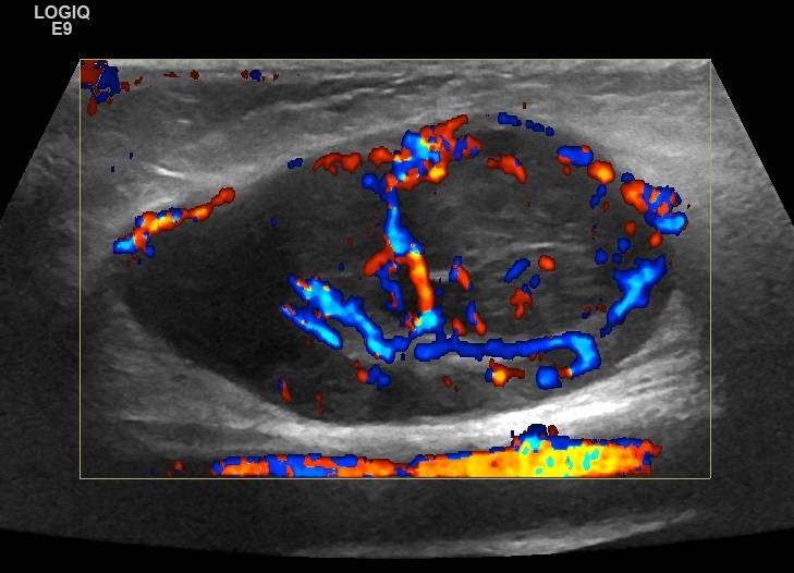
A lymph node on color Doppler showing the chaotic, scattered flow of a node taken over by tumor, with vessels entering around the edge. This is the pattern a melanoma scan looks for in a draining node. The machine label is its own.
The lymph nodes are where ultrasound does its strongest work in melanoma. A melanoma spreads first along the lymph channels to the nodes that drain its part of the body, the groin for a leg, the armpit for an arm, the neck for the face. Catching that spread early changes the treatment and the outlook. The nodes sit a centimeter or two under the skin, in the exact reach a high-frequency probe reads best. The drainage follows a known map. A melanoma on the calf drains to the groin, one on the forearm to the armpit, one on the cheek to the neck and the parotid. A clinician scanning for spread knows which basin to read for a given lesion. The probe goes to the nodes that lesion drains to, the first place a spreading melanoma lands.
A healthy node has a look the probe knows. It is an oval with a bright center, the fatty hilum, and a thin darker rim of working tissue around it. Blood enters through the hilum and spreads from the center outward. A clinician scanning a node reads that shape and that flow as the signature of a node doing its normal job. The size alone tells little. A node can be large from an old infection or a recent one, with its shape and its center intact. A clinician reads the shape and the flow before the size, since a normal-shaped node with a clear center reassures even when it is big. The inside of the node carries the answer, more than its width does.
A node taken over by melanoma changes that look in ways the scan picks up. The oval rounds out toward a circle. The bright fatty center shrinks or vanishes as tumor fills the node. The darker rim thickens unevenly. Blood starts entering around the edge of the node, the central flow lost, a chaotic pattern color Doppler shows plainly. A node carrying any one of these signs earns a closer look. A node carrying several points strongly toward spread. The change builds in a sequence the scan can stage. Tumor settles first under the outer rim of the node, thickening it in one spot. It crowds the fatty center next, pushing it aside. A node far along shows a rounded shape with no center left, filled through with tumor. Reading where a node sits along that sequence tells the clinician how far the spread has gone.
The reading beats the fingers by a wide margin. A small deposit of melanoma sits inside a node that still feels normal to the hand, undetectable by palpation until it grows large. The probe reads the inside of the node, the shape and the flow and the fading hilum, long before the node swells enough to feel. That head start is the reason ultrasound has a fixed place in melanoma care. The gain shows in the numbers a clinic tracks. Catching a node deposit early, when it is small, opens treatments that work best before the spread grows. A scan that reads the nodes at each visit moves the catch earlier than waiting for a node to swell into something the hand can find. The earlier read is the whole point of putting the probe on the nodes.
The scan also guides the needle that settles the question. A node that looks suspicious on ultrasound can be sampled on the spot, the probe steering a fine needle into the abnormal part of the node for a few cells. The result confirms or clears the suspicion without an open operation. A clinic that scans the nodes and samples the doubtful ones builds a staging picture far richer than an examination by hand alone. The needle sampling closes the loop without surgery. A few cells drawn from the suspicious part of a node, read in the lab, confirm or clear the spread the scan flagged. A clinic that pairs the nodal scan with this sampling settles many questions in the clinic room, sparing a patient an operation to find out. The probe finds the doubt. The needle answers it.
Watching for a return
Melanoma is followed for years after it is treated. Ultrasound carries much of that watch. The cancer can come back in three places the probe reads well: in the scar where it was removed, in the skin between the scar and the nodes as an in-transit deposit, and in the regional nodes themselves. A scan over these areas at follow-up reads each one in turn. Each site has its own look on the scan. A recurrence in the scar shows as a dark nodule growing back where the tumor was removed. An in-transit deposit shows along the lymph path as a small mass in the skin or the fat. A nodal return shows the same node changes that staging looks for. A surgeon sweeping all three at a visit reads the whole territory a melanoma travels.
An in-transit deposit shows as a small dark nodule in the skin or the fat along the path the lymph takes toward the nodes. These deposits sit too deep to see and too small to feel when they are young. The probe finds them when they are still a few millimeters across, the size at which treatment has the best chance. A surgeon following a high-risk melanoma sweeps the skin between the scar and the nodes for exactly these. The sweep is quick and covers a defined path. The skin and the fat between the old scar and the draining basin take a minute or two to read end to end. A surgeon repeats that path at each follow-up, building a sense of the patient’s normal so a new nodule stands out the moment it appears. The reading is a routine the visit can carry without much added time. A deposit the size of a grain of rice, set deep in the fat, would pass unnoticed by hand for months. The probe brings it to light at that size.
The follow-up scan turns a worry into a reading. A patient treated for a thick melanoma carries a real chance of return. The years of watching weigh on them. Each visit, the nodal scan reads as a reassurance or an early warning, both of them concrete. The probe gives the follow-up something to measure, visit after visit, in place of waiting for a lump to declare itself.
One instrument in a longer pathway
Ultrasound is one tool among several that carry a melanoma from a spot on the skin to a cancer under control. The dermatologist’s eye and dermoscope find it. The pathologist stages it. The surgeon removes it. The probe sits among these, measuring the depth that shapes the surgery, reading the nodes that stage the spread, and watching the skin for a return. A clinic that uses it for those three jobs adds a layer of information to a team effort, on equipment that travels to the bedside and runs from a phone. The probe’s place in melanoma is settled by what it reads that nothing else reaches at the bedside. The depth before the operation, the node before it swells, the deposit before it can be felt: each is a reading the surface exam and the hand cannot give. A clinic that adds the scan to its melanoma pathway sees a layer of the disease that stayed hidden before, on a tool it already carries for the rest of its skin work.
Common questions about ultrasound and melanoma
Can ultrasound diagnose a melanoma?
No. The diagnosis is made on the surface and in the lab, by the clinician’s eye, the dermoscope and the biopsy a pathologist reads under the microscope. Ultrasound works below that, on a melanoma already diagnosed or strongly suspected. It measures how deep the tumor reaches and checks the lymph nodes for spread. The probe says nothing about the surface pigment pattern that names the cancer.
How does ultrasound measure the depth of a melanoma?
A high-frequency probe reads the tumor as a dark mass set into the brighter skin and measures from the surface down to its base. That gives the surgeon an estimate of the thickness before the operation, when the margins and the node plan are being drawn. The pathologist’s Breslow depth, read off the removed tumor, remains the figure that stages the cancer. The scan is a planning guide ahead of that.
Why is ultrasound used on the lymph nodes?
Because a melanoma spreads first to the nodes that drain its area. The probe reads a node far better than the fingers. It shows the shape, the fatty center and the blood flow of a node, the features that change when tumor moves in. A small deposit sits inside a node that still feels normal. The scan catches the change long before the node swells enough to feel. The probe can also guide a needle to sample a doubtful node.
What does a suspicious lymph node look like on the scan?
It loses the signs of a healthy node. The oval rounds toward a circle, the bright fatty center shrinks or disappears. The darker rim thickens unevenly. Blood starts entering around the edge of the node, the central flow gone, a pattern color Doppler shows. Any one of these earns a closer look. Several together point toward spread and call for sampling.
Can ultrasound find a melanoma that has come back?
Yes, in the places it returns. The probe reads the scar where the tumor was removed, the skin between the scar and the nodes where an in-transit deposit can settle, and the regional nodes themselves. It finds a deposit when it is still a few millimeters across, the size at which treatment has the best chance. This is why nodal ultrasound is built into the follow-up of a higher-risk melanoma.
Does ultrasound replace the skin exam and the biopsy?
No. The skin exam and the dermoscope find the melanoma. The biopsy confirms it. Ultrasound adds the depth, the nodes and the surveillance that the surface tools cannot reach. Each tool answers the question it suits. A clinic uses the probe alongside the exam and the biopsy, as one instrument in the team that carries a melanoma through to treatment.
About the Author
Julien Mercier
Senior R&D Engineer · Medical Ultrasound Transducer Development
Senior R&D Engineer with an M.S. in Applied Physics and over 15 years of experience in medical ultrasound transducer development, specializing in the design verification and performance testing of high-frequency imaging transducers. Currently leading the development and verification of the company’s next-generation high-frequency linear-array transducer, responsible for imaging performance evaluation and reliability analysis in preclinical testing. Brings extensive hands-on experience in piezoelectric element tuning, beamforming parameter optimization, and system-level performance testing.