A high-frequency probe measures the layer of fat under the skin as a number. It reads the thickness of that layer to a fraction of a millimeter. The eye and the pinch can only guess at the same thing.
What the fat looks like on the scan
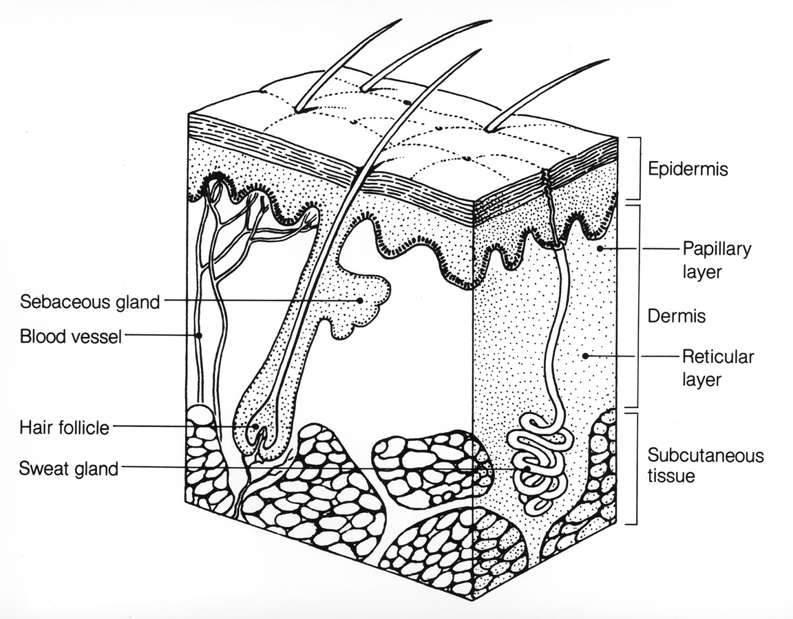
A cross-section of the skin: the epidermis and dermis on top, the subcutaneous tissue below, drawn as the lobules of fat the probe reads as a darker band. This is an anatomical diagram. It is not an ultrasound image. The labels are part of the original.
The fat under the skin has a clear look on ultrasound. It sits as a darker band below the bright lines of the skin. Bright lines of fibrous tissue run through it, the septa that hold the fat in place. The lobules of fat between them read as an even gray. The muscle below shows its own pattern under a bright sheet of fascia. The probe reads the whole stack from the skin down to the muscle in one image.
The fat is not one block. A sheet called the superficial fascial system runs through it and divides it into two layers. The layer above this sheet holds finer lobules packed tightly. It gives the body much of its surface shape. The layer below holds coarser lobules with looser packing. It carries the bulk in many areas. The probe shows the dividing sheet as a bright line partway down the fat. Reading the two layers apart matters to a clinic, because a treatment usually works on one of them. A device that cools the deep layer leaves the surface contour alone. A laser that tightens the superficial layer changes the shape a patient sees. The scan shows which layer is thick and which one a treatment is reaching.
The depth the probe reaches suits this work. At 18 to 24 MHz it sees about a centimeter in fine detail. Over a thin area, the cheek or the inner arm, the whole fat layer sits in range. Over a thick belly or flank the probe reads the upper layers in fine detail and shifts its settings to reach the rest. The layer aesthetic work acts on is the one near the surface, the layer the probe reads best.
Edema, scar and the changing fat
The fat does not always read the same way. When fluid builds in it, from swelling after a treatment or from a lymphatic problem, the lobules push apart and dark lines of fluid appear between them. The pattern takes on a look some call cobblestone. The probe tells this swollen fat from a layer that is thick on its own. One is fat. The other is fat with fluid in it.
This counts after a body-contouring treatment. A patient who swells in the days after a session can look as though the fat grew. The scan separates the swelling from the fat underneath it. A reading taken once the swelling settles gives the true thickness. A clinic that knows the difference avoids treating water as though it were fat. The clinic that reads the swelling correctly waits for it to settle before it measures. A number taken while the fluid is up reads too high. A reading at the wrong moment misleads as surely as no reading at all.
Scar tissue reads its own way too. Fat that was injured, by surgery or by an earlier injection, can hold bands of scar that show brighter than the fat around them. The probe maps these bands. That helps a clinician plan around tissue that will behave differently under a needle.
Putting a number on it
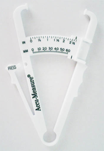
A skinfold caliper, the pinch tool a fat-layer scan replaces. It squeezes a fold of skin and fat and reads the width, so the squeeze itself biases the number. Ultrasound reads the layer without pinching it. The brand name and scale are part of the original photo.
The measurement is the point of the scan. The clinician freezes the image and drops two markers, one on the underside of the skin and one on the muscle fascia. The screen reads the distance between them. That distance is the fat thickness at that spot. The reading carries down to a fraction of a millimeter.
The number holds up to scrutiny. Studies of the technique report readings accurate to around two tenths of a millimeter at a single site. When different observers measure the same spots, their readings sit within about a millimeter of each other in almost every case.
What makes the reading trustworthy is the discipline around it. This is where ultrasound pulls ahead of the older tools. A caliper pinches a fold of skin and fat between two arms and reads the width, so the squeeze itself changes the answer, by a different amount on a soft belly than a firm thigh. Ultrasound reads the layer where it lies, with no fold to pinch and no compression to bias the result, as long as the hand keeps the probe light on the skin. The technique asks the clinician to hold the probe square to the surface, to press just enough for contact, and to return to the same marked spot on each visit. A reading taken with heavy pressure flattens the fat and reads it thin. A reading taken at a slightly different spot picks up a different thickness. Get those two habits right, the light touch and the fixed spot. Then the numbers from one day line up against the next inside the margin the method allows. A clinician used to ultrasound learns this in a short stretch of practice. The reward is a figure a clinic can stand behind, one that means the same in March as it does in June.
The reading sorts patients before it tracks them. A clinic offering body contouring measures the fat at the spots a patient wants treated. The number sets a starting point. It also separates the patient with a fat layer to treat from the one whose concern is loose skin, which a fat treatment will not change. That patient is steered toward a skin-tightening option, not a fat-reduction one. The scan tells the two apart before a course begins, when the choice still costs nothing.
Devices ask different things of the fat. A cooling head needs a layer thick enough to draw up into it. A heating device needs a margin of fat above the muscle. The scan reads whether the fat at the planned spot fits the device the clinic plans to use. A scan that finds the fat too thin for the planned device spares the patient a course that cannot work. That is a harder conversation to have after three paid sessions than before the first one. The reading turns a hopeful sale into an honest plan.
The marked grid
Reproducible numbers come from a marked routine. A clinician marks the spots to be measured on the skin, often a small grid over the area a patient wants treated. Each mark gets its own reading. The marks go on a chart or a photo, so the same points come back on the next visit. A measurement floats with the spot it was taken at, and the marks hold the spot still.
The probe stays square to the skin for each reading. A tilt stretches the fat in the image and reads it too thick. A press flattens it and reads it too thin. The clinician learns a light touch held square to the skin and repeats it the same way each time. The grid and the steady hand together give numbers that can be trusted against each other across months.
Tracking what a treatment does
The measurement repeats, so it follows change over time. Fat-reduction treatments work slowly. A course of cooling, of radiofrequency, or of an injection that dissolves fat shows its effect over weeks to months. The patient sees a vague difference in the mirror. The probe puts a figure on it. The clinician measures the same marked spots before the course and again after it.
The change shows up as a figure on the chart.
A drop of several millimeters in the fat layer is a result the patient can see beside the first reading. A layer that did not move points to a treatment that missed its depth or fell short. The timing of the second reading matters too. A cooling treatment shows its full effect over two to three months as the body clears the treated fat, so a reading taken too early catches a change that has not settled.
This turns a soft promise into a measured one. A patient asks whether a treatment worked. The honest answer is a pair of numbers, the fat before and the fat after. That stands on firmer ground than a clinician’s impression or a patient’s hope. It also lets a clinic spot the treatments that earn their place. The numbers protect the clinic as well. A patient who claims the treatment did nothing meets a chart with the fat before and after, or an honest reading that little changed. Either way the conversation rests on a measurement, not on two memories that disagree.
Reading the common spots
The fat under the chin is a frequent target. An injection that dissolves fat treats it. The area sits close to structures the needle has to respect, the platysma muscle and the salivary glands among them. The scan shows the fat layer below the skin and the muscle of the neck beneath it. It reads how thick the submental fat is and where the safe layer ends. A pad of fat thick enough to treat shows on the screen before the first injection. A thin pad over a prominent muscle points the clinician toward a different plan. A reading here keeps an injection in the fat and off the structures around it.
The abdomen, the flank, the thigh and the upper arm each carry a fat layer a patient may want reduced. The thickness runs from a few millimeters to several centimeters across these areas. The probe reads the upper part of a thick layer in fine detail and reaches the rest with a setting change. A clinic marks the spots a patient cares about, measures each one, and follows it over a course of treatment. The marks let the next visit land the probe on the same place, so the second number means something against the first. The thickness a clinic measures on a thigh says nothing about the flank, so each treated area carries its own baseline and its own follow-up. A single number for the whole body would hide the differences that decide a plan.
The breast and the back hold fat a clinic sometimes reads as well. Fat grafting to the breast leans on a measure of the recipient tissue. A back full of fibrous tissue resists a fat-reduction device differently from a soft abdomen. The probe reads the layer in each spot on its own terms, since a number from the belly says little about the thickness on the flank. Each treated area gets its own baseline and its own follow-up.
Fat grafting
Fat grafting moves a patient’s own fat from one area to another, the face or the buttock among them. The scan reads the donor site before the harvest, showing how much fat sits there and how deep it runs, so the clinician plans the harvest against a real thickness. A donor area too thin for the volume needed shows on the screen before the procedure starts. The scan also reads the recipient site over time. Some of the grafted fat survives. Some is reabsorbed. Measuring the fat layer at the recipient site before the graft and again months later shows how much volume held. That figure tells a patient what to expect from the next graft. It also tells the clinician how much to overfill the first time to allow for the loss. The grafted fat that survives stays as living tissue. The rest is gone by the later scan. Knowing the survived fraction lets a clinic quote a realistic result, not a hopeful one. It also lets the patient decide whether a second graft is worth the cost.
Cellulite and the septa
The dimpled look of cellulite comes partly from the septa, the fibrous strands that run through the fat. Some of these strands pull the skin down toward the deeper tissue. The fat between them pushes up. The result is the uneven surface a patient notices on the thigh or the buttock. The skin dimples where a strand holds it down and bulges where none does. The probe shows which dimples sit over a taut strand and which sit over a thicker pad of fat, a difference that decides whether cutting the strand will help.
Ultrasound shows these strands and the fat around them. It can show a septum running straight down to the skin at a dimple. A treatment that aims to release those strands gains a target the clinician can see, in place of working by feel through the skin. The same scan checks afterward whether the strand was cut. A dimple that stays after a treatment can be scanned again to find the strand the first pass missed.
Why a handheld probe fits this
The fat a clinic treats sits in the first centimeter under the skin. A high-frequency handheld probe reads that centimeter in fine detail at the point of care. An MRI or a CT scan reaches the whole body and far deeper, at a cost in money, time and access that a routine fat measurement does not justify. The surface layer is where the handheld probe resolves the fat as finely as anything, in the treatment room, in the minute the question comes up.
The probe runs from a phone or a tablet the clinic already owns. The clinician measures the fat in the same visit as the consultation, marks the spots, and saves the images with the patient’s notes. The next visit pulls up the first reading and lays the new one beside it. The whole record lives on the device the clinic carries from room to room.
The saved images carry weight beyond the next visit. A clinic that records the fat before and after a paid course can show a patient the result in numbers, can answer a later question with a stored image, and can weigh one device against another across many patients. None of that comes from a memory of how the skin looked. The probe turns a treatment a clinic sells into a measured service it can stand behind.
What the number does not tell
The fat thickness at a spot is a local reading. It is not a measure of whole-body fat or of health. A thin reading at one site sits beside a thick one at another. The scan answers one narrow question: how much fat sits here, in this layer, at this spot. The bigger questions of weight and health belong to a scale and a blood test, not to a probe that reads one spot.
The reading rests on a steady hand and the same spots each time. A probe pressed hard flattens the fat and reads it thin. A spot measured loosely on one visit and tightly on the next gives a change that is not real. The method earns its reliability from consistent technique. A clinic gets the value by measuring the same way every time.
The rest belongs to other tools. Weight, body composition and metabolic health each have their own measures.
Common questions about fat layer ultrasound
How accurate is ultrasound for measuring fat thickness?
Studies of the technique report readings accurate to around two tenths of a millimeter at a site, with different observers landing within about a millimeter of each other in almost every case. A change of a couple of millimeters between visits is a change that stands above the noise of the method.
Is ultrasound better than a caliper for body fat?
For a layer at a spot, yes. A caliper squeezes a fold of skin and fat. The squeeze changes the reading by a different amount on soft and firm areas. Ultrasound reads the layer where it lies, with no fold to pinch. It also separates the superficial and deep fat, which a caliper cannot do.
Can the scan show whether a fat-reduction treatment worked?
Yes, by comparing readings. The clinician measures the same marked spots before a course and again after it. A drop of several millimeters in the fat layer is a result the patient can see beside the first reading. A layer that did not change points to a treatment that missed.
What is the superficial fascial system on the scan?
It is a sheet of tissue that runs through the fat and splits it into a superficial layer and a deep one. The probe shows it as a bright line partway down the fat. The split counts because many treatments act on one layer. The scan shows which layer is being reached.
Does the fat reading tell me my body fat percentage?
No. The scan gives the fat thickness at a single spot. That is a local number, useful for planning and tracking a treatment in that area. It is not a measure of whole-body fat or of health, which other tools handle.
How much training does the measurement take?
A clinician used to ultrasound learns it in a short time. The skills are holding the probe square to the skin with a light touch and measuring the same marked spots each visit. Consistent technique is what makes the numbers from one day line up against the next.
Can ultrasound help with a double-chin treatment?
Yes. The fat under the chin is a common target for an injection that dissolves fat. The scan shows the thickness of that fat and the muscle and structures below it. It keeps the injection in the fat layer and off what sits beneath. It also tracks the change over the course of treatment.
About the Author
Julien Mercier
Senior R&D Engineer · Medical Ultrasound Transducer Development
Senior R&D Engineer with an M.S. in Applied Physics and over 15 years of experience in medical ultrasound transducer development, specializing in the design verification and performance testing of high-frequency imaging transducers. Currently leading the development and verification of the company’s next-generation high-frequency linear-array transducer, responsible for imaging performance evaluation and reliability analysis in preclinical testing. Brings extensive hands-on experience in piezoelectric element tuning, beamforming parameter optimization, and system-level performance testing.