Transrectal ultrasound of the prostate is a scan that puts a probe a short way into the rectum to read the prostate gland that sits against the rectal wall, mainly to guide a biopsy of it. The prostate lies just beyond the front wall of the rectum, an inch from a probe placed inside, close enough to read in fine detail and to steer a needle into.
Why the rectal route
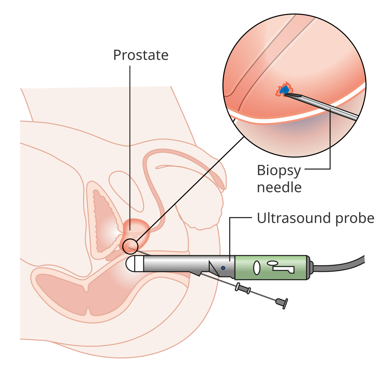
A prostate biopsy under transrectal ultrasound: the probe in the rectum reads the gland just ahead, with the biopsy needle guided through into the prostate, shown here in the inset.
The prostate is a small gland set deep in the pelvis, below the bladder and wrapped around the start of the urethra. It sits directly against the front wall of the rectum, with only that thin wall between it and the inside of the bowel. A probe placed in the rectum rests against the gland from behind, an inch or so away, with nothing in between to blur the view. No other route reaches the prostate so closely. The gland’s position is fixed and known. It sits below the bladder, ahead of the rectum, the urethra running down through its middle, the seminal vesicles tucked behind its upper edge. That arrangement puts the back of the gland, the part a probe in the rectum meets first, right against the rectal wall. A scan from there reads the prostate from the side cancers tend to favor, the peripheral zone at the back.
That closeness is what the rectal route is for. A probe an inch from the gland runs at a high frequency, the kind that draws fine detail over a short reach, and reads the prostate sharply from end to end. The same gland seen from the lower belly sits too deep, behind the bladder and the pubic bone, for a clear picture. The rectal route trades a moment of discomfort for a view of the prostate nothing else matches. The numbers behind the closeness are the same as for any internal scan. A probe an inch from the gland runs in the higher range, where a millimeter of tissue reads as a millimeter on the screen. The peripheral zone, the seminal vesicles and the capsule that wraps the gland all read clearly from that range. The detail the rectal route gives is the reason it is the route the prostate is read through.
The route also lines the probe up for the needle. A biopsy of the prostate has to place a needle into the gland. The rectal probe sits exactly where a needle can be guided through it into the prostate just ahead. A guide on the probe sets the needle’s path on the screen, so each core is taken under direct view. The rectal route is the one that both reads the gland and aims the needle into it.
The probe is shaped for the route. An endocavity transducer made for the rectum is slim, with its array set to look forward and to the sides into the gland, and a channel or a clip to carry the biopsy needle. The whole instrument is built to sit in the rectum, read the prostate ahead, and guide the needle in. The route and the probe are made for each other. The needle guide is the piece that turns reading into biopsy. A bracket clips along the probe and holds the needle on a fixed line, that line drawn onto the screen as a dotted track from the probe into the gland. The urologist lines the track up with the spot to sample and fires. The guide fixes the path, so every core is aimed before it is taken.
Transrectal prostate ultrasound at a glance
Route
a probe in the rectum, against the gland
Why this route
the prostate sits an inch away, behind the rectal wall
Main job
guiding a biopsy of the prostate
Systematic biopsy
around 12 cores spread across the gland
Prostate volume
length × width × height × 0.52
The probe
a slim endocavity transducer, IPX7-sealed for cleaning
What the prostate looks like on the scan
On the scan the prostate reads as a rounded gland, walnut-sized in a younger man and larger with age, its outline clear against the tissue around it. The gland has zones the scan can tell apart, the peripheral zone toward the rectum where many cancers begin, and the inner transition zone that enlarges in older men. Reading those zones, and the line between them, is part of how the gland is assessed. The two zones read a little differently, and a reader learns both: the peripheral zone, the outer shell toward the rectum, an even mid-grey when healthy and the part watched closely; the transition zone within, growing nodular and enlarged with age. The zone a finding sits in shapes what it is likely to mean, which is why the line between them is read with care.
A cancer in the prostate sometimes shows as a darker, hypoechoic patch in the peripheral zone, though many cancers are not visible on the grey scan at all. The scan reads the size of the gland, its shape, any obvious lesion, and the way it sits against the bladder and the seminal vesicles behind it. Much of what the scan does, in the end, is set the map the biopsy needle works across, whether or not a lesion stands out on the picture. The grey picture has a known blind spot. The reader works around it. Many prostate cancers look just like the tissue around them on ultrasound, neither darker nor brighter, invisible on the scan alone. For that reason the biopsy does not rely on seeing the cancer; it samples the gland systematically, trusting the map more than the picture. The scan’s job is to read the gland and place the needle; spotting every cancer by eye is beyond it.
Guiding the biopsy
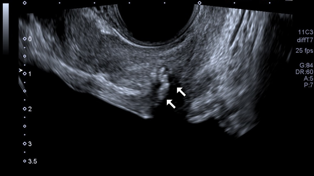
A prostate on a transrectal scan, the curved probe at the top reading the gland from the rectum. The white arrows mark small calcifications, the prostatic stones that are a common incidental finding.
Guiding the biopsy is the main work of transrectal ultrasound. With the probe against the gland and a needle guide clipped to it, the path the needle will take is drawn on the screen. The urologist fires a spring-loaded needle along that path to take a thin core of tissue. Each core is taken under the live picture, the needle seen entering the gland, and dropped into a pot for the pathologist. How a TRUS-guided prostate biopsy is performed, step by step, is the procedure this guidance is built for. The biopsy needle is a spring-loaded device that takes a slim core in a fraction of a second. Loaded into the guide, it is advanced to the edge of the target and fired, a fine notch in its tip filling with a thread of tissue on the way through. The core, a few millimeters of gland, drops into a labelled pot for the pathologist. A set of these threads, read under the microscope, is what a diagnosis is built from.
The cores are spread across the gland on a systematic plan. Rather than sampling one spot, the urologist takes a set of cores from named regions of the prostate, base, middle and apex, on both sides, to sample the whole gland evenly. Spread this way in a systematic set of cores, the biopsy reads the prostate broadly, an approach a single-center study of more than a decade of biopsies found significantly raises the cancer detection rate over the sparser schemes it replaced. The reasoning behind sampling the gland in twelve cores is the systematic map at the heart of the biopsy.
An MRI of the prostate has changed where the needle goes. When an MRI has marked a suspicious area, that picture can be brought together with the live ultrasound so the needle is steered straight to the marked spot, a targeted core taken from the area likeliest to hold cancer. MRI and ultrasound fusion for a targeted biopsy joins the detail of the MRI to the live aim of the ultrasound. It is often done alongside the systematic cores.
The route to the gland is itself a choice the biopsy makes. The needle can pass through the rectal wall into the prostate, the transrectal way, or through the skin of the perineum, the transperineal way, each guided by the same rectal probe. The transperineal route avoids carrying bowel organisms into the gland, and many centers have moved toward it for that reason. The probe reads and aims the same way for either path the needle takes. The two routes differ in where the needle crosses into the gland. The older transrectal path sends it through the rectal wall, the short way in. The transperineal path, through the cleaned skin between the legs, keeps bowel organisms out of the gland. Many centers have moved to it for the lower infection it brings. The same rectal probe reads and guides for either one.
Through all of it the picture is live. The urologist watches the needle on the screen from the moment it enters, tracking the bright line of it into the gland and out, core after core. Keeping the needle in view is what makes a deep needle pass a controlled one, each core placed where the plan calls for it. The guidance turns the biopsy from a blind sampling into a mapped, watched procedure. That live view is what makes the count of cores safe to take. A dozen needle passes into a gland an inch across are safe only because each is watched on the screen, placed and checked before the next. The urologist sees the needle clear the bladder and the urethra, sees it enter the zone the plan calls for, sees the gland after. The number of cores the diagnosis needs is reachable because every one is taken in view.
Reading the size of the gland
The scan also measures how big the prostate is, a number that matters in its own right. The volume is worked out from three diameters of the gland, its length, its width and its height, multiplied together and by about 0.52, the factor that treats the gland as an ellipse. That volume helps read a raised PSA, since a large gland makes more PSA on its own, and it guides the treatment of an enlarged prostate. Measuring the prostate volume on the scan turns three quick calipers into a figure the rest of the assessment leans on. The volume earns its place through PSA density. Dividing the PSA level by the gland’s volume gives a density that reads a raised PSA in the light of the gland that made it, since a big benign prostate makes more PSA on its own. A high PSA from a small gland reads as more worrying than the same PSA from a large one. The volume the scan measures is what lets that density be worked out at all.
When the scan is done, and on whose advice
A transrectal prostate scan and biopsy usually follow a sign that the prostate needs a closer look. A raised level of prostate-specific antigen in the blood, or a prostate that feels abnormal on a finger examination, is the common trigger, the reason a man is sent for the scan that may end in a biopsy. Assessing a raised PSA with ultrasound is the step that turns a blood result into a look at the gland itself. The trigger is rarely a symptom of cancer, since early prostate cancer is usually silent. It is a number on a blood test, the PSA, or a firmness the examining finger feels, that sends a man down this path before he feels anything wrong. The scan and the biopsy that follow are how a quiet sign is run to ground. Catching the cancer in its silent stage is the point of acting on the PSA.
How the biopsy is done follows the advice of the urological bodies, who set out who should be biopsied and how. Their guidance has shifted in recent years toward using an MRI before the biopsy and toward the transperineal route, changes a clinic reads from the current recommendations. The shift has been real over the last decade. An MRI before the biopsy, once uncommon, is now widely advised, used to find a target and sometimes to spare a man a biopsy he does not need. The route has moved toward the perineum for the lower infection it carries. A clinic reads the current guidance to keep its practice in step, since the recommendations change when the evidence does. The detail of each body’s position is one to read on its own.
The European guidance is set by the European Association of Urology. What the EAU says on transrectal biopsy with ultrasound sets out the European position, which a clinic reads alongside the American one to see where the field stands.
The wireless probe for the work
A handheld wireless probe brings the whole of this to the bedside. A slim endocavity transducer made for the rectum, a micro-convex end-fire probe that looks forward into the gland, runs from a phone or a tablet and reads the prostate and guides the needle without a cart in the room. A urology clinic doing biopsies on its own list reaches for a probe it can keep close, the picture on a screen at the couch. Choosing a wireless transrectal probe comes down to the frequency, the needle guide, the seal for cleaning and the wireless link.
The cleaning the probe needs shapes the choice as much as the picture. An internal probe is disinfected to a high level between patients, so it has to carry a watertight seal that survives the cleaning, the same standard the transvaginal probe meets. A probe bought for prostate work is read for that seal alongside its imaging, since a probe that cannot be cleaned to standard cannot be used at all. The needle guide is read as closely as the picture. A probe for prostate work has to carry a guide that holds the needle on a true line and shows that line on the screen, since the whole biopsy is aimed along it. A guide that fits the needle the clinic uses, and that reads cleanly on the app, is part of what a buyer checks. The probe, the guide and the app are chosen together for the biopsy they will do.
Put together, transrectal ultrasound is the close, guided reading the prostate is assessed and biopsied through. It reaches the gland from an inch away, maps the cores across it, steers the needle to a target the MRI marked, and measures the size that the rest of the workup leans on. On a handheld probe at the couch, cleaned to standard between patients, it carries the diagnosis of prostate cancer from a raised blood test to a tissue answer. What began as a number on a blood panel ends, by this route, as a set of cores a pathologist can read. The scan reaches the gland, the guide aims the needle, the systematic map covers the prostate, and the MRI fusion points to what the grey picture cannot show. On a handheld micro-convex probe at the couch, cleaned to standard between men, transrectal ultrasound is the close, guided reading the diagnosis of prostate cancer turns on.
Common questions about transrectal prostate ultrasound
Why is the prostate scanned through the rectum?
Because the prostate sits directly against the front wall of the rectum, an inch from a probe placed inside. From there the probe reads the gland sharply at a high frequency and lines up a needle to biopsy it, neither of which a scan from the lower belly can do well, since the gland sits too deep behind the bladder and the pubic bone. The rectal route is the close, direct way to the gland.
What is the scan mainly used for?
Guiding a prostate biopsy. The probe reads the gland and draws the needle’s path on the screen, so cores of tissue are taken under direct view. The scan also measures the volume of the prostate and reads its zones and any obvious lesion. Much of what it does is set the map the biopsy needle works across, whether or not a cancer is visible on the grey picture.
How many cores are taken in a prostate biopsy?
A systematic biopsy takes around twelve cores, spread from the base, middle and apex on both sides to sample the whole gland evenly. Sampling the gland broadly this way is the standard for diagnosing prostate cancer. When an MRI has marked a suspicious area, extra targeted cores are taken from that spot, often alongside the systematic set.
What is MRI-ultrasound fusion biopsy?
It is a biopsy that brings an MRI of the prostate together with the live ultrasound. A suspicious area marked on the MRI is overlaid on the live scan, so the needle is steered straight to it for a targeted core. Fusion joins the detail of the MRI, which reads soft tissue well, to the live aim of the ultrasound, which guides the needle in real time.
How is the prostate volume measured?
From three diameters of the gland on the scan, its length, width and height, multiplied together and by about 0.52, the factor that treats the gland as an ellipse. The volume helps read a raised PSA, since a larger gland makes more PSA on its own, and it guides the treatment of an enlarged prostate. It is three quick caliper measurements that feed the rest of the assessment.
Does a transrectal probe need cleaning like other internal probes?
Yes. A probe used in the rectum is disinfected to a high level between patients, the same standard as a transvaginal probe, so it has to carry a watertight seal that survives the cleaning. A probe bought for prostate work is checked for that seal alongside its imaging, since a probe that cannot be cleaned to standard cannot be used.
About the Author
Julien Mercier
Senior R&D Engineer · Medical Ultrasound Transducer Development
Senior R&D Engineer with an M.S. in Applied Physics and over 15 years of experience in medical ultrasound transducer development, specializing in the design verification and performance testing of high-frequency imaging transducers. Currently leading the development and verification of the company’s next-generation high-frequency linear-array transducer, responsible for imaging performance evaluation and reliability analysis in preclinical testing. Brings extensive hands-on experience in piezoelectric element tuning, beamforming parameter optimization, and system-level performance testing.