How Transrectal Ultrasound TRUS Guided Prostate Biopsy Is Performed
A transrectal ultrasound-guided prostate biopsy is the procedure that takes small cores of tissue from the prostate, using a probe placed in the rectum to see the gland and steer a needle into it. A urologist orders it after a raised PSA or an abnormal feel on a finger examination, to settle whether the prostate holds cancer. The cores it takes go to a pathologist. The reading of them under the microscope is the answer the whole procedure exists to reach. The steps below are how that biopsy is carried out, from the preparation a man makes at home to the result he waits for afterward.
Before the day of the biopsy
A few preparations come before the biopsy itself. The urologist prescribes a short course of antibiotics to cover the procedure, since passing a needle through the rectal wall can carry bacteria toward the gland. A blood-thinning medicine is usually paused for several days beforehand, on the doctor’s instruction, to lower the risk of bleeding. An enema clears the lower rectum on the morning of the biopsy so the probe reads against a clean wall. None of this is onerous. Many men carry on normally until the day. The antibiotic carries real weight among these steps. A needle through the rectal wall can carry gut bacteria toward the gland, so a course timed around the procedure guards against an infection. The exact drug follows local guidance, sometimes chosen from a swab of the rectum taken beforehand. A man takes it as directed and finishes the course.
The man is told what the day will hold. The biopsy is an outpatient procedure, done in a clinic room in well under half an hour, with no general anesthetic and no overnight stay. He signs a consent form that sets out the common after-effects and the small risks before anything begins. Arranging a lift home is sensible for peace of mind. Many men feel well enough afterward to drive themselves. The clinic gives clear instructions for the days on either side. The man learns when to pause and restart any blood thinner, how to take the antibiotic, and what the enema involves. He is asked about allergies and about any heart valve or implant that might change the antibiotic plan. Knowing the steps ahead settles much of the worry a biopsy brings.
The biopsy in figures
Time in the room
well under 30 minutes
Anesthetic
a local periprostatic block, no general
Needle
18-gauge spring-loaded core, through a probe-mounted guide
Cores taken
about 12, spread across the gland
Blood afterward
in urine and semen for days to a few weeks
Infection risk
a few percent, by the transrectal route
On the table: position and the first scan
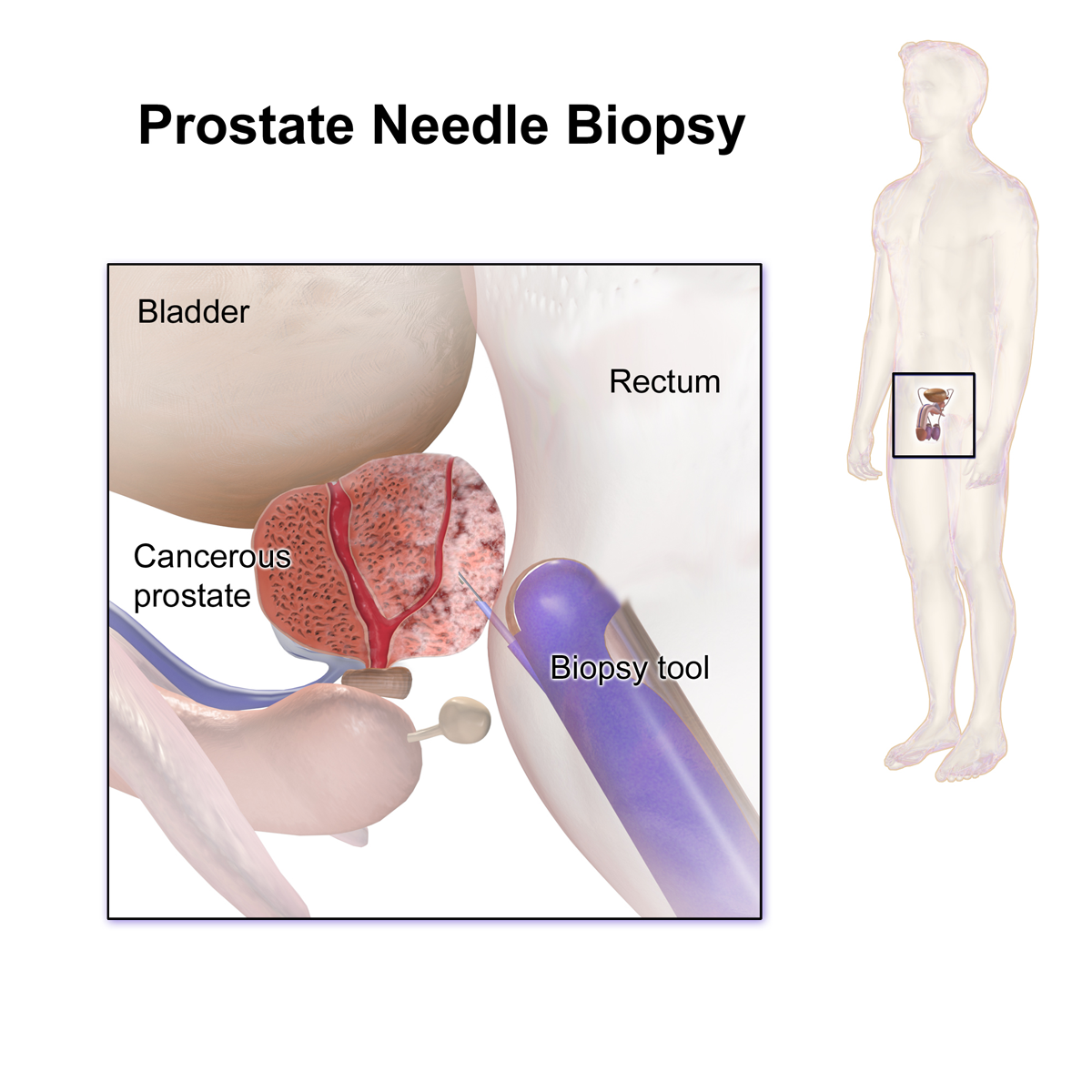
A prostate needle biopsy: the biopsy tool sits in the rectum against the prostate, the bladder above it, the needle reaching the gland. The small inset marks where the prostate lies in the body.
The biopsy begins with the man lying on his side. He lies on his left, knees drawn up toward the chest, the position that opens the rectum to the probe and steadies the gland in front of it. The urologist starts with a finger examination of the prostate, feeling its size and any firm area before the probe goes in. The probe follows, slim and well lubricated, covered with a clean sheath, sliding in only as far as the gland. A man feels the pressure of it more than any pain at this stage. The side-lying position is chosen with care. Drawing the knees up flattens the angle between the rectum and the prostate, so the probe reaches the gland with the least pressure. A drape keeps the man covered throughout. A nurse stays alongside to talk him through each step.
The first job of the probe is to read the gland, before any needle is loaded. The urologist sweeps through the prostate in two planes, measuring its three diameters for a volume and looking over its zones. A dark area in the outer zone is noted carefully, since it may mark a spot that earns an extra core later. The seminal vesicles behind the gland and the bladder above it are checked at the same time. This survey builds the map that the rest of the procedure works from. The volume it measures has a use of its own. Three diameters of the gland, multiplied together and by a set factor, give a size that helps read the PSA, since a larger gland makes more PSA on its own. The reading also notes the shape of the gland and how it meets the bladder neck. All of this is set down before the first core.
The picture on the screen guides everything that follows. The probe shows the gland in real time, with the bright wall of the rectum just in front of it and the bladder sitting above. The urologist confirms these landmarks and settles where the cores will be taken. Nothing touches the gland until the picture is clear and the plan is set in mind. The whole biopsy is steered by what shows on that screen. A frozen frame can be saved to the record, marking where a core was taken. The image is part of the documentation the biopsy leaves behind.
Comfort is looked after through this opening part. The probe is no wider than an examining finger. The survey scan takes only a minute or two to complete. A man notices fullness and pressure, with no sharp pain so far. The anesthetic that makes the cores themselves painless is the next step.
Numbing the gland
The cores are taken under a local anesthetic placed exactly where the gland’s nerves gather. The urologist injects a small amount of lidocaine at the base of the prostate, where the nerves enter beside the seminal vesicles, with a little more toward the apex. According to the StatPearls review of image-guided prostate biopsy, this periprostatic nerve block, given at the base and the apex, relieves the pain of the biopsy by blocking a sensitive somatic branch of the pudendal nerve. A few minutes are allowed for the block to take full hold. Only then does the first core follow. The block changes the experience entirely. The cores register as pressure and a click once it has taken hold. The injection itself feels like a small pinch deep inside, over in a moment. A urologist who takes time over the block is rewarded with a still, comfortable patient for the cores.
Taking the cores
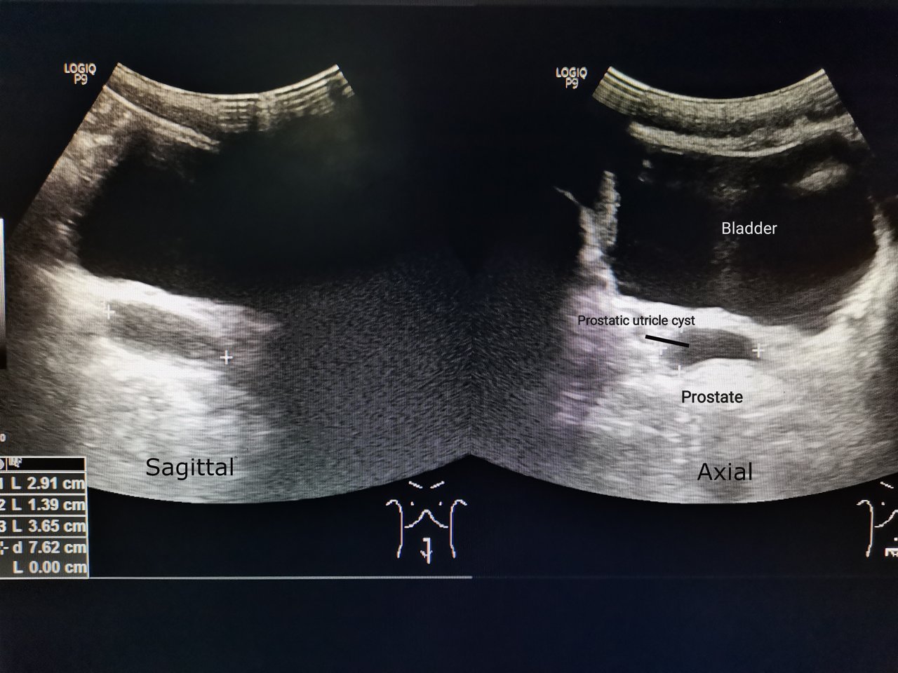
A prostate on transrectal ultrasound, in the sagittal and axial planes, the kind of live picture the urologist reads during the cores. The labelled spot is a prostatic utricle cyst, a benign midline finding.
Each core is taken with a spring-loaded needle that fires in a fraction of a second. The needle rides in a guide clipped onto the probe, so its path appears on the screen as a fixed line before anything fires. The urologist lines that guide line up with the spot to sample and presses the trigger. The needle darts in and back out faster than the eye can follow it. It leaves carrying a thin thread of tissue caught in a notch near its tip. The needle is a standard core-biopsy device. It is spring-loaded, fired by a trigger the urologist controls, set to travel a fixed short distance into the gland. The speed is what makes each pass nearly painless under the block. A practiced hand places core after core in a steady rhythm.
The core itself is a thread of gland about a centimeter long. A small notch near the needle tip opens, fills with a sliver of tissue on the way through, and closes around it. That thread drops into a labelled pot, kept apart from the others so the pathologist knows which part of the gland it came from. Each core is a separate record of one named region of the prostate. Keeping them in order is part of how the gland is mapped. The pots are labelled by the region each core came from. Some clinics keep each core in its own pot. Others group them by side or by level. The pathologist reads them in that order, building a picture of where any cancer sits, since a finding means little without knowing where in the gland it lies.
The cores are spread across the gland to a set plan. The urologist takes a series of them from named regions, the base, the middle and the apex on each side, so the sampling covers the whole prostate evenly. Where the scan or an earlier MRI marked something suspicious, an extra core is aimed straight at it. This spread is what gives the biopsy its reach across a gland where cancer often hides from the grey picture. The detailed pattern of where each core is taken is a subject in its own right.
Through all of it the picture stays live on the screen. The urologist watches the needle enter the gland with each firing, core after core, checking one before moving to the next. Watching the needle is what keeps a dozen passes into a small gland controlled and safe. The man hears a sharp click at each core and feels a brief pressure under the anesthetic. The live image is what turns the biopsy from a blind sampling into a watched one. Seeing the needle is what keeps the cores where they belong. The urethra runs down the middle of the gland. The nerves run along its sides. The urologist steers each pass clear of both, watching the bright line of the needle reach its mark and withdraw.
The number of cores is more than just a few. A standard biopsy takes around a dozen, each a quick firing of the needle, the whole set finished in a few minutes. The clicks come close together once the block has taken hold. The cores together are what let the pathologist read the gland, since any one alone samples too little. The set as a whole is the specimen the diagnosis rests on. A larger gland, or an MRI with several targets, can call for a few more than the dozen. The urologist takes what the gland and the plan ask for.
Through the rectum or the perineum
This biopsy takes the transrectal route, the needle passing through the wall of the rectum to reach the gland just beyond it. The probe and the needle share the one passage, which is what makes the procedure quick and needs no skin incision. The short distance from the rectal wall to the prostate is why the route reaches the gland so directly. It is the long-standing way a prostate biopsy has been done.
A second route has grown more common in recent years. The transperineal route passes the needle through the cleaned skin between the scrotum and the anus, with the probe still in the rectum to read the gland. Passing outside the bowel, it carries fewer of the infections that can follow a needle through the rectal wall. Many centers have moved toward it for that reason. The scan and the needle guide work the same way whichever skin or wall the needle crosses. The shift toward the perineum has a clear driver. Infections after a transrectal biopsy are uncommon. They can be serious when they do take hold. A route that keeps the needle out of the bowel removes that path for bacteria, which is why many guidelines now lean toward it for men at higher risk.
The choice of route rests with the urologist, by training and by the man’s own situation. Both reach the same gland and read it with the same probe in the rectum. A man is told which route his biopsy will take and the reason for it. The steps of numbing, firing and collecting the cores run much the same on either path. A man with a question about the route can raise it beforehand. The reasoning is straightforward to explain.
Afterward
Recovery from the biopsy is quick. The man rests a few minutes, passes urine to confirm the flow is fine, and goes home within the hour. No stitches are needed. The small needle tracks close on their own. Normal activity resumes within a day or two on the urologist’s advice. Men commonly describe the experience as easier than they had feared. A short rest in the clinic is usual before leaving. The man sits for a few minutes, passes urine to confirm the stream is clear, and dresses. He is given written notes on what to expect and the numbers to call. A light day is sensible, with a return to full activity once any soreness settles.
Some after-effects are expected and harmless. Blood in the urine, in the semen, and from the back passage is common for days to a few weeks, settling on its own. The blood in the semen tends to look dark and to last the longest of the three, which is normal and no cause for worry. A man is warned of all this in advance so it does not alarm him when it appears. Drinking plenty of water in the days after helps the urine clear. The flecks of blood can come and go over the weeks. This is the gland settling down.
A few signs call for prompt attention. A fever, shaking chills, or trouble passing urine after the biopsy can mean an infection is taking hold. Any of these needs same-day medical help without delay. Heavy bleeding that does not settle is the other reason to seek care. These problems are uncommon under good antibiotic cover. Every man is still told plainly to watch for them.
The result is what the man is waiting for. The pathologist examines each core under the microscope, looking for cancer and grading any that is found. The report comes back over several days to a week or two. The urologist then goes through it with the man and explains what it means. That reading, core by core, is the answer the biopsy was carried out to reach.
Where the procedure sits
The biopsy is one step along a longer path. It follows a raised PSA or an abnormal examination, and often an MRI that points to where a cancer might lie. The cores it takes are what turn a suspicion into a firm diagnosis, with a grade that guides what comes next. Nothing short of tissue settles the question the way a biopsy does. The grade the pathologist assigns is what shapes the next decision. A low-grade finding may lead to careful monitoring, with treatment held back for the time being. A higher grade points toward treatment chosen to the man’s situation. The biopsy turns a number and a suspicion into something a urologist and a man can plan around.
A handheld ultrasound brings the whole of this into the clinic room. A slim transrectal probe run from a tablet reads the gland and guides the needle, with no cart standing at the couch. The picture, the guide line and the cores are the same as on any larger machine. The procedure travels to wherever the probe and a steady, trained hand can go. That portability is changing where the biopsy can happen. A urology clinic without a fixed ultrasound room can still offer it, the scanner kept in a drawer between patients. The cleaning the probe needs between men is the same high standard as for any internal probe. The picture, the block and the cores follow the steps set out here, wherever the couch happens to be.
Common questions about the biopsy procedure
Does a prostate biopsy hurt?
The cores are taken under a local anesthetic, a periprostatic nerve block placed where the gland’s nerves gather, so the firing of the needle is felt as no more than a brief pressure. A man hears a sharp click at each core. The probe in the rectum gives a sense of fullness through the procedure. Men generally find it far easier than they expected once the block has taken hold.
How long does the procedure take?
The biopsy itself takes only a few minutes once the anesthetic has taken hold. The whole visit, including the scan and the settling time, runs well under half an hour. It is done in a clinic room with no general anesthetic. The man goes home within the hour. There is no overnight stay.
How many cores are taken?
A standard biopsy takes around a dozen cores, spread from the base, the middle and the apex on each side so the whole gland is sampled evenly. Where an MRI or the scan marked a suspicious spot, an extra core is aimed there. Each core is a thin thread of tissue about a centimeter long. The cores together are what let the pathologist read the gland.
Is blood afterward normal?
Yes. Blood in the urine, in the semen, and from the back passage is common for days to a few weeks and settles on its own. The blood in the semen tends to look dark and to last the longest. Drinking plenty of water helps the urine clear. A man is warned of all this in advance so it does not alarm him.
What are the warning signs after a biopsy?
A fever, shaking chills, or trouble passing urine can mean an infection is taking hold, and any of these needs same-day medical attention. Heavy bleeding that does not settle is the other reason to seek help promptly. These problems are uncommon under good antibiotic cover. Every man is told plainly to watch for them before he goes home.
When do the results come back?
The pathologist examines each core under the microscope, looking for cancer and grading any that is found. The report usually comes back over several days to a week or two. The urologist then goes through it with the man and explains what it means. That reading is the answer the biopsy was carried out to reach.
About the Author
Julien Mercier
Senior R&D Engineer · Medical Ultrasound Transducer Development
Senior R&D Engineer with an M.S. in Applied Physics and over 15 years of experience in medical ultrasound transducer development, specializing in the design verification and performance testing of high-frequency imaging transducers. Currently leading the development and verification of the company’s next-generation high-frequency linear-array transducer, responsible for imaging performance evaluation and reliability analysis in preclinical testing. Brings extensive hands-on experience in piezoelectric element tuning, beamforming parameter optimization, and system-level performance testing.