12 Core Prostate Biopsy How To Sample Under Ultrasound Guidance
A twelve-core prostate biopsy takes twelve small cores of tissue from set points across the gland, mapped so the whole prostate is sampled evenly. The number and the placement are not random. They follow a template that catches cancer in a gland where it often hides from the grey picture. Each core has its own named place. The set of them maps the prostate region by region.
Why the whole gland is sampled
The reason for twelve cores starts with what ultrasound cannot show. Many prostate cancers look the same as the tissue around them on the grey scan. A gland with a real cancer can look completely normal on the picture. The scan cannot point the needle at a tumor it cannot see. The sampling has to do the work the picture cannot. This is the hard truth at the center of the biopsy. A man can carry a real cancer behind a normal-looking scan. The needle goes in by a plan drawn before the scan even starts. The whole design of the twelve-core biopsy follows from this one fact about the grey picture. A blind biopsy of one or two spots would be a gamble against those odds. The plan removes the gamble by sampling broadly. The twelve cores buy coverage across the whole gland.
So the biopsy does not rely on seeing the cancer. It samples the whole gland by a fixed plan, trusting the map. Spread evenly across the prostate, the cores catch cancer by covering the ground where it tends to grow. The sampling stands in for a picture the scan cannot give. A dozen cores across the gland is a broad net thrown over hidden disease. The logic is the logic of searching a field by walking it in rows. No single step finds the lost thing. The pattern of steps, end to end, is what covers the ground. The twelve cores walk the gland in just that way.
The peripheral zone is where the plan concentrates. Cancer of the prostate usually begins in this outer shell, the part toward the rectum. The template weights its cores toward the peripheral zone for that reason. The sampling follows where the cancer is likely to be. The plan is a map of probability across the gland. The weighting is not a guess. Decades of biopsies have shown where the cancers turn up. The template puts its cores where the odds run highest, in the peripheral zone toward the back of the gland. The plan carries that learning in its shape.
Even coverage is the goal of the template. The cores are placed to cover the gland from base to apex and from side to side. No large region of the prostate is left untouched by the plan. The aim is a fair sample of the whole gland in twelve threads of tissue. A gap in the map is a place a cancer could hide. Even coverage is what closes those gaps. A core at the base, a core at the middle, a core at the apex, on each side, leaves no large stretch unsampled. The cores sit far enough apart to stay separate, with little ground left unsampled between them. A fair map has no dark corners. The design has been tested against thousands of glands. A template that left a region thin would let cancers slip through unfound. The twelve-core grid earned its place by catching what looser sampling skimmed over.
The twelve-core map
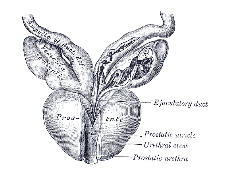
The prostate seen from behind, the surface a transrectal probe faces through the rectal wall. The twelve cores are spread across the gland this view shows, from the base down to the apex on each side.
The twelve cores fall into a grid across the gland. The prostate is read in three levels from top to bottom, the base, the middle, and the apex. Each level is sampled on the left side and on the right. Each side, at each level, gives a medial core and a lateral one. The grid is the same in every standard biopsy. Reading the gland in levels keeps the sampling orderly. The base sits up near the bladder. The apex sits down at the tip, by the muscle that controls flow. The middle lies between them. Naming the three levels gives the urologist a fixed frame to work the cores across.
That grid comes to twelve. Three levels, two sides, two cores at each place gives the count, six on the left and six on the right. The names follow the grid, a right-base-lateral core, a left-apex-medial core, on through the set. Each name fixes one spot on the gland. The table below sets the twelve out in full. The naming is plain once the grid is clear. Side, then level, then medial-or-lateral names each of the twelve. A note of left-base-medial points to one exact place. The pathologist reads the same names off the pots. The shared naming is what lets a finding be placed back on the gland. The names travel on the request form and on every pot. A lab tech, a pathologist, and a urologist all read the same labels. The naming keeps the map intact from the couch to the report.
The twelve-core systematic template
Level of the gland
Left side
Right side
Base (top, near the bladder)
medial core + lateral core
medial core + lateral core
Middle
medial core + lateral core
medial core + lateral core
Apex (tip, farthest from the probe)
medial core + lateral core
medial core + lateral core
Total
6 cores
6 cores
The lateral cores carry much of the weight. The far edges of the peripheral zone hold cancers the older sampling left out. Aiming cores out toward the sides reaches that ground. The lateral, apically-directed cores are the heart of the modern template. Leaving them out is what made the old scheme miss disease. The peripheral zone wraps around the back and the sides of the gland. The lateral cores angle outward to reach its far edges. Those edges are the corners a straight-back sampling skims past. The reach to the side is the single biggest gain of the modern template.
The apex gets careful attention in the plan. The tip of the gland sits farthest from the probe in the rectum. The apex is a common home for cancer and an easy region to under-sample. Cores angled into the apex reach a place a careless biopsy skims. A good template lands on the apex squarely. Reaching the apex takes a deliberate angle. The needle comes up toward the tip from below, the probe tilted to set the guide on it. A core taken short of the apex leaves the tip itself unsampled. The careful operator makes sure the apical cores reach the end of the gland.
The result is a fair map of the gland. Twelve cores, named and placed, cover the prostate from end to end and side to side. The pattern holds the same from one gland to the next. That sameness lets one biopsy be compared to another, and a repeat to its first. The map is what a systematic biopsy means. Sameness is the quiet strength of the template. Two urologists working the same plan sample the same gland the same way. A repeat biopsy a year on lands its cores where the first set went. The fixed map turns a single biopsy into a record that later scans can be read against. On active surveillance, a man may have the same template repeated over years. Each round lands on the same named regions. The fixed plan lets one year be set beside the last with confidence.
Why twelve
The number twelve came from improving on an older scheme. The first systematic biopsies took six cores, the sextant pattern, sampling down the middle of each side. The six-core plan left out the lateral edges, and cancers there went unfound. Adding six laterally-directed cores to the old six made the twelve-core template. According to a study comparing the two schemes, the extended twelve-core biopsy raised the cancer detection rate by roughly twenty percent over the sextant. The American Urological Association recommends the twelve-core scheme with its apical and far-lateral cores. The jump from six to twelve was a real gain in cancer found. The lateral cores reached ground the sextant never touched. The cost was six more quick passes of the needle under the same block. The trade was settled long ago. Twelve became the standard a systematic biopsy is built around.
Placing each core under the picture
Each named core is placed under the live scan. The probe shows the region the plan calls for. The urologist lines the needle guide up with that spot on the screen. The bright line of the needle confirms the place a moment before the core is taken. The picture turns a name on a plan into a real position in the gland. Nothing is taken by feel alone. The screen shows the needle path before the trigger, a dotted line into the chosen region. The operator nudges the probe until that line sits where the plan wants the core. The picture makes each of the twelve a placed shot.
The picture keeps each core where the map wants it. The urethra runs down the middle of the gland. The nerves run along its sides. Both are seen on the screen and kept clear of the needle. Each core lands in its named region, checked under the live picture, so the dozen passes stay accurate from first to last. Watching also keeps the passes safe. The bladder above and the wall of the rectum stay in view through each core. The operator sets the needle clear of them every time. The live picture is what lets a dozen passes into a small gland stay controlled. A short run of clicks marks the twelve cores taken in turn under the block. The whole grid is sampled in a few minutes at the couch. The picture guides each pass to its named place.
Keeping the cores in order
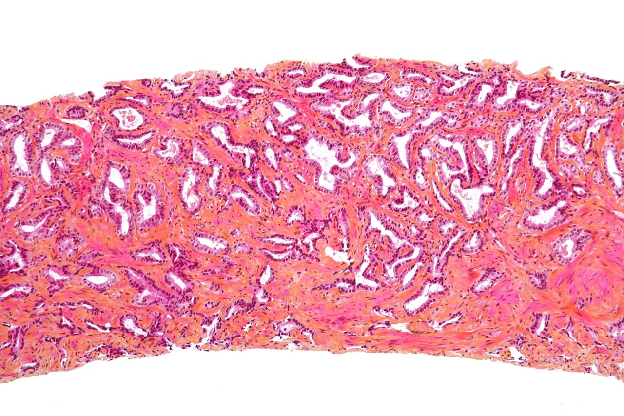
A prostate biopsy core under the microscope, the curved thread of tissue one needle pass yields. The pathologist reads each of the twelve like this, naming which holds cancer and how much.
The cores are kept apart by the region each came from. Each core goes into its own labelled pot, named for its place in the gland. The label travels with the core to the pathologist. The order is part of the information the biopsy carries. A core without its place tells far less. The handling is careful from the moment the core leaves the needle. Each thread goes straight into its own labelled pot. The pots are kept in the order of the plan. A mix-up of pots would scramble the map the biopsy is built to give. The order is guarded as closely as the cores.
The labelling lets a finding be placed on the gland. A cancer in the right-apex cores tells the team where in the prostate it sits. The map of positive cores carries the side, the level, and the spread of the disease. A finding means much more once its place is known. The named cores turn a yes-or-no into a map. A pathologist reads each pot on its own. A report comes back core by core, naming which held cancer and how much. The grade and the length of cancer in each core are set down by name. The team reads that report as a map of the gland, region by region. The count of positive cores carries its own weight. A few positives in one region read as contained disease. The named report gives the team the count and the spread at a glance.
The map carries into the treatment that may follow. A cancer found in one or two cores on one side points to a smaller, contained disease. The pattern of positive cores feeds the plan for surgery or radiation. The careful labelling at the biopsy pays off long after the needle is put down. A well-mapped biopsy keeps giving. The pattern shapes the choice of treatment. Cancer found in just one core, on one side, points to a limited disease the team can plan around. The map of positives is read into the choice between a focused path and a broader one. The pattern guides how far the treatment must reach.
Targets, the edges, and the handheld probe
The twelve-core map is the floor a biopsy starts from. When an MRI has marked a suspicious area, extra cores are aimed there on top of the twelve. The targeted cores and the systematic twelve go together in one sitting. The map stays the base under any added targets. The template is the start a biopsy builds on. The targeted cores answer a different need. An MRI may flag one spot the systematic grid could thin out. A few cores aimed straight at it sample that spot closely. The grid still runs underneath, covering the rest of the gland. The two sets read the prostate together in one sitting.
Some glands call for more than the twelve. A large prostate, or a man with a high suspicion and an earlier clear biopsy, may have a saturation set of twenty cores or more. The extra cores follow the same logic, spread to cover ground the twelve might thin out. A bigger gland, harder to cover, takes more cores to sample fully. The number grows with the gland and the suspicion. A saturation biopsy maps the gland more finely. More cores, spaced closer, leave smaller gaps between them. The plan is the same in spirit, even coverage by a named grid. A clinic reaches for it when a standard set has come back clear and the suspicion holds.
The front of the gland is the hard ground. The anterior prostate sits farthest from a transrectal needle. A standard twelve-core set can skim past it. A cancer hiding at the front may need cores aimed especially forward. The template is a strong start a careful operator extends where a gland needs it. The anterior gland is the classic blind spot of the transrectal route. The needle reaches it across the deepest run of tissue, at the steepest angle. A standard set can leave the front thinly sampled. An operator who suspects an anterior cancer adds cores aimed well forward.
A handheld ultrasound takes the whole map to the bedside. A slim transrectal probe run from a tablet shows each region the plan calls for and guides the needle to it. The twelve named cores are placed the same way in a clinic room. The map needs the picture. The picture rides in a probe the size of a marker pen. The handheld setup changes nothing about the map. The same twelve named cores are placed by the same plan. The tablet shows each region as the probe sweeps to it. A urologist runs the systematic biopsy in a clinic room with the scanner in one hand. The cleaning and the needle guide come with the probe. A urology clinic can run its own list without a cart in the room. The whole twelve-core method fits on a couch and a tablet.
The twelve-core template is the backbone of the prostate biopsy. It samples the gland evenly, weights its cores where cancer begins, and reaches the apex and the lateral edges the old scheme left out. It maps every positive core to a place on the gland. On a handheld probe in the clinic, it brings the systematic reading of the prostate to wherever a man is seen. A systematic biopsy is the prostate read by a plan, region by region, under a live picture. Each core is named, placed, and mapped back to a spot. The map and the picture together are how the gland is sampled well. That is the whole of a systematic biopsy, a plan and a picture and twelve named threads of tissue. It finds cancer a scan cannot show by covering the gland that hides it. The method travels wherever the probe can go.
Common questions about the twelve-core biopsy
How many cores does a standard prostate biopsy take?
Twelve. The cores are taken from a fixed template, three levels of the gland, the base, the middle, and the apex, sampled on each side with a medial core and a lateral one. That comes to six cores on the left and six on the right. The number and placement follow a fixed plan, set before the biopsy begins.
Where are the twelve cores taken from?
From a grid across the whole gland. Each of three levels, base, middle, and apex, is sampled on the left and the right, with a medial and a lateral core at each. The lateral and apical cores reach the edges where cancer often sits. The plan weights its cores toward the peripheral zone, the outer shell where prostate cancer commonly begins.
Why twelve cores and not six?
The older six-core sextant scheme sampled down the middle of each side and left out the lateral edges, where many cancers sit. Adding six laterally-directed cores made the twelve-core template, which raises the cancer detection rate by roughly twenty percent over the sextant. The twelve-core scheme is the one the American Urological Association recommends.
What happens if an MRI found a target?
Extra cores are aimed at the marked area on top of the systematic twelve. The targeted cores sample the suspicious spot directly. The twelve-core map still runs alongside them, covering the rest of the gland the MRI did not flag. The two sets go together in one sitting.
Why does labelling each core matter?
Each core goes into its own pot, named for the region it came from, so a cancer can be placed on the gland. The pattern of positive cores tells the side, the level, and the spread of the disease. That map feeds the plan for surgery or radiation later. A finding means much more once its place in the gland is known.
About the Author
Julien Mercier
Senior R&D Engineer · Medical Ultrasound Transducer Development
Senior R&D Engineer with an M.S. in Applied Physics and over 15 years of experience in medical ultrasound transducer development, specializing in the design verification and performance testing of high-frequency imaging transducers. Currently leading the development and verification of the company’s next-generation high-frequency linear-array transducer, responsible for imaging performance evaluation and reliability analysis in preclinical testing. Brings extensive hands-on experience in piezoelectric element tuning, beamforming parameter optimization, and system-level performance testing.