MRI-ultrasound fusion is a way of taking a prostate biopsy that brings an MRI picture of the gland together with the live ultrasound, so the needle reaches a spot the MRI marked. The MRI finds and grades a suspicious area the grey ultrasound cannot see. Fusion carries that target onto the live scan during the biopsy. The needle then samples the exact spot the MRI flagged. The technique joins the reading power of the MRI to the live aim of the ultrasound.
What fusion means
Fusion joins two pictures of the same gland. One is the MRI, taken earlier, with any suspicious area marked on it. The other is the live ultrasound, running on the probe during the biopsy. Fusion lines the two up so a point on the MRI shows as a point on the live scan. The marked target then has a place on the screen the urologist watches. The needle goes to that place. The technique is, at heart, a way of putting the MRI’s finding onto the live picture. The two pictures are taken at different times on different machines. Fusion is the software work of making them agree on where each point of the gland sits. A point clicked on the MRI lights up the same point on the live scan.
The MRI is the source of the target. A radiologist reads the MRI before the biopsy and marks any area that looks like cancer. That mark, with its PI-RADS score, is the target fusion carries. The grey ultrasound on its own would pass straight over it. Fusion lets the live scan borrow the MRI’s eye for the length of the biopsy. The target the radiologist drew becomes a spot the needle can reach. The radiologist’s mark covers a small region of the gland. Fusion carries that whole flagged area onto the live scan. The needle samples within it, core by core.
The live ultrasound is the source of the guidance. The probe shows the gland in real time and steers the needle the way it always has. Fusion lays the MRI target onto that live picture. The urologist aims at the overlaid target under the moving scan. The reading power of the MRI joins the live aim of the ultrasound in one step. The needle follows the picture to the marked spot. The urologist never stops watching the live scan. The MRI target rides on top of it, a coloured outline over the moving grey picture. The needle is driven into that outline.
Why fuse at all
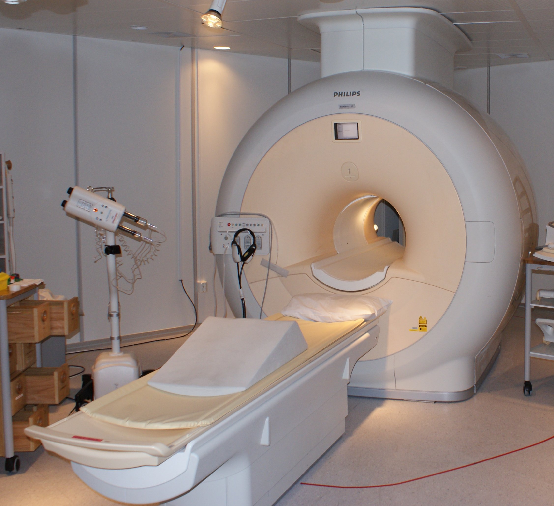
An MRI scanner. A fusion biopsy begins here, with a scan that finds and marks a suspicious area of the prostate before the needle is brought near.
The reason for fusion is the blind spot of plain ultrasound. A cancer can blend into the tissue around it, lost to the grey scan. The MRI reads it on a different kind of picture, one tuned to soft tissue. Fusion is the way to act on that reading. It steers the needle to the marked spot under the live scan. The eye that finds the cancer and the hand that samples it are joined. Plain ultrasound has guided biopsies for decades without seeing the cancer at all. Fusion is what finally lets the needle aim at the tumor the MRI found. The guesswork of a blind sample gives way to a placed shot. The change is as much about confidence as about yield. A urologist aiming at a marked target knows where the needle is going. The blind spread asked the gland to give up its secret by chance.
Fusion does its best work on the significant cancers. The MRI tends to flag the tumors that matter, the ones that need treating. Steering cores into those flagged areas catches them directly. According to a systematic review and meta-analysis, MRI-fusion targeted biopsy detects more clinically significant cancer than the systematic scheme alone. The targeting raises the yield of the cancers a man needs found. It puts the cores where the disease is likeliest to sit. The cancers the MRI flags tend to be the larger, higher-grade ones. Those are the cancers that change how a man is treated. Catching them is the point of the whole workup.
Fusion also spares some men over-diagnosis. A systematic biopsy can stumble onto tiny cancers that need no treatment. Aiming at the MRI target finds fewer of these incidental ones. The man is steered toward the cancers that change his care. Finding less of what does not matter is a real gain of the targeted approach. A biopsy that troubles fewer men over harmless disease is a kinder one. Many small prostate cancers would never have caused harm in a man’s life. A biopsy that leaves them unfound spares him needless worry and treatment. The targeting helps it leave them alone.
A targeted biopsy takes fewer cores, each one better aimed. A handful of cores can come from the flagged spot alone. Each lands where the cancer is likely to be. The yield per core runs higher than a blind spread gives. The targeting puts the needle where it counts. A few well-placed cores can do the work of many scattered ones. Fewer cores also makes for a gentler biopsy. A man takes fewer needle passes for the same answer. The recovery is as easy as any biopsy.
The MRI comes first
Fusion begins with an MRI taken before the biopsy. A multiparametric scan reads the prostate in several ways and scores any suspicious area on the PI-RADS scale. A radiologist marks the lesion and records its place in the gland. That marked lesion is the whole input fusion works from. With no target on the MRI, there is nothing to fuse to. The quality of the targeted biopsy rests on the quality of the MRI behind it, and a careful read of the scan is the first step of a good fusion biopsy. The MRI is read and reported days before the man comes for his biopsy. By the time he is on the couch, the target is already drawn and waiting. The biopsy only has to reach it.
How the two pictures are joined
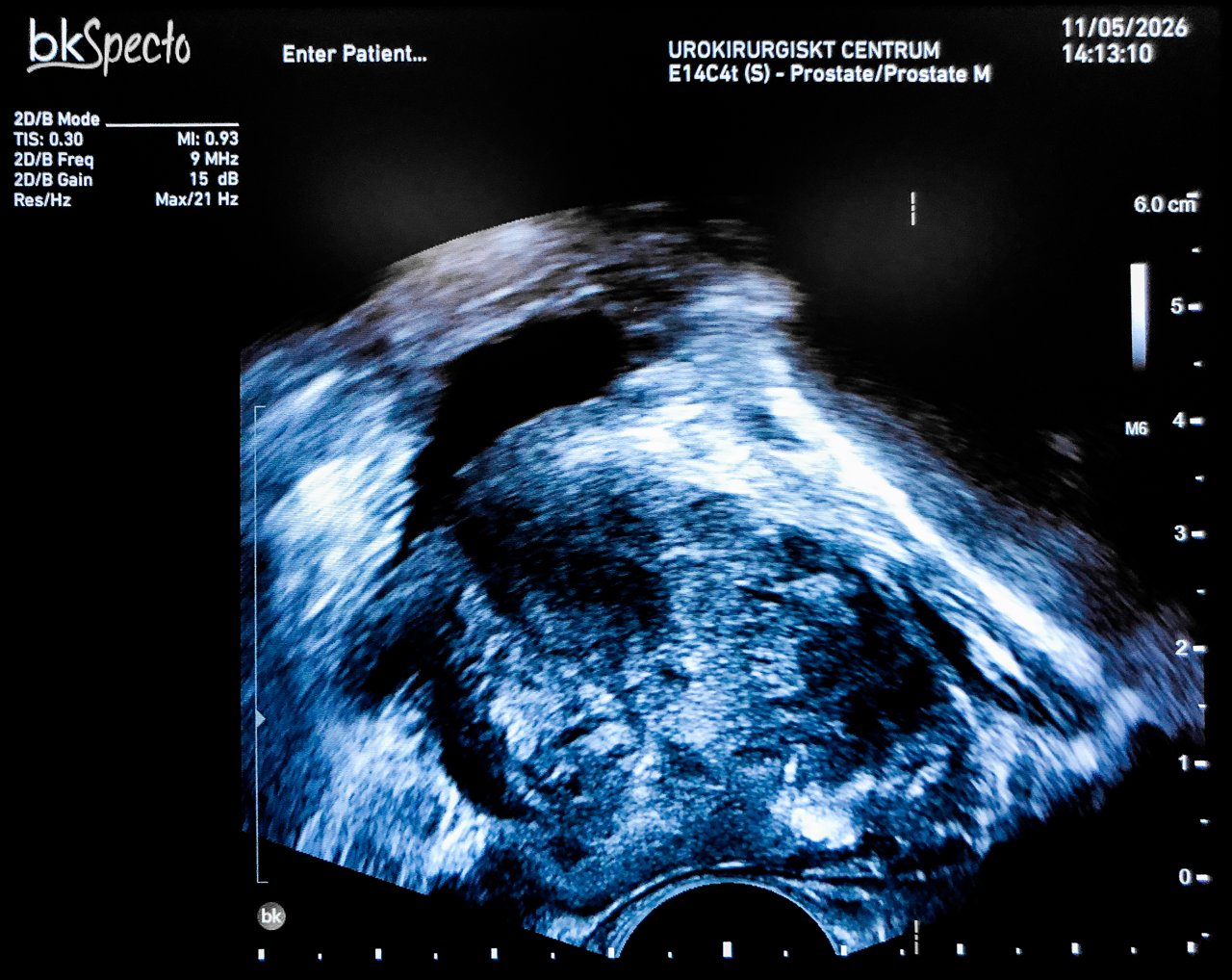
A prostate on a transrectal ultrasound, the kind of live picture a fusion biopsy overlays the MRI onto. The needle is steered through this picture to the target the MRI marked.
There are three ways to bring the MRI and the ultrasound together. The simplest is cognitive fusion. The urologist studies the MRI, notes where the lesion sits, and aims the live ultrasound at that region by eye. No special equipment is needed for it. The accuracy rests on the operator’s reading of the MRI and a steady mental map of the gland. Cognitive fusion costs nothing beyond skill. A practiced urologist can place cores well by it. The accuracy falls off for a small lesion the eye struggles to hold in place. Cognitive fusion is where many clinics start. It needs only a urologist who reads MRI well. The step up to software fusion buys accuracy the eye cannot match.
Software fusion is the second way. A computer registers the MRI to the live ultrasound and overlays the two on one screen. A sensor on the probe tracks its position, so the overlay follows the probe through the scan. The marked target shows on the live picture as the urologist works. The needle is steered to the target the software holds in place. The table below sets the three ways side by side.
PI-RADS score and the chance of significant cancer
PI-RADS
What it means
Significant cancer found
1 to 2
significant cancer is unlikely
low
3
equivocal, a judgement call
about 1 in 5
4
a suspicious lesion
about half
5
very suspicious
about 9 in 10
In-bore biopsy is the third way. The biopsy is done inside the MRI scanner itself, the needle guided by the live MRI. No ultrasound fusion is needed for it, since the MRI guides the needle directly. The accuracy of in-bore guidance is high. The cost is the scanner time and the room it ties up for one biopsy. Few clinics can give an MRI scanner over to biopsies all day. In-bore biopsy gives the truest aim, since the MRI sees the needle go in. The scanner time keeps it rare. Few men get an in-bore biopsy outside a research center.
Software fusion is the common middle path. It brings much of the accuracy of in-bore guidance into an ordinary ultrasound room. The man lies on the couch as for any transrectal biopsy. The software does the joining of the two pictures behind the scenes. A clinic with the system runs targeted biopsies on its own list, with no scanner booked. The everyday targeted biopsy is a software-fusion biopsy. The man notices no difference from a standard transrectal biopsy. The probe, the position, and the cores are the same. The fusion happens on the screen the urologist watches.
Registration is the heart of all three. The MRI and the ultrasound have to be lined up so a point on one is the same point on the other. A good registration puts the needle within a few millimeters of the target. The urologist checks the alignment against landmarks before the cores are taken. The fusion is only as good as the registration under it. A careful line-up is what turns an overlay into an accurate aim. Registration is checked at the start and watched through the biopsy. A landmark on both pictures, the edge of the gland or the urethra, confirms the line-up. A drifted overlay is corrected before the next core. The few millimeters of accuracy are what the whole technique buys. A core a centimeter off the target samples healthy tissue. The care taken over registration is care taken over the result.
Targeted and systematic together
Targeted cores are rarely taken alone. The usual plan takes a few cores from the MRI target and a systematic set across the rest of the gland. Together they sample both the flagged lesion and the ground around it. The combination finds more cancer than either set alone. The targeted cores add precision to the systematic map, and the map guards the gland the MRI did not flag. Neither set carries the whole job alone. Together the targeted and systematic cores leave little of the gland unread. The combined pattern is the fuller answer. The targeted cores are taken first, with the registration at its freshest. The systematic set follows across the gland. The whole biopsy is one sitting under one block.
The combined plan is the current standard for a man with a positive MRI. The targeted cores answer the MRI’s question directly. A systematic set still runs alongside them, in case a cancer sits where the MRI read clear. A biopsy that does both covers the gland as fully as it can. The two approaches read the prostate more completely side by side than either does on its own. Doing both closes the gaps a single approach would leave open. A cancer the MRI overlooked still falls in the systematic net. The combined plan is the safer reading of the gland.
What fusion needs to work
Fusion leans on a good MRI. A scan that misses the cancer, or a reader who marks the wrong spot, leaves the targeting aimed at nothing useful. The targeting is only as good as the picture it borrows. A weak MRI is the commonest reason a targeted biopsy comes up empty. The whole technique stands on the scan behind it. A good MRI program is the quiet condition for good fusion. A clinic invests in the imaging before the biopsy can deliver. The needle reaches only what the scan first found.
Registration error is the other weak point. The MRI and the ultrasound must be lined up exactly. A small error in that alignment moves the target off its true place in the gland. The prostate can also shift or deform under the probe, pulling the overlay out of true. A careful operator re-checks the registration through the biopsy and corrects it as needed. The error is usually small in skilled hands. A few millimeters of drift can still move a core off a small lesion. The closer the aim, the more a small error matters. Modern systems show the likely error on the screen. A urologist can judge whether the aim is tight enough to trust. The honest operator re-registers when the line-up looks loose.
Motion is a constant challenge. The gland shifts a little under breathing and under the pressure of the probe. The software follows the probe and holds the target in place. The operator works smoothly to keep the alignment steady. Good fusion holds the target through the small movements of a real biopsy. A rushed or heavy hand is what lets the aim drift. Steady pressure and a calm pace keep the gland where the overlay expects it. The operator takes the targeted cores with care. A moment of patience holds the aim true.
Fusion works alongside the systematic biopsy. It adds a targeted set on top of the systematic cores. A negative MRI does not rule out cancer on its own. A man with a worrying PSA may still need a systematic biopsy with no target to aim at. Fusion is a powerful addition to the toolkit, used wherever an MRI has given it something to aim at. Fusion does not make the systematic biopsy obsolete. It sits on top of it for the men with a target. The systematic map stays the foundation under the targeted cores. A man with a clear MRI may still have a systematic biopsy when the suspicion runs high enough. The MRI guides the needle where it can. The plan covers the gland either way.
Fusion on a handheld probe
A handheld ultrasound can carry fusion to the bedside. A slim transrectal probe runs from a tablet. Fusion software can run on the same device. The MRI loads onto the tablet before the biopsy. The target overlays on the live scan in the clinic room. A urologist runs a targeted biopsy with no fixed fusion suite around him. The whole setup is a probe, a tablet, and the loaded MRI. The cleaning and the needle guide travel with the probe. A urology clinic can run a fusion list of its own.
The portability widens who can offer a targeted biopsy. A clinic without an in-bore setup can still fuse an MRI to the live scan. The man has his targeted biopsy in an ordinary room. The MRI was done earlier and loaded onto the system. The handheld probe brings the targeting into the clinic, and a urologist works it with the scanner in one hand. The man gets the same targeted biopsy a large center offers. The MRI was read elsewhere and carried in on the system. The advanced part of the work was done before he arrived. A small clinic and a large center can offer the same targeted biopsy this way. The MRI travels as a file. The fusion runs on the tablet in the room.
MRI-ultrasound fusion is the prostate biopsy aimed by the MRI’s eye. It carries a target the grey scan cannot see onto the live picture the needle follows. It raises the yield of the cancers that matter. On a handheld probe in the clinic, it brings targeted sampling of the prostate to wherever a man is seen. The method began in big academic centers with fixed fusion rigs. A handheld probe carries the same idea into a clinic room. The MRI’s eye and the needle’s reach travel together now.
Common questions about fusion biopsy
What is MRI-ultrasound fusion biopsy?
It is a prostate biopsy that brings an MRI of the gland together with the live ultrasound, so the needle reaches a spot the MRI marked. A radiologist marks a suspicious area on the MRI first. Fusion overlays that target onto the live scan during the biopsy. The needle is then steered to the exact spot the MRI flagged.
Why use fusion at all?
The grey ultrasound cannot see a cancer that blends into the tissue around it. The MRI reads it on a soft-tissue picture and marks it. Fusion lets the needle act on what the MRI saw. It detects more clinically significant cancer than the systematic scheme alone, and it finds fewer of the tiny, harmless ones that need no treatment.
What are the ways to fuse the two pictures?
There are three. Cognitive fusion has the operator read the MRI and aim the live scan at the lesion by eye. Software fusion uses a computer to overlay the MRI on the live scan, with a sensor tracking the probe. In-bore biopsy is done inside the MRI scanner itself, guided by the live MRI. Software fusion is the common everyday method.
Is the systematic biopsy still done with fusion?
Usually, yes. The standard plan takes a few targeted cores from the MRI lesion and a systematic set across the rest of the gland. The targeted cores answer the MRI directly. The systematic cores guard against a cancer the MRI did not flag. The two together find more than either set alone.
How accurate is the targeting?
A good registration puts the needle within a few millimeters of the target. The accuracy rests on lining the MRI up with the live scan exactly, and on the gland staying still under the probe. A careful operator re-checks the alignment through the biopsy. The whole result is only as good as the MRI and the registration behind it.
Can fusion be done on a handheld machine?
Yes. A slim transrectal probe runs from a tablet, and fusion software can run on the same device. The MRI loads onto the tablet before the biopsy. The target then overlays on the live scan in the clinic room. A urologist can run a targeted biopsy in an ordinary room, with no fixed fusion suite.
About the Author
Julien Mercier
Senior R&D Engineer · Medical Ultrasound Transducer Development
Senior R&D Engineer with an M.S. in Applied Physics and over 15 years of experience in medical ultrasound transducer development, specializing in the design verification and performance testing of high-frequency imaging transducers. Currently leading the development and verification of the company’s next-generation high-frequency linear-array transducer, responsible for imaging performance evaluation and reliability analysis in preclinical testing. Brings extensive hands-on experience in piezoelectric element tuning, beamforming parameter optimization, and system-level performance testing.