





































Small Animal Veterinary Ultrasound Handheld Dog Cat
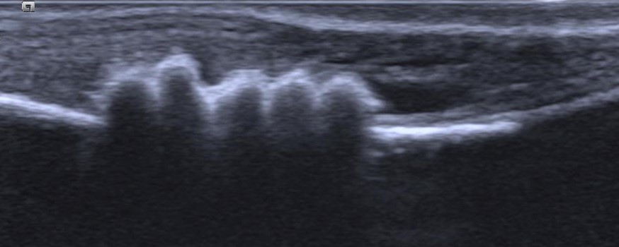
Small-animal ultrasound is the dog-and-cat clinic’s workhorse scan, a handheld probe that turns a vague illness into a clear look at the organ behind it. It runs in the consult room many times a day, on whatever the morning brings through the door. A dog or a cat is small, its organs lying close under the skin and within easy reach of the sound, so a pocket scanner draws the whole organ cleanly. The answer is on the screen before the owner has stood up to leave.
The second exam
What sets this branch apart is where the scan sits in the visit. It is not a test the vet orders and waits days to read. The probe comes off the shelf during the consultation, the moment the hands and the stethoscope have gone as far as they can. A lump the fingers found becomes a picture of what it is made of. The belly that felt tense gives up the one organ behind the trouble. The patient is small, the scan quick, so the whole thing happens in a single visit: the animal still on the table, the owner still watching, the finding and the plan landing together. That fusion of exam and imaging, in one pair of hands across one short visit, is what made the handheld probe as ordinary in a dog-and-cat clinic as the otoscope.
That habit grew out of the economics of a small-animal practice. A referral to a specialist costs the owner both time and money. A sick pet cannot always wait for the appointment to come around. A probe the clinic owns and reads on the spot erases that delay, so the diagnosis and the plan stay under one roof and inside one fee. The picture from a pocket unit is good enough for the questions a first-opinion practice puts to it, day in and day out. That match of tool to need, more than any leap in image quality, is why the dog-and-cat clinic turned ultrasound into a daily instrument.
What the belly gives up
The bulk of the work is abdominal. A vague illness, off food or vomiting or a blood result that came back wrong, sends the patient to a scan that reads the organs in turn. A full abdominal sweep follows a set order, so nothing gets skipped. Each organ is weighed for size, shape, edge, and the grain of its tissue. The order holds the same shape every time, the bladder first, then the kidneys, the liver and spleen, the adrenal glands, and the loops of gut, so a clinic reads each patient by one fixed habit and skips nothing in a hurry.
The bladder gives the cleanest read. Urine sits black on the screen, so stones in the bladder show at once as bright caps with dark shadows falling away beneath them. The blocked tom cat that cannot pass urine is read here in seconds.
The kidneys keep their own record. A healthy one holds a clean bean shape and a sharp inner line. A kidney in failure blurs that line, shrinks and scars, or swells when a stone backs urine up behind it. Read against each other and against the bloodwork, the two separate a sudden crisis from the slow loss of age. A kidney ballooned into a thin sac of urine points straight at a blockage lower down, a stone or a growth pinching the outflow. That single sight turns a vague case of a cat drinking far too much into a clear next move.
Higher up, the liver and spleen are read for their size and for any mass breaking their even grain, the gut for a thickened wall or a swallowed object sitting behind its own gas shadow. These are the pictures that move a dog from the consult room to the surgery table the same afternoon. A swollen, lumpy spleen, a liver studded with dark spots, a node grown to the size of a grape, each is a flag the eye can raise in the time it takes to listen to a chest.
The probe also steers the needle. A node, a mass, or a pocket of fluid can be sampled while the screen watches the tip go in, so a diagnosis comes from a few cells drawn with no blade. The detail of each of these reads lives a level down, on the page for what to scan in a dog and cat abdomen.
What you did not go looking for
A quick scan also turns up what no one was looking for. A liver nodule found while chasing a vomiting dog, a bladder stone seen on a routine pregnancy check, a swollen spleen behind a vague off-colour cat: the probe reads the whole region it sweeps, every organ that falls under the beam. Some of those chance finds are the real diagnosis, hiding behind the complaint that brought the animal in. Others are harmless, the lumps and cysts a healthy belly carries; chasing them only pulls an owner into tests the animal never needed. Reading the picture means knowing which incidental marks to follow and which to leave alone.
Deeper in the belly
Back in the abdomen, organs past the routine four reward a careful look. The gallbladder can pack with a stiff sludge that stacks into the shape of a kiwi fruit, a pattern that warns of a wall about to burst and shows long before the dog turns yellow. A stone or a swollen duct blocking the bile reads here too. A thickened, bright wall flags an inflamed gallbladder a surgeon may have to take out.
The adrenal glands, small and tucked beside the big vessels, carry an endocrine story. A gland swollen on both sides points to an overactive pituitary driving the whole system. A lump on one side alone points to a tumour of the gland itself. The split matters, since the two are treated nothing alike. The probe is often the first to tell them apart. An adrenal mass turned up by chance, while the scan was chasing something else, is a common and awkward find.
The spleen and the gut round out the deep sweep. A diseased spleen swells into a huge, lumpy organ, or twists on its stalk into a red emergency. The probe reads its size and grain in a single pass. In the gut, the neat layered wall of a healthy bowel thickens and blurs where disease takes hold. A length of bowel telescoped into the next throws up a ringed target the eye learns to spot. These deeper reads turn a vague, painful belly into a labelled map.
Free fluid in the belly is a find of its own. A dark rim of fluid around the organs can be blood from a ruptured spleen, urine from a torn bladder, pus from a burst gut, or the clear seepage of a failing heart or liver. The probe finds it in seconds and guides a needle to draw a sample. The colour and content of that sample often name the emergency before any blood result is back.
Lymph nodes are read across the body for the same reason. A node swollen behind the knee, under the jaw, or deep in the belly can be reacting to a nearby infection or filling with the spread of a cancer. The probe measures it, reads its shape and its blood flow, and guides the needle that tells reaction from spread. For a dog with a known tumour, a sweep of the draining nodes is part of working out how far it has gone.
The eye and the neck
Not every scan looks at the belly or the chest. When a cloudy cornea or a chamber full of blood hides the inside of an eye, a small high-frequency probe laid on the closed lid reads what no light can reach. It finds a lens knocked off its moorings, a retina peeled away from the back wall, a mass crowding the space behind the globe. For a sudden blindness with no view in, that picture often decides between an eye that can be saved and one that has to come out. It also measures the eye for the lens implant a cataract operation will need.
The neck carries its own targets. In an older cat losing weight for no clear reason, the probe hunts the thyroid for the small nodule behind a runaway metabolism. A swelling under the jaw might be a salivary gland gone wrong, a duct blocked by a stone, or a firm node left by infection or spread. The probe tells the soft from the solid there, and reads how deep it sits, before any needle goes near.
The male patient and the moving parts
A male dog brings the prostate into the picture. An old entire dog grows a big prostate of even texture that presses on the gut and the urethra. An angry, uneven one, pocketed with fluid or studded with bright flecks, warns of infection, a cyst, or a cancer. The probe reads its size against the dog’s frame and guides a needle into the right pocket for a sample, a structure a gloved finger cannot reach or read on its own.
The same flat probe that reads tendons in a racehorse reads them in a dog. A working dog or an athletic breed strains the tendons and muscles of its legs. The probe shows the dark gap of a tear in the bright weave of a healthy tendon. The screen reads the nature of a leg swelling, a torn muscle, a clot, or a tumour. For the lame dog with no fracture on the X-ray, the soft-tissue read often finds the cause.
The screen earns the call
A scan does not replace a careful pair of hands. It tells those hands what they were only feeling for.
Litters and the womb
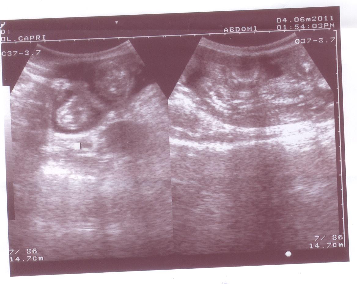
A pregnant bitch comes in with two questions. Has she caught, and are the pups alive. From about day twenty-five the sacs and their flickering hearts answer both, weeks before any swelling shows. A canine pregnancy scan is timed for that early yes and for a late look at fetal heart rates, since a rate sliding low warns of a litter in trouble that may need a caesarean. Later still, the size of the pups read against the size of the bitch flags the oversized single that will never pass on its own.
Counting the litter is the weak spot. A breeder is told so plainly: the pups overlap and hide one another near term, so a firm number belongs to an X-ray. A queen’s pregnancy runs the same path on a smaller, finer uterus. The scan catches the trouble early, a dead fetus or a pregnancy that has stalled, while there is still time to act.
Not every swollen womb holds a litter. In an unspayed female off her food and drinking hard after a season, the scan hunts for pyometra, a uterus filling with pus, easy to take for pregnancy until the probe finds no sacs inside it. That one difference is the line between a course of tablets and a surgery the same night.
The heart on the table
A murmur on the stethoscope sends the patient to the echo probe. A focused heart scan answers the bedside questions quickly: is the muscle squeezing, are the chambers stretched, is there fluid pressing the heart shut. It is a short fixed routine of a few views, learned without a fellowship. For the next hour’s decision it stands on its own. The views come from a handful of fixed windows low on the chest wall behind the elbow. Once the hand learns where to sit, a useful study takes a couple of minutes. A cardiology service goes deeper when a case earns it.
The commonest heart trouble in dogs is a quiet one. The mitral valve thickens and stops sealing, so a thread of blood slips back through the valve. Small breeds carry the leak into old age. The probe sees it on colour flow, with the left chambers swelling over months to take the load. Reading how far that has gone is what times the first tablet.
Cats hold an overgrown heart muscle out of sight until the day a clot breaks free or the chest fills with fluid. The wall grows inward until the chamber is a slit that cannot fill. A scan measures that wall while the cat still looks well, which for some owners is the only warning they get.
Big breeds run the other way, into a dilated, weak heart, its chambers ballooned, its walls thinned, its squeeze down to a feeble ripple. On the screen it barely moves, often beside the belly fluid of a circulation backing up behind it. Each of these conditions opens onto its own page for the detail.
The cat is the harder patient
A cat asks for a different approach than a dog. It is smaller, quicker to stress, and built to hide how ill it is until late. A frightened cat breathes in fast shallow gulps. A tightened belly smears the picture the scan needs. The fixes are patience and a quiet room, a towel wrapped snug, a low light, sometimes a mild sedation that drops the animal into a state still enough to read. Rushing a cat buys a blurred study and a worse-tempered patient.
The cat’s heart adds its own difficulty. It beats nearly twice as fast as a person’s, so the muscle and valves blur on a slow frame. A still picture loses what a live loop holds. A handheld unit set up for cats freezes a cleaner frame and runs the loop back to read the squeeze. The same care that gets a clean cat scan is the one that catches the thick-walled heart these animals hide so well.
From the chest outward
The probe does not stop at the diaphragm. Laid on the chest, it reads the lungs through the gaps between the ribs. A lung filling with fluid throws up bright vertical streaks, the same signs read in people for heart failure and pneumonia. Fluid around the lung shows as a black band that lets it float and flap. None of this asks the gasping animal to lie flat or hold a breath, which is why an animal in no state for an X-ray will still give up a lung picture taken gently from the side.
After a road accident the same trick turns urgent. The fast abdominal sweep after trauma checks a few set spots where blood gathers. A dark stripe in any of them answers in under a minute whether the animal is bleeding inside. Run again an hour later, it catches a bleed that was not there before, in time to act on it.
A lump’s character
Owners find lumps. The fingers alone cannot say which ones matter. A lump under the skin, read with a high-frequency probe, gives up its depth, its edges, what runs through it, and enough of its make-up to steer whether it is watched, sampled, or cut away. Depth counts as much as nature here, since a lump tucked against the body wall or pushing in from inside is read for how far it reaches before anyone picks up a blade.
A few have a look the probe knows on sight: the even sliding patch of a fatty lipoma, the messy fluid pocket of an abscess, the round dark swelling of a node. None of it stands in for a sample sent away. It does sort the lumps that can wait from the one that wants a needle today.
The exotic patient
The clinic table takes more than dogs and cats. A rabbit gone quiet with a still gut, a reptile with a stuck egg, a ferret with a swollen adrenal gland, each is read on the same probe, with the high-frequency end doing the work on such small bodies. Rabbits, birds, and reptiles ask the operator to know that species’ own normal cold, which is the real skill behind the easy part of holding a probe.
Watching a case change
A scan read once is a snapshot. The handheld probe does more when the same patient is read again. A belly checked today and again tomorrow shows whether free fluid is filling or draining, whether a blocked gut is moving or stuck fast. A heart started on a new drug is read a week on to see its stretched chambers settle. A pregnancy is followed across its weeks. Because the scan costs only minutes and no second trip, a vet reaches for it as a way of watching a problem move from one day to the next.
That habit changes how a case is worked. A finding no longer waits for a referral and a second appointment. It lands in the same visit. The next visit tracks it. An owner who watched the first scan grasps the follow-up without a long explanation. The picture becomes a running record of the illness.
When the quick look is not enough
A focused scan trades thoroughness for speed. A small early tumour can hide in an organ that reads normal at a glance. A gut lesion can sit in a loop full of gas the sound cannot cross. A heart murmur may need the full measured study a pocket scan does not run. The handheld answers the common question quickly and flags the case that needs a longer, deeper look on a better machine.
Fitting a probe to the caseload
A clinic choosing its first handheld unit lets the caseload set the spec. A practice working on bellies, the odd pregnancy, and a few hearts wants one strong high-frequency probe and a screen large enough to follow a moving heart. What to weigh before buying is the work the clinic already does week to week. A long feature list rarely matches it.
Image quality on the small structures is the thing to test, since a soft picture of a cat’s kidney buries the early disease the scan exists to find. Weight, and a screen readable in a bright room, count for a tool lifted twenty times a day. The unit that fits is the one that ends up in a hand on every other case. A trial scan of a real patient, run before any money changes hands, shows more about a unit than a printed page of specifications ever will.
What clinics ask about scanning dogs and cats
What is the commonest reason to scan a dog or cat?
The abdomen, by a wide margin. A vague illness such as vomiting, going off food, or a bad blood result sends the patient to a belly scan that reads the liver, kidneys, bladder, spleen, and gut in one sitting.
Can a handheld scanner do a real heart study on a dog or cat?
It does a focused one. It answers the bedside questions of squeeze, chamber size, and fluid around the heart. It screens for the slow valve leak of small dogs, the overgrown cat heart, and the stretched heart of big breeds. A full cardiology workup still adds detail a pocket scan does not reach.
How early can ultrasound confirm a pregnancy in a dog?
From around day twenty-five after mating, when the sacs and beating hearts appear. Ultrasound is the early, alive-or-not test. For a firm count of how many pups are coming, an X-ray closer to term does it better.
How does ultrasound differ from an X-ray in a dog or cat?
They answer different questions. Ultrasound reads soft tissue, the inside of organs, free fluid, and the moving heart. An X-ray maps the skeleton, the gas pattern of the gut, and the overall shape and size of things. Plenty of cases use both.
Is ultrasound useful in rabbits and other exotics?
Yes, often urgently. A rabbit with a still gut, a bird or reptile with a stuck egg, or a ferret with a swollen adrenal gland is read with a fine probe, as long as the operator knows that species’ normal anatomy.

About the Author
Julien Mercier
Senior R&D Engineer · Medical Ultrasound Transducer Development
Senior R&D Engineer with an M.S. in Applied Physics and over 15 years of experience in medical ultrasound transducer development, specializing in the design verification and performance testing of high-frequency imaging transducers. Currently leading the development and verification of the company’s next-generation high-frequency linear-array transducer, responsible for imaging performance evaluation and reliability analysis in preclinical testing. Brings extensive hands-on experience in piezoelectric element tuning, beamforming parameter optimization, and system-level performance testing.