





































Canine Feline Subcutaneous Mass Ultrasound Handheld Veterinary
A subcutaneous mass scan reads the lump under a dog’s or cat’s skin for what the fingers cannot feel, whether it holds fluid or solid tissue, how deep it runs, and what blood feeds it. An ultrasound probe laid on the lump shows its inside on the screen in a minute. The scan guides the needle that samples it and steers the vet toward what the lump is likely to be.
What the scan reads in a lump
A lump under the hand is a closed box to the fingers, a swelling the touch can press and roll, its inside shut to the fingers. The scan opens it. Laid on the skin over the swelling, the probe turns the blind bump into a picture of what it holds, four things read off that picture, each one steering what happens next. The whole read takes a minute on a calm animal, the four questions answered before the owner has finished describing how the lump first appeared.
The first read is the content. The screen shows whether the lump holds fluid or solid tissue, the one fact that decides whether a needle drains it or samples it. A pocket of fluid passes the sound clean through and shows as an even, dead black, a shade the reader learns to know at a glance once the eye has met a few. Solid tissue, grained with the texture of its packed cells, returns a busy grey the reader knows just as fast. That one split, the dark from the grey, sets the rest of the read in motion, deciding the kind of needle the lump will meet.
The second read is the edge, where the trouble shows. A border that fades into the flesh around it, the mass sending fingers off into healthy tissue, is the look of something that invades, a lump a vet treats with respect long before the lab speaks. The reader runs the probe the whole way round before judging an edge clean.
The third read is the depth. The probe shows how far down the lump reaches, whether it sits free in the fat under the skin or runs down to grip the muscle and the sheet of tissue below. A lump anchored to what lies beneath it is a graver thing than one that slides loose over it. The probe rocks the lump to test it, a swelling that glides over the deep tissue still loose, a deeper read called for the moment it drags the muscle along with it.
The fourth read is the blood. A touch of colour Doppler lights the vessels running through the lump. A mass flushed with flow draws the eye, the busy blood supply a clue, read with the rest, to a tumour to hurry to the lab. The colour is read on every lump the scan meets, a few seconds of Doppler added to the grey picture, the blood supply a clue the reader never leaves unread.
Fluid or solid
The split between fluid and solid is the one the scan makes first and best. A fluid lump shows an even black pocket, the sound sailing clean through it to brighten the tissue behind. It gives under the probe when pressed, the squash of a fluid that nothing solid would show.
A needle drains that fluid lump to nothing, the swelling often gone for good and the worry with it. The same needle, set to a solid lump, draws a sample of its tissue, a single tap under the probe settling which kind a lump is and acting on it in one pass. The scan rarely leaves that first question in doubt, fluid and solid looking as unlike on the screen as water and stone.
The common lump: the lipoma
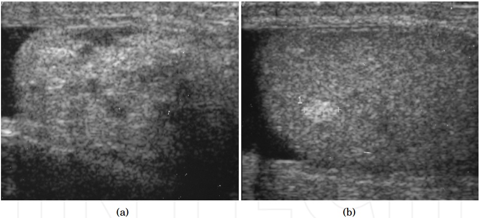
The lump a vet meets more than any other is the lipoma, a soft mound of fat under the skin. It reads on the screen with a look of its own, a mass run through with fine bright lines, the fat drawn out in streaks that follow the lie of the skin. It sits pale, an even grey throughout, walled in a thin capsule, with no busy blood inside.
That streaked, quiet look is reassuring, the picture of a fat lump that does little harm. The scan leans toward a lipoma when it sees it, the read confirmed by a needle that draws clear fat onto the slide. A lipoma sampled gives sheets of plain fat cells, the cleanest answer in all of lump work, and the owner goes home reassured the same day.
A lipoma can still surprise. One grown deep can press between the muscles, a few turn out to be the firmer kind that creeps along the tissue planes. The scan reads the depth and the spread before the lump is written off as the harmless fat it usually is. A plain lipoma earns no more than a note in the record and a watch for any change, the scan returned to only if the lump starts to grow or to harden under the hand.
The cyst and the abscess
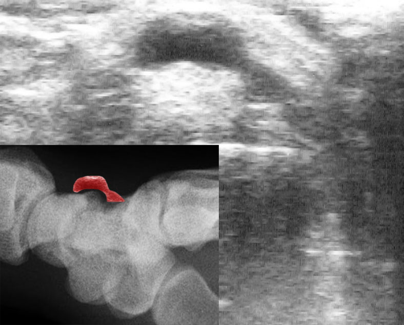
A fluid lump asks one more question of the scan: clean fluid, or fluid gone bad. A simple cyst holds clear fluid in a smooth-walled pocket, an even black on the screen, a brightening behind it where the sound passed through. It drains to a clear or pasty fluid and often troubles the animal no further. The brightening behind a cyst, the sound racing on through clear fluid, is a sign the reader leans on to call a pocket fluid for certain before a needle confirms it.
An abscess is fluid turned to pus, busier on the screen than a clean cyst. The pocket fills with floating muck and swirling debris, the wall thick and angry, a rim of hot blood lit around it on the Doppler. The animal flinches as the probe presses a pocket that a quiet cyst would let pass. An abscess found is an abscess drained and flushed under the probe, the emptied pocket checked for any solid wall left hiding in a corner that a simple squeeze would miss.
A blood-filled lump, a hematoma, reads as a third kind, its insides shifting over the days from solid-looking clot to dark fluid. The scan tells the three apart by their insides and their walls, the read pointing the vet toward a drain, a flush, or a watchful wait. The story of the lump helps the read, a swelling that rose overnight after a knock pointing the reader toward a hematoma.
The wrong call on a fluid lump costs little time. A pocket tapped under the probe gives up its fluid to the eye and the slide, the question of cyst against abscess settled by what the needle draws. A swing in the wrong direction, a cyst that turns out an abscess or a lump that turns out solid, costs only the minute it takes to read the slide.
The needle still has the last word
The scan reads a lump’s shape and its blood. Its true name waits on the slide.
How deep it goes
The depth of a lump is half of what makes it dangerous. A mass reaching down to grip the muscle, or running along the sheet of tissue beneath it, is the graver problem, the kind that takes a wider cut and leaves more behind. The hand on the skin reads only the top of such a lump, the bulk of it running on into depths the fingers never reach.
The scan reads that depth where the hand only guesses. The probe follows the lump down through the layers, marking where it stops, whether a clean plane of tissue still runs under it or its base has fused to the muscle below. A mass fixed to what lies under it, moving as one with the muscle when the probe rocks it, has grown roots, the deepest worry the read turns up. That one test, the glide or the drag of the lump under the rocking probe, tells a surgeon more about the work ahead than the size of the lump ever could.
That reading shapes the surgery before the first cut. A surgeon shown the true reach of a lump plans a margin around it that the skin alone would never reveal, taking the spread the scan found, wider than the lump the hand could feel.
Depth tells the vet which lump is the bigger job. A mass rooted to the muscle and spread along the planes beneath needs a planned surgery, wide margins, and sometimes a deeper scan before the knife to map its full reach. The reach the scan maps is the reach the surgeon plans to.
The blood feeding it
A growing tumour builds its own blood supply. The vessels it lays down to feed itself show on the colour Doppler, a wash of flow through a mass that a quiet lump never carries. The pattern of those vessels, their number and their tangle, leans the read one way or another well before the slide is read. A dense tangle pushing right through the middle of a lump is the pattern that worries a reader, weighed with care against the rest of the picture.
A lump barely touched by colour is the kind a vet worries over less. The reading is never taken alone, a fast-growing mass and a flush of disordered flow together raising the suspicion that hurries a lump to the lab and the knife. The Doppler adds a layer the grey picture alone would miss.
The blood read carries its limits. Colour alone calls no lump, the flow able to mislead in either direction, so it is weighed with the shape, the depth, and the speed of growth before it counts for anything. The colour is one vote among several, counting heavier when a worrying shape and a fast growth vote the same way.
Guiding the needle
The scan earns its keep above all in the needle it guides. A lump sampled blind is a needle pushed on faith, the tip landing where it lands, sometimes in the dead centre of a mass where the tissue has died and tells the lab nothing. The probe ends that guesswork. It shows the needle on the screen as it enters the lump, the tip steered into the living tissue at the edge of the mass where the cells still read true, past the dead core that a blind pass so often draws. A vessel lit on the Doppler is a thing the needle is steered around, the guided tap sparing the bleed a blind one into a vascular mass can start. The reader watches the bright point of the needle the whole way in, laying it where the sample will count and drawing back where the picture warns. A still hand and a clear screen turn the riskiest tap into a routine one, the needle laid exactly where the picture says it should go. A lump that gave a useless smear blind gives a clean answer once the probe guides the needle, the difference often deciding whether a second visit and a second needle are needed at all.
The guidance matters more the deeper and the trickier the lump. A mass pressed against a big vessel, sitting deep between the muscles, or hard to pin under a thick coat is the one the probe makes safe to reach, where a blind needle would court a bleed or a miss.
The same guidance serves the bigger biopsy. A core of tissue taken with a wider needle, read under the probe as it cuts, comes from the part of the mass the reader chose, the living edge that names the tumour, away from the necrotic middle that wastes the test. The thread of solid tissue it brings back is one the pathologist can section and stain, a fuller answer than the scatter of cells a fine needle draws.
A drain runs under the probe too. A pocket of fluid is emptied to the last of it with the needle watched on the screen, the wall checked for any solid lump hiding in a corner that the fluid had masked. A pocket that fills again after a clean drain is read for the solid lump or the leaking vessel behind it, the scan turning a failed drain into a fresh question. The probe stays on the lump while the fluid leaves, the pocket watched down to its last and the wall behind the needle drawing together.
The needle and the probe work as one tool. The scan finds the spot, the needle reaches it, and the slide reads what comes back, a chain that turns a blind lump into a named one in a single sitting. The owner often leaves with the sample already on its way to the lab, the lump read, sampled, and routed onward in the time of a single visit.
The great pretender
Some lumps wear a harmless face. The mast cell tumour is the one a vet never trusts on looks, a cancer that can pass for a fatty lump, a wart, or an insect bite to the hand and even to the scan. It is the reason a lump is rarely written off on feel alone. A hard lump, a soft one, a lump up overnight, a lump that sat for a year: any of them can hide a mast cell tumour, which is why a needle outranks a hunch on every one.
The scan reads the same shape and blood on a mast cell tumour as on something benign, no certain mark setting it apart. The needle is what unmasks it, which is why the rule on any new or changing lump is to read it and sample it before ever trusting it. A lump an owner could swear was last year’s same fatty bump is read and sampled anew, the cost of the check far below the cost of a missed cancer.
What the scan cannot do
The scan does not name the tumour. It reads a lump as fluid or solid, deep or loose, quiet or flush with blood, and it narrows the field down to a short list of what the lump might be. The cell type, the grade, the question of cancer or not, all wait on the slide and the pathologist. A lump well read is a lump well sampled, a diagnosis still a step beyond it.
It misses the smallest changes inside a mass that looks plain. A subtle cancer in a lump that reads benign can slip past the grey picture and the colour both, which is why a worrying history outranks a reassuring scan. A lump that grows fast, bleeds, or sits on a breed prone to trouble earns the needle whatever the screen shows.
The scan reads the lump it can reach. A mass buried deep, or one a thick coat and a restless animal blur, gives a dimmer picture that hides its finer marks. The reader clips, wets, and steadies before trusting a hard read. A poor study earns a poor grade, the lump rescanned on a calmer day before any weight is laid on a blurred picture.
The scan is one read among several. The hand on the lump, the story of how it grew, the breed and the age of the animal, the slide from the needle: each adds to the call, the picture on the screen weighed in with the rest. A scan trusted alone against a worrying history is a scan misused.
Where the handheld fits
The handheld scanner brings this read into the consult room, onto the lump while the owner is still in the chair. A pocket probe laid on the swelling answers the first questions in a minute, fluid or solid, deep or shallow, fed by blood or not, and turns a vague worry over a lump into a clear next step. An owner who walked in afraid of a lump walks out with a plan for it, the scan done while the animal sat on the table between them.
The scan sorts the lumps that need hurrying from the many that can wait. A firm, flush, deep, or fast-growing mass is read, sampled, and moved quickly toward the specialist and the surgeon, the scan setting that pace in the room before the lab has spoken. The pace the scan sets is half its value, a worried owner walking out with a clear next step on the day the lump was found.
The grading and the naming stay with the lab and the referral. The handheld does the first read and points the needle, the cytology and the histology carrying the diagnosis the screen can only hint at. The reading and the sampling happen in the one room on the one visit, the handheld folding two steps into a single short sitting. It is the fast first look that decides how hard and how soon to chase a lump.
From a lump to a plan
A subcutaneous mass scan turns a lump the hand can only feel into a picture the vet can act on. It reads what the lump is made of, how deep it runs, and what blood feeds it, and it guides the needle that names it. Each of those reads on its own would change little, the value sitting in the four together, a lump weighed for its content, its edge, its depth, and its blood in a single pass.
The read changes the road a lump travels. A mass read deep, fixed, and flush with blood is sampled at once and carried fast toward the surgery that has the best chance while the lump is still small. The road forks early on what the scan reads, and taking the right fork at the first visit is what spares a dangerous lump the months of watching that let it grow.
The whole of it costs the animal a clipped patch and a few quiet minutes on the table, and it buys a lump a name and a plan far sooner than the hand alone ever could. A bump the fingers could only call something to watch becomes, in a minute, a thing the vet can name a plan for.
Common questions about a lump scan
Can an ultrasound tell what a lump on my dog or cat is?
It tells a great deal short of the final name. The scan reads whether the lump is fluid or solid, how deep it runs, and what blood feeds it, and it narrows the likely causes. The cell type and the question of cancer come from a needle sample read under the microscope, which the scan guides.
How does ultrasound tell a fatty lump from something serious?
A lipoma reads as a soft mass run through with fine bright streaks, pale, quiet, with little blood inside. The marks that raise concern on any lump are a border that invades the tissue around it, a reach down to the muscle, a flush of disordered blood. The scan narrows the field, and the needle settles it.
Why use ultrasound to guide the needle for a lump?
A blind needle can land in the dead centre of a mass and draw a useless sample, or surprise a vessel and start a bleed. The scan shows the needle entering the lump, steering it into the living tissue that reads true and around the vessels best left alone, so the first sample more often gives a clear answer.
Does every lump on a pet need scanning and sampling?
A new lump, a growing one, or one that bleeds or bothers the animal earns a look and usually a needle. Some cancers, the mast cell tumour above all, can pass for a harmless lump on feel and even on the scan, so the safe path on any lump in doubt is to read it and sample it.
Can a handheld scanner read a lump in the consult room?
Yes, and it suits the job. A pocket probe laid on the lump answers the first questions in a minute and guides the sampling needle on the spot. Grading the tumour and naming it stays with the laboratory, the step the handheld points the lump toward.

About the Author
Julien Mercier
Senior R&D Engineer · Medical Ultrasound Transducer Development
Senior R&D Engineer with an M.S. in Applied Physics and over 15 years of experience in medical ultrasound transducer development, specializing in the design verification and performance testing of high-frequency imaging transducers. Currently leading the development and verification of the company’s next-generation high-frequency linear-array transducer, responsible for imaging performance evaluation and reliability analysis in preclinical testing. Brings extensive hands-on experience in piezoelectric element tuning, beamforming parameter optimization, and system-level performance testing.