





































Canine Dilated Cardiomyopathy DCM Ultrasound Diagnosis Handheld
Dilated cardiomyopathy weakens a dog’s heart muscle, stretching the main pumping chamber wide until the pump can no longer keep up. An ultrasound is how the disease is found. The scan reads the slack chamber and the feeble squeeze straight off the screen, often years before the dog shows a sign. Catching it that early is the whole point of the scan, since the silent disease holds a treatable window before it tips into failure.
The disease behind the picture
Dilated cardiomyopathy is a disease of the heart muscle itself. The wall of the left ventricle, the chamber that drives blood to the body, loses the strength to contract. A failing pump moves less blood with each beat. The body asks for more. The chamber stretches to hold a larger load and to squeeze harder against the shortfall. The disease takes hold over months or years, the muscle weakening cell by cell until the chamber can no longer hide the loss.
That stretch is a trap. A wider chamber takes more force to empty. The weakened muscle has none to spare. The heart dilates further, the squeeze fades further, and the spiral runs one way only. The scan reads the heart at whatever point on that slope the dog has reached.
In many dogs the cause is never named, an idiopathic disease written into the breed. In some it follows a fault in the diet or a worn-out muscle from another illness. The scan reads the same dilated heart whatever drove it. The cause may be inherited, written into a stricken breed, or it may lie in a diet of the wrong sort or a key nutrient run low, the kind a change of food may undo.
The shape of a dilated heart
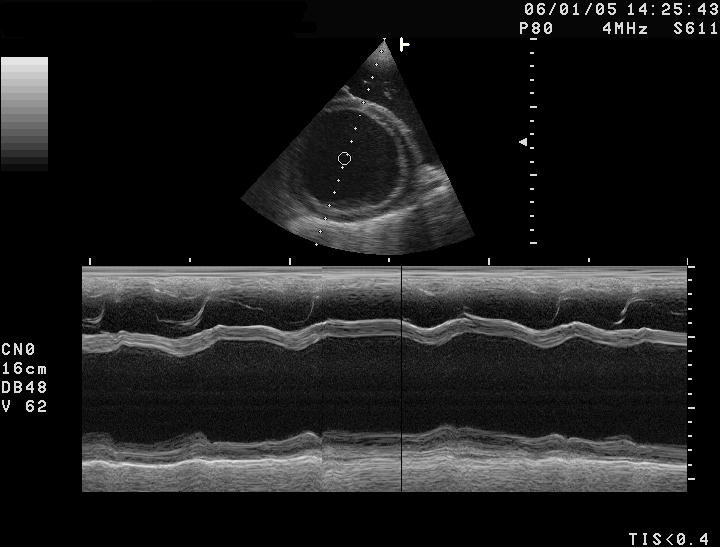
The dilated heart has a look a trained eye knows at a glance. The left ventricle reads large, its walls bowed out into a rounded chamber that has lost the bullet-point taper of a healthy heart.
The walls run thin around that wide chamber. The muscle has stretched thin lining a larger space, with no more tissue to do it, so the wall reads thinner than it should for the size of the dog. The thinning and the widening go together, two faces of the same overstretched muscle. A thin wall around a wide chamber is a core mark of the disease.
The squeeze tells the rest. The scan measures how far the chamber narrows from its widest to its tightest, the contraction reading any heart scan takes. In a dilated heart that narrowing is feeble, the walls barely moving at all, the inward drive of a healthy beat all but gone. The feeble motion shows to the eye before any number is taken.
The left atrium swells behind the failing ventricle, the pressure backing up into it. A valve stretched on its widened ring leaks, adding a backward jet that loads the atrium further. The scan reads the swollen atrium as the warning that fluid is close to backing into the lungs. A left atrium grown large is the line between a disease still quiet and one about to break into the open.
Each of these signs leans on the others. A large round ventricle, thin walls, a weak squeeze, a swollen atrium: read together on a breed known for the disease, they spell a dilated heart with little room left for doubt. A single borderline measurement on its own says far less. The reader weighs the whole picture together. A lone figure settles nothing.
Putting numbers to the heart
Numbers grade the dilated heart the eye has named and track it over time. The scan freezes the ventricle at its widest and at its tightest and measures across the chamber, the two figures set against the dog’s body weight so a wolfhound and a spaniel are each judged on their own frame. The figures go into the record at every visit, turning a single snapshot into a line the disease can be tracked along.
A full reading leans on several measures taken together. The width of the ventricle at the end of filling and at the end of the squeeze gives the size and the change between them. The fraction by which the chamber narrows, the shortening, falls in a dilated heart far below the band a healthy dog holds. The gap between the mitral valve and the ventricle wall at the valve’s nearest approach grows once the chamber swells and the valve stops opening fully. That gap gives another angle on the failing pump. The roundness of the chamber is read too, the long taper of health gone, the chamber rounding toward a ball. Each figure is set against the published range for the dog’s size and, where it exists, for the dog’s breed, since a measurement normal for a Great Dane would be gross in a terrier. No single number makes the diagnosis. The pattern across all of them, on a heart that already looks dilated to the eye, is what carries it.
A measurement at the edge of normal is read with care. A heart caught mid-change can sit in a grey zone where one scan cannot call it until a second scan months later shows the chamber drifting the wrong way.
The numbers earn their keep above all in the rescan. On a dog still showing nothing, a chamber a millimetre wider and a squeeze a few points weaker than last year is the disease on the move, still early enough to slow. Read once, with no earlier scan beside them, the same numbers say only half as much.
The measurements mean little taken once and trusted blindly. A chamber caught off the proper plane reads smaller than its true width, a foreshortened view flattering a failing heart. The reader works to the standard views, the probe squared to the long axis, so one year’s number can be set honestly against the next.
The silent years
The cruelty of this disease is how long it hides. A heart can be dilating and weakening for years inside a dog that runs, eats, and plays as though nothing is wrong. The body has reserve to spend, and it spends it quietly until the reserve runs out. The cough or the faint that finally brings the dog in often comes a year or more into a silent decline.
That long quiet phase carries its own name in the clinic, the occult or preclinical stage. The changes are already on the scan, the chamber widening and the squeeze slipping under a dog that looks perfectly well. A scan run in those years is the one that finds the disease in time to act on it.
When the silence breaks
The first outward sign holds off until late, then strikes without mercy. A dog that seemed fine collapses, or drowns slowly in the fluid filling its lungs, or drops dead the moment the heart loses its rhythm. By then the disease has run far along its course. The scan exists to find the heart long before that day.
A dilated heart is seen long before it is felt
For years the scan sees a change the dog cannot feel.
Screening the breeds at risk
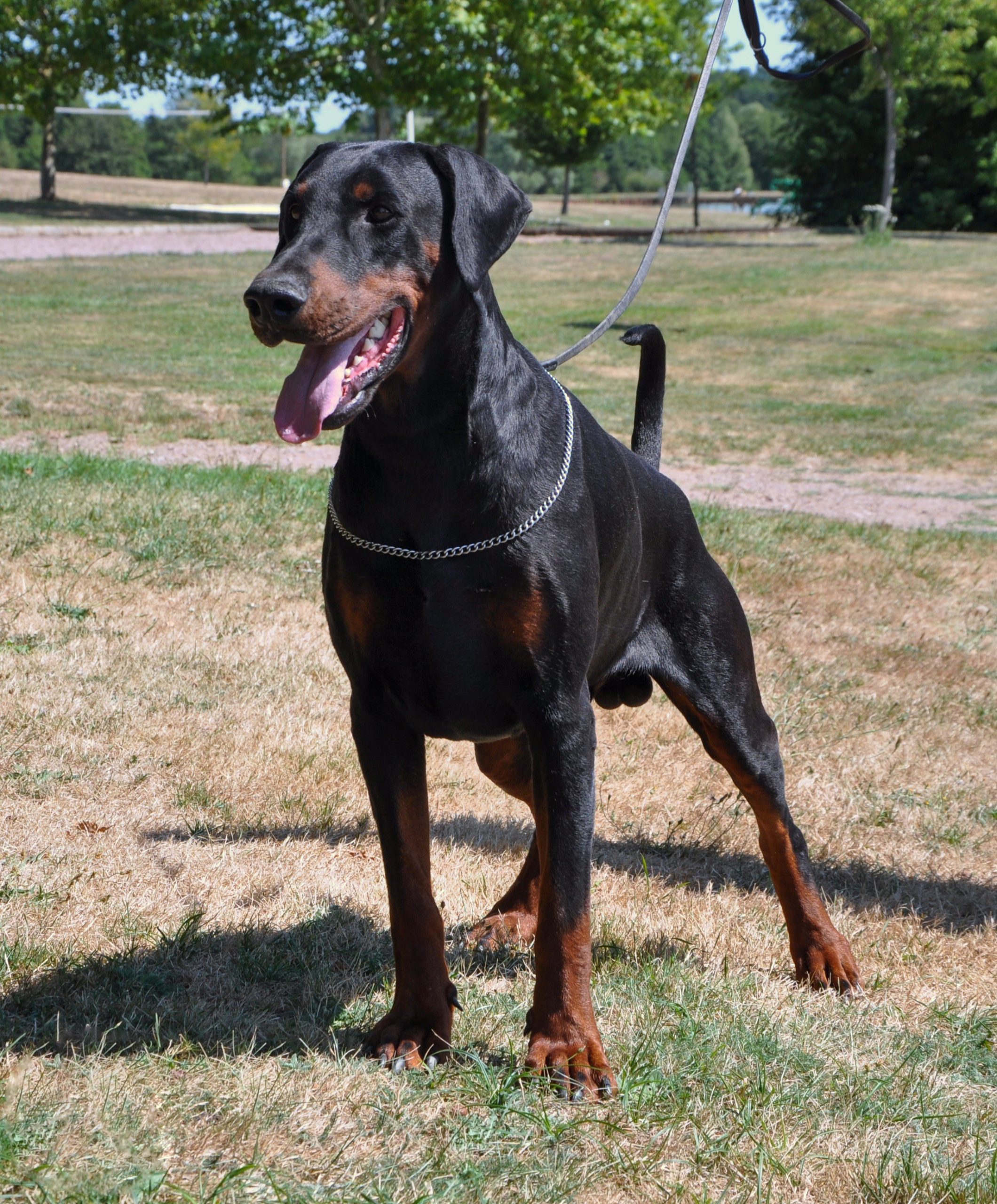
A handful of breeds carry this disease far more than others. The risk runs in the bloodline, handed down through families, so a breed and a pedigree carry as much weight here as anything on the screen. The Doberman, the Great Dane, the Boxer, the Irish Wolfhound, the Cocker Spaniel: each line runs a known risk, so a heart scan on a well dog of these breeds is screening, done before any sign of a complaint.
Screening starts young and repeats. A Doberman is scanned from around its third year, before any sign, the heart measured and noted as a baseline. The scan is repeated each year over the dog’s life, since a clean heart at one screening says nothing about the next. One look is never enough to clear a breed. A dog with a parent or a litter-mate found with the disease is watched the closer still, the family history sharpening the odds it carries.
The yearly scan reads the same handful of measurements each time, the chamber width and the strength of the squeeze, set against the last scan and against the range for the breed. A chamber that has widened or a squeeze that has slipped from one year to the next is the disease declaring itself, caught at a point where treatment still buys time. The slow slide shows in the trend across years, caught before it ever reaches the surface.
The screening scan does not work alone. A reading of the heart’s rhythm over a full day runs alongside it, since the rhythm fault of this disease often shows before the chamber does. The two together catch more dogs than either alone.
The breeds and their forms
The disease does not wear the same face in every breed. The Doberman runs the classic course, years of silence and then a sudden death, the rhythm stopping the heart, the chamber sometimes still only mildly changed the day it stops. The scan and the rhythm reading are both watched closely in this breed across its life.
The giant breeds carry their own version. The Great Dane and the Irish Wolfhound dilate young and slip early into a rhythm gone fast and out of order, the heart enlarging on a scale that matches the dog. A wolfhound that has tipped into that rhythm is watched hard, since this rhythm so often brings on heart failure.
The Boxer follows a path of its own, a disease that scars the heart muscle and breeds dangerous beats, sometimes with little of the dilation the name would lead one to expect. A scan in a Boxer is read knowing the chamber may look better than the heart in its true state.
The Cocker Spaniel marks a gentler corner of the disease. A dilated heart in this breed often answers to taurine, the muscle firming once the shortfall is filled, the chamber pulling back over months. A Cocker found with a dilated heart has its taurine checked before the case is ever called hopeless.
The rhythm fault
Many dogs with this disease throw extra beats long before the chamber gives them away. The weakened muscle fires off rhythm, scattering early beats through the day that slip past a brief clinic check.
A day-long recording on a portable monitor catches them. The tally that counts is the one over the whole day, since a quiet hour in the clinic hides a heart misfiring at home. A count of these stray beats above a set number over the day, or a rising count across two recordings in a year, marks the disease as surely as the scan does, sometimes sooner. In the Doberman especially, the rhythm is read as carefully as the chamber. A dog flagged by its rhythm is fitted for a longer watch, sometimes a monitor worn for days, and started on the drug that steadies the beat before the worst can happen.
The stakes of the rhythm are their own. A heart scattering enough bad beats can stop without warning. A dog can drop dead with a chamber still only mildly changed. The rhythm reading is what flags that danger, and what steers a dog toward the drugs and the watching that guard against it. A normal rhythm reading does not clear a dog for good. The bad beats come and go, a quiet recording one month giving way to a stormy one the next, so the rhythm is checked again on the dogs most at risk.
When the heart gives out
The silent years end when the failing pump can no longer keep up. Fluid backs up behind the weak left heart and seeps into the lungs. The dog comes in struggling for breath, drowning slowly from the inside. The scan that reads the wet lung and the dilated heart together names the cause in minutes.
The rhythm often gives way at the same time. A heart this stretched can slip into a racing rhythm with no order to it, the atria quivering, no longer pumping, a state that steals the last of the heart’s output. The scan reads the wide atria where this rhythm takes hold. The giant breeds are the ones it strikes hardest.
By this stage the work turns from finding the disease to holding it back. Drugs to strengthen the beat, to clear the fluid, and to steady the rhythm carry the dog through. The scan tracks how the heart answers them. The early scan is what spares a dog from reaching this point with no warning at all. A dog whose disease was found years before reaches this stage, if at all, already on treatment and already watched, the crisis softened or held off.
The line between a dog watched quietly at home and a dog fighting for breath in an oxygen cage can come down to which heart was scanned in time. The early scan is what buys the dog the good years that come before this stage.
The diet question
A wave of this disease has been traced to the food bowl. Certain diets, many of them grain-free and built around peas, lentils, and other pulses, have turned up again and again in dogs found with dilated hearts. A health agency raised the alarm on the link, with the work to pin down the cause still under way.
Taurine sits near the centre of it. This building block matters to the heart muscle, its shortage able to weaken the pump in dogs that run low. In some breeds and on some diets a dog runs short of it, the shortage enough to dilate the heart. A blood test for it runs alongside the scan when the diet falls under suspicion. Not every dog on a suspect diet falls ill, and not every dilated heart traces to the bowl, so the diet is weighed as one thread among the breed, the genes, and the rest.
The diet-driven kind carries a rare hope. When a diet is the cause, the heart can recover some of its strength once the food is changed and the shortfall corrected, the chamber pulling back and the squeeze firming over months. The scan tracks that recovery, the rare case where a dilated heart turns back toward normal. A dilated heart rarely sees that turn, the damage by then too deep, so the recovery of a diet-driven heart stands out as the exception it is.
Proving a diet caused a dilated heart is rarely clean-cut. A dog eats one food for years, and a heart found weak on the scan could trace to the bowl, to the breed, or to both at once. The diet is changed, the taurine corrected, the heart rescanned, a chamber that pulls back over the months that follow pointing at the food after the fact.
Reading more than the picture
The scan does not read the heart alone. A blood test for a marker the strained heart muscle releases climbs when the chamber is under load, a high reading on a screened dog pointing the scan toward a heart that earns a closer look. A second marker tracks muscle damage, rising when the heart is hurting.
Genes are read too in some breeds. A test for the faults known to drive this disease in the Doberman tells an owner the risk a dog carries before its heart shows a thing. A dog that carries the fault is watched the closer, its scans started early and read with extra care. The gene test is a one-time check, a cheek swab or a blood draw read once and good for the dog’s life.
None of these stands in for the scan that reads the heart itself, the chamber and the squeeze. The blood and the genes point to which dogs need that scan, and how often. The three together catch the disease earlier than any one of them alone.
What catching it early buys
Finding the disease in its silent years pays off, because the silent years can be treated. A drug that helps the weakened muscle squeeze and eases the load on it, started in the preclinical stage, holds off the day the heart tips into failure. The early start adds time to the dog’s life. Those are good years, won by a scan that looked before there was anything to feel.
The evidence behind this is why screening earns its place. A trial in Dobermans found the disease early on the scan showed a longer time to failure and a longer life on the drug than without it. For a dog that felt nothing wrong the day the scan found its heart, that gain was counted in months, sometimes longer. The scan that finds the occult heart is what opens that door.
The findings shape the plan. A heart caught early goes onto the drug and into a schedule of rescans. A rhythm scattering danger gets its own treatment and its own close watch. The scan turns a breed’s known risk into a managed disease. A disease watched from its first stirrings is one a dog can live a long while alongside.
How the handheld fits
The handheld scanner brings this screening into the consulting room and onto the breeder’s floor. The pocket unit goes where the dogs are, the kennel, the show ring, the farm, well away from the cart-based machine in the referral hospital. A vet reads the chamber and the squeeze on a pocket unit, picks out the heart that has begun to dilate, and flags the dog that needs the full study and the specialist. The whole check takes only minutes in the consulting room.
A breeder running a line through a screening programme can read a yard of dogs in a morning on a pocket unit, flagging the few that need the drive to a specialist. The handheld settles the screening question and refers the rest. Grading the disease, timing the drug to the stage, and ruling out the look-alikes is the work of a complete echo in a cardiologist’s hands, the work the pocket scan points the dog toward. On a healthy-seeming dog, that heart is the hardest finding to come by.
What can mimic a dilated heart
Not every large weak heart is this disease. A heart that a long-leaking valve has worn out dilates and weakens in much the same way. The scan reads the valve and the history to tell the two apart. A heart starved of its blood supply, or hurt by a drug or a toxin, can weaken alike; the scan reads the pattern and sends the vet hunting the cause. A heart driven too long by a fast rhythm can dilate, then recover once the rhythm is brought under control. Each of these leaves its own trail for the vet to follow.
The breed and the story carry weight here. A dilating heart in a young Doberman with nothing else to explain it points hard at the inherited disease. The breed and the story shift where the same picture points. The vet reads the heart against the dog it sits in, the picture never weighed on its own.
The window the scan opens
The years of quiet that dilated cardiomyopathy gives before it strikes are exactly what a scan fills with a warning. It reads the widening chamber and the fading squeeze on a dog that still seems well, and it hands the vet the time to act before the heart gives out.
What the early scan offers is time, the years a treated dog lives before the heart gives way, all of it bought on a quiet day when nothing yet seemed wrong.
Common questions about canine dilated cardiomyopathy
How is dilated cardiomyopathy diagnosed in a dog?
An ultrasound of the heart is the test that confirms it. The scan shows a left ventricle grown large, round, and thin-walled, its squeeze weak, often with a swollen left atrium behind it. A reading of the heart’s rhythm over a full day runs alongside, since the rhythm fault of the disease can show before the chamber changes.
Why scan a dog that seems healthy?
The disease hides for years in a silent phase, the heart dilating and weakening across years in which the dog still runs and eats well. A scan in those years finds it in time to treat. The at-risk breeds, the Doberman, the Great Dane, the Boxer, the Irish Wolfhound, are screened from young and rescanned each year.
Can dilated cardiomyopathy be treated if it is caught early?
Yes, and catching it early is the point. A drug that supports the weakened muscle, started in the silent stage, pushes back the day the heart tips into failure, the life that follows running longer. That window opens only when a scan finds the occult heart.
Is dilated cardiomyopathy linked to diet?
Some cases are. Certain diets, many grain-free and built around pulses, have turned up in dogs with dilated hearts. A shortage of the building block taurine can weaken the muscle in dogs that run low. When diet is the cause, a change of food can win back some of the heart’s strength, one of the few forms of this disease that ever turns back toward normal.
Can a handheld scanner screen for dilated cardiomyopathy?
Yes, for the screening question. A vet reads the chamber size and the strength of the squeeze on a pocket unit, picking out the heart that has begun to dilate. Grading the disease and timing the treatment is the cardiologist’s full echo, the step a handheld scan is there to point the dog toward.

About the Author
Julien Mercier
Senior R&D Engineer · Medical Ultrasound Transducer Development
Senior R&D Engineer with an M.S. in Applied Physics and over 15 years of experience in medical ultrasound transducer development, specializing in the design verification and performance testing of high-frequency imaging transducers. Currently leading the development and verification of the company’s next-generation high-frequency linear-array transducer, responsible for imaging performance evaluation and reliability analysis in preclinical testing. Brings extensive hands-on experience in piezoelectric element tuning, beamforming parameter optimization, and system-level performance testing.