





































Canine Feline Bladder Stones Ultrasound Handheld Veterinary
A bladder stone shows on ultrasound as a bright spot that throws a black shadow behind it. That one pairing, a hard echo over a dark streak, names a stone of any size and any make-up, including the kinds a plain X-ray would miss. A handheld probe laid on a dog’s or a cat’s lower belly finds the stones, counts them, and reads the blocked animal whose stone has turned an ache into an emergency.
The bright stone and its black shadow
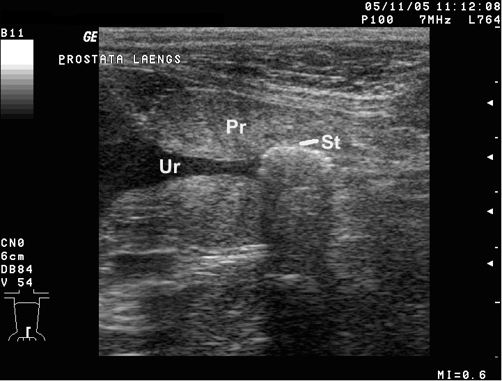
Urine sits black on the screen, a clean dark pool that shows up anything solid afloat in it. A stone is a lump of hard mineral, so it bounces the sound straight back as a bright white mark, sharp against the black. A bone-hard surface is what does it. Soft things afloat in the urine, blood and shed tissue, let the sound pass straight through, leaving no such mark. The brightness on its own would not prove a stone, since plenty of things in a bladder glow white. The proof is what lies behind it.
Sound cannot pass through a stone. It stops dead at the hard surface. The space beyond reads as a black column running down to the bottom of the screen, the shadow the stone casts. This clean dark streak is the true signature. The brighter the mark and the sharper the shadow under it, the surer the read, so a vet leans on the shadow as the surer of the two signs.
Colour Doppler adds a last check. Switched on over a stone, it lights the rough mineral surface with a scatter of mixed colour, a flicker called the twinkling sign. A faint stone with a weak shadow will often twinkle bright, the artifact catching what the grey picture nearly missed.
How the scan is run
The bladder is the easiest organ in the belly to read. A stone is among the easiest finds in it. The animal lies on its back or its side, a small patch of the lower belly clipped and wetted with gel, the probe set low over the bladder. A few minutes is all a clear bladder asks of the vet, the organ sitting right under the lower belly wall at the easiest depth for a probe to reach.
A full bladder reads best. Urine stretches it into a clean dark window. A stone inside stands out plainly against the black. A bladder emptied just before the scan folds down on itself, its walls falling together, the creases swallowing a small stone. A vet who can will scan before the animal is let out, or fill the bladder back up to look again.
The whole bladder is swept, every wall and the floor of it. The probe runs from the neck at the front to the dome at the back, down both walls and across the floor, since a stone can hide anywhere, a second often waiting behind the first. The number found changes the plan, so the count is made with care.
Why the animal came in
An animal does not complain of a stone. It shows the trouble in other ways. An owner brings it in for signs that could mean a dozen things. The scan is what turns those signs into a stone, or rules one out. A scan early in the work-up spares the animal a fortnight of treatment aimed at the wrong thing.
Blood in the urine is the loudest sign. A dog passing pink or red water, a cat leaving bloody spots on the bedding, has a bladder that something is rubbing raw, a stone the commonest cause. The animal squats and strains, over and over, to pass a dribble, the bladder wall left sore and irritable.
The signs read the same as a plain infection. That is the trap. A bladder infection and a bladder stone both bring blood, straining, and a sore bladder. The two travel together so often that the stone is what seeded the infection. A course of antibiotics quiets the signs for a while, the stone sitting there all along, so the scan is run to look past the signs to the cause that an infection alone would never explain.
Some stones say nothing at all. A bladder can carry one for months in silence, the trouble found only when the animal is scanned for something else. A quiet stone is watched, since the day it shifts to the outflow is the day it turns dangerous.
Sand, gravel, and the moving floor

Not every stone is a single pebble. A bladder can hold a slurry of fine grit, a sand so fine that no single grain throws a shadow of its own. This sediment settles to the lowest point and lies there as a bright layer, a flat white line drawn across the dark pool. Fine crystal in the urine, the raw material a stone is built from, can show as this same haze well before any true stone has formed.
The way to read it is to move the animal. A dog rolled from its back onto its side sends the sand pouring to the new low point, the bright layer shifting under the watching probe. That slide is the proof. A bright patch that stays put when the animal turns is something growing from the wall. A layer that runs to the bottom like sugar in a glass is grit, loose in the urine and free to pass. A bladder full of grit often means a stone is forming, or that one has just broken up, so a sandy scan is read as a warning to watch the bladder closely.
The blocked animal
A stone that leaves the bladder and lodges in the urethra turns a manageable problem into a race against the clock. The animal strains, passes little or nothing, and grows sick within hours, the trapped urine backing up with nowhere to go. Every hour the block holds, the kidneys behind it suffer the more, so the clock the scan starts runs fast.
The male is the one at risk. He has a long urethra that narrows to a tight channel, tightest of all in the tom cat. A stone a female would pass slips into it and jams there. A blocked tom cat, crouched and straining over an empty tray, is one of the genuine emergencies of small-animal work. The probe reads the over-full bladder, stretched tight as a drum, and follows its neck down toward the stone wedged in the outflow. A female blocks far less often, her wider urethra letting all but the largest stones through, so a blocked female is itself a flag for a big stone or a mass.
The scan does more than find the stone. It reads how full and how stretched the bladder has grown, whether the wall is thickening under the strain, whether urine has begun to back up into the kidneys above. That whole picture sets how fast the animal has to reach treatment.
A blocked bladder left too long bursts, or poisons the blood through the kidneys behind it. The scan that catches the block early is the one that saves the animal.
The cat that blocks without a stone
A blocked cat is not always a cat with a stone. The narrow feline urethra clogs as readily with a plug of mucus and crystal, a soft paste that sets in the outflow, or clamps shut on its own in spasm and swelling. The scan tells these apart from a true stone. The telling changes the treatment.
A stone shadows hard and twinkles bright. A plug of mucus and crystal throws only a faint shadow or none, a soft grainy mass low in the outflow. A urethra clamped tight shows no mass at all, only an over-full bladder above a tight neck that holds no stone. Each blocks the cat the same way. Each is read off a different picture.
The commonest trouble in a cat’s lower water has nothing to do with stones. It is an inflamed, irritable bladder, the syndrome vets call feline idiopathic cystitis. The wall reads thickened, the urine cloudy with debris, not a stone in sight. A scan that finds no stone in a straining cat has ruled in a different illness. The empty search is itself the answer.
What a stone is made of
The scan finds a stone and reads its size, its number, and its place. What it cannot read for certain is what the stone is made of. The make-up matters as much as anything, since it sets whether the stone can be dissolved away or has to come out through a cut.
Four kinds cover the bulk of what a dog or a cat grows. Struvite is the commonest of them. It builds up where an infection sweetens the urine. It can often be dissolved over a few weeks on a diet that turns the urine acid, the antibiotics clearing the infection that fed it in the first place. Calcium oxalate is just as common. It is the stubborn one. No diet shifts an oxalate stone, so one that troubles an animal has to be taken out. Urate stones grow in Dalmatians and in dogs with a liver shunt, held down by a special diet and a drug that lowers the waste they form from. Cystine stones come of an inherited fault carried in a handful of breeds. They too can sometimes be coaxed away before a knife is reached for. The screen cannot tell these four apart by sight. The stone itself, once passed or lifted out, goes to a laboratory that names the mineral and guides the plan against the next one.
Until the laboratory reports, a stone is treated by the company it keeps. An infected dog with the right kind of urine earns a trial at dissolving the stone. A breed that grows the stubborn oxalate goes more often straight to removal. The signs and a simple urine test point the way long before the stone itself is ever in hand.
The scan is what watches a dissolving stone shrink. A struvite stone put on its diet is scanned every few weeks, the bright mark growing smaller from one look to the next. A stone that holds its size after a fair trial is one no diet will move. The plan turns toward surgery.
Size and number steer the choice as well. A single small stone may flush out or pass on its own with help. A bladder packed with large stones, grinding against an inflamed wall, is a bladder a surgeon empties. A stone the size of a pea troubles a cat far more than the same stone in a big dog, the cat’s plumbing the less forgiving of the two.
The shadow is the stone
A bright mark is a maybe. The shadow behind it settles the question.
The stones a film can miss
An X-ray is the older way to hunt for stones, and for the common hard kinds it works well enough. A struvite or an oxalate stone holds enough mineral to throw a clear white shape on a plain film. The trouble lies with the stones that do not.
Urate and cystine stones carry less mineral. The smaller of them can show only faintly on an X-ray, or slip past it altogether. A bladder full of urate or cystine stones can read as clear on an X-ray. The film never picks those two kinds up. Ultrasound reads them all the same way, off the shadow behind the mark, the make-up no barrier to the sound. This is the reason a scan is reached for when the X-ray and the signs refuse to agree.
What else throws a bright spot
A bright mark in a bladder is not always a stone. A clot of blood can settle on the floor and glow. A tumour can grow from the wall and glow bright, its surface rough. A clump of mineralised debris can ape a stone closely. The probe sorts them out by the company each keeps.
A stone shadows, twinkles, and slides to the new low point when the animal is rolled. A blood clot throws no clean shadow. It breaks up or drifts away over hours. A tumour grows out of the wall, holds its place when the animal moves, and carries a blood supply of its own that the Doppler lights up. A stone has no blood running through it. Read together, these tells keep a real stone from being missed and keep a tumour from being mistaken for one.
Gas in the bladder throws a bright mark of its own, left behind by a catheter or a gas-forming infection. Gas floats up to sit at the roof of the bladder. Its shadow comes back dirty and streaky. A vet reads the high position and the messy shadow and knows the bright mark for gas.
The hardest case is a stone sitting on a mass, the one hiding the other beneath it. A bladder that will not come clear once the obvious stone is gone earns a careful second look. More than one bladder cancer has first shown itself only after the stone above it was lifted out.
Following the trail upstream
Stones are not the bladder’s alone. The same bright-and-shadow mark turns up in the kidney, a stone lodged in the funnel where the urine gathers before its run down. A kidney stone often sits quiet for years, found by accident when the animal is scanned for an unrelated problem. A large one can fill the whole collecting space, a great branching cast the probe reads at a glance.
The danger is a stone that travels. A kidney stone that drops into the narrow ureter, the slender tube down to the bladder, can jam there and dam the kidney behind it. The scan reads the swollen kidney, its centre ballooned with urine that cannot drain, and tracks down the ureter toward the stone doing the damage. The stone itself may be no bigger than a grain, the kidney it has dammed swollen huge above it. A blockage caught in its first days is a kidney saved from dying.
The urethra carries the last stretch. A stone caught in the male’s outflow is hunted along the urethra from the bladder neck down, the first spot the scan checks in any animal that has blocked. A male cat that has blocked once will often block again, so its lower water is watched for the rest of its days.
What the picture changes
A scan turns a vague urinary case into a plan with edges. Blood in the urine, straining at the tray, a dog licking and restless: the scan reads whether a stone lies behind it, where that stone sits, and how many there are. What was a guess becomes a map the vet can act on.
The numbers steer the route. A stone the animal has no hope of passing, a bladder awash with gravel, a male blocked at the outflow: each one points a different way, toward a diet, a flush, or a surgeon’s table. The scan hands the vet the facts the choice gets made on. A stone lodged at the outflow jumps the queue ahead of all of it, the one finding that cannot wait.
It reads, too, the bladder the stone has been hurting. A wall worn thick by years of grinding stones, a pocket of angry lining, the leftover debris of an old infection: these ride along with the stone and shape both how the animal is treated and how well it mends afterward.
After treatment the scan checks the work. A bladder scanned a few weeks past surgery or a course of diet shows whether it has fully come clear, or whether a missed fragment has already begun to grow itself back into a stone. A single fragment left behind seeds the next bladder full of them, so the after-check matters nearly as much as the treatment.
Keeping the next stone away
A stone removed is not a stone prevented. The fault that grew the first one, an infection, a breed’s inherited chemistry, a diet that loads the urine, sits ready to grow another. The make-up named by the laboratory is what points to the cause, and to the guard against it.
The greater part of the guard is in the food bowl. A diet matched to the stone keeps the urine dilute and shifts its chemistry away from the mineral that formed. Plenty of water drunk keeps the bladder flushed. A bowl of wet food, or water poured over dry, does as much as any prescription by keeping the urine weak. A struvite former is kept clear of the infections that seed the stone. An oxalate former is fed to hold its mineral in solution.
The scan is the watch that follows. A pet known to grow stones is scanned now and then across the years, the bladder read for the first small bright fleck before it grows into a stone that blocks or bleeds. Caught while it is still grit, a recurrence is far easier to head off.
Where a stone hides from the probe
The scan is not flawless at the task. A stone smaller than a grain of rice may glow without a shadow and slide past a hurried look. A stone tucked behind a loop of gas-filled gut, or sitting low in the urethra under the pelvic bones, rests in a corner the probe strains to reach at all.
Gas is the steady enemy here, the same as everywhere in the belly. A bladder read in a rush, or one emptied just before the scan and folded down on itself, can hide a stone away in its folds, lost until the bladder is filled again. The honest scan marks down what it could not see clearly and brings the animal back for a fuller look.
Disease that has thickened the bladder wall hides a stone best of all, the bright mark of the stone blurring into the bright wall around it. A vet who knows the wall is diseased reads it twice over, working the gain hard to part the stone from the tissue it rests against.
The shadow falls and the stone is found
A bladder stone gives itself away the moment the dark shadow falls in behind the bright mark. The scan finds it, sizes it, counts it, and reads the animal whose water it has put in danger.
Common questions about bladder stones
How does ultrasound show a bladder stone?
A stone reads as a bright spot on the dark pool of urine, with a clean black shadow running down behind it. That pairing of a bright mark over a dark shadow is the signature of a stone. It marks a stone of any make-up, even the kinds a plain X-ray can miss.
Can ultrasound tell what a stone is made of?
No. The scan reads a stone’s size, number, and place. Naming the mineral takes a laboratory, working on the stone itself once it is passed or removed. The make-up matters, since it decides whether a stone dissolves on a diet or has to be taken out.
Why is a male animal with a stone an emergency?
A male has a long narrow urethra, narrowest in the tom cat, where a stone leaving the bladder can lodge and dam the flow of urine. A blocked animal strains, passes nothing, and grows sick within hours. The scan reads the over-full bladder and the stone in the outflow. The animal needs treatment without delay.
Can bladder stones be treated without surgery?
Some can. A struvite stone, the kind an infection builds, can often be dissolved over a few weeks on a special diet with antibiotics. A calcium oxalate stone will not dissolve and usually has to be removed. The scan follows a dissolving stone as it shrinks from one look to the next.
Does ultrasound find stones an X-ray cannot?
Yes. Urate and cystine stones carry less mineral and can show only faintly on a plain X-ray, or hide from it. Ultrasound reads every stone the same way, off the shadow behind it, so the make-up is no barrier. A scan is the answer when the X-ray and the signs do not agree.

About the Author
Julien Mercier
Senior R&D Engineer · Medical Ultrasound Transducer Development
Senior R&D Engineer with an M.S. in Applied Physics and over 15 years of experience in medical ultrasound transducer development, specializing in the design verification and performance testing of high-frequency imaging transducers. Currently leading the development and verification of the company’s next-generation high-frequency linear-array transducer, responsible for imaging performance evaluation and reliability analysis in preclinical testing. Brings extensive hands-on experience in piezoelectric element tuning, beamforming parameter optimization, and system-level performance testing.