Transvaginal Endocavity Ultrasound Handheld Dual Head
A transvaginal endocavity ultrasound puts a slim probe a short way inside the vagina to read the pelvic organs from close range. The uterus and the ovaries sit deep in the pelvis, beyond the clear reach of a probe on the lower belly. A probe placed beside them, an inch or two away, reads them in fine detail. On a handheld system the same wireless handle carries this endocavity head alongside a curved one for the belly, so one device covers the scan from outside and the scan from within.
Why closeness beats reach
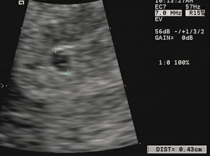
A four-millimeter gestational sac on a transvaginal scan, the calipers measuring it. A sac this small reads clearly from close range, weeks before a belly scan would find it. The labels and the measurement are the scanner’s own.
The pelvis is hard to see from the outside. The uterus and the ovaries lie deep behind the bladder and the bowel. A probe on the lower belly looks at them across five to fifteen centimeters of tissue. Gas in the bowel scatters the sound along the way. The picture that reaches the screen has softened over that distance. A full bladder helps by pushing the bowel aside, at the cost of a patient holding discomfort through the scan. The distance is the whole trouble. Sound loses detail with every centimeter it travels through tissue. The deep pelvis sits at the far end of a belly probe’s useful range. The bladder, the bowel and the fat between the skin and the uterus each blur the picture a little more. By the time the echo returns, the fine edges of a small structure have washed out.
Closeness changes the picture. A probe in the vagina sits one to five centimeters from the uterus and the ovaries, with no bowel in between. The short path lets it run at a higher frequency, the kind that draws fine detail. The same organs that softened on the belly scan come back sharp from within. Proximity does for the pelvis what raw frequency does for the skin. The frequency is the lever. The short path inside lets an endocavity probe run high, around six to twelve megahertz. That high frequency spends itself on fine detail, the resolution the pelvic organs reward. A belly probe, reaching deeper, runs lower and trades that detail for the penetration it needs to get there. The numbers tell the trade in plain terms. A probe at twelve megahertz draws a sharper line than one at four, over a shorter reach. The pelvic organs sit within the short reach the high frequency allows, so the probe gives up nothing it needs by running high. The trade falls the right way for this work.
The gain shows in what the probe resolves. A small structure, an early sac, a single follicle, a thin endometrial line, comes clear only from close range. The clinic that needs to measure these small things reaches inside to do it. The detail is the reason the route exists at all. The measurements depend on it. A clinician dates a pregnancy by a sac a few millimeters wide, times an IVF cycle by follicles measured to the millimeter, judges a cervix by a length read to a tenth of a centimeter. Each of these asks for detail that only the close view supplies. The numbers a fertility unit and an obstetric clinic run on come from the probe that sits nearest the organ.
The rule is plain. The closer the probe sits, the finer the picture it draws.
What the inside view is for
The earliest weeks of pregnancy are the probe’s clearest use. A gestational sac of two to three millimeters shows around five weeks from the last period, often before a belly scan finds anything. The yolk sac follows near five and a half weeks, a fetal pole with a heartbeat by about six to seven weeks. Measuring the embryo from crown to rump dates the pregnancy closely in these early weeks. The work of reading an early pregnancy at five to eight weeks rests on this close, early view. The early scan answers the questions that matter then. It confirms the pregnancy sits in the uterus, the first thing to settle. It finds the heartbeat that marks a viable pregnancy. It dates the pregnancy from the crown-rump length, the measurement at its truest in these weeks. A scan a week apart shows the sac and the pole growing on schedule, the sign of a pregnancy on track.
The same probe runs a fertility cycle. It counts the growing follicles in each ovary and measures them one by one, the numbers that time the trigger and the collection. Counting and measuring follicles through an IVF cycle rests on the resolution from within, since follicles sitting only millimeters apart need the close view to be told apart. The cycle runs on the counts. Early in stimulation the clinician counts the small follicles in each ovary, the number that sets the dose. The probe measures the leaders day by day, the diameters that say when the eggs are ready. A follicle near eighteen to twenty millimeters signals the trigger. The whole timing of the cycle, the trigger and the collection, turns on numbers the probe reads from within.
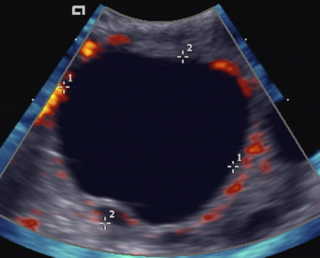
A mature ovarian follicle on a transvaginal scan, calipers across it, with color marking the blood flow at its rim. A follicle measured this way through an IVF cycle times the trigger and the collection. The labels are the scanner’s own.
The probe measures the cervix in a pregnancy at risk. Its length, read from within, flags a cervix shortening too early, the sign that a pregnancy may come before its time. Measuring the cervix when a pregnancy is at risk of coming early needs the precise length the close view gives. The length is a number with a threshold. A cervix measured short, under about twenty-five millimeters in mid-pregnancy, marks a raised risk of an early delivery. The reading guides whether a stitch or a progesterone treatment goes in. Read from within, the length comes clean and repeatable, the same spot measured the same way at each visit.
The probe also guides a needle. During egg retrieval for IVF, it steers the needle into each follicle to draw out the egg, watching the tip the whole way. Guiding the needle during egg retrieval joins the imaging to the procedure, the same probe finding the target and watching the needle reach it. The guidance is what makes the retrieval safe. A needle guide clips to the probe, setting the path the needle takes onto the screen as a line. The clinician lines that path up with a follicle and advances, watching the tip enter and the follicle empty under the needle. Each ovary holds several follicles, emptied one after another on the live picture.
Outside pregnancy the probe reads the lining of the uterus, the ovaries and the structures around them. A thickened endometrium, an ovarian cyst, a fibroid pressing on the cavity: each reads in detail from close range. The probe earns a place in the gynecology clinic the same way it earns one in the fertility unit, by reading small things clearly. The gynecology list keeps the probe busy between fertility cases. A patient with abnormal bleeding gets the endometrium measured for thickness, the reading that sorts who needs a sampling. An ovarian cyst gets its walls and its contents read for the features that mark it simple or complex. A fibroid gets mapped against the cavity it may distort. The pelvic floor reads from here too. A probe placed at the opening images the muscles and the bladder neck, the structures a continence clinic assesses. The same close access that serves the uterus serves the floor below it. The probe covers a range of pelvic work from one position.
When to reach for it
The inside route is a deliberate choice. A scan starts on the belly for the broad view, the whole uterus and both ovaries in one sweep. The probe moves inside when the belly view falls short, in early pregnancy, on a retroverted uterus, on a patient whose body habitus defeats the outside scan. Knowing when a scan must move from the belly to inside is a clinical judgment the sonographer makes case by case. A few situations call for it almost always. An early pregnancy scan goes inside, since the structures are too small to read from the belly. A retroverted uterus tips away from the belly probe and toward the endocavity one. A patient carrying weight on the abdomen puts the organs out of the belly probe’s reach. A clinician reads these signs at the start and picks the route the case needs.
How the two routes compare
Each route answers the same organs in its own way: the belly scan for the wide field and the easy approach, the inside scan for the close detail. How the transvaginal and transabdominal views compare, point by point, sets out how a clinic pairs the two across its obstetric and gynecologic work. The pairing has a logic. The belly scan opens the exam with the wide survey of the whole pelvis. The inside scan then closes in on what the survey flagged, the detail of a single structure. Leading with one and following with the other reads the pelvis whole and close in one sitting.
Often a scan uses both, the belly view to orient and the inside view to measure the detail it finds. A sonographer who reads both builds the fuller picture. The handheld dual head makes the switch a matter of changing the probe on the same app. The switch costs no time to speak of. The belly probe comes off, the endocavity head goes on, and the app loads the right preset. A scan flows from the wide view to the close one in the same visit, on the same screen. The patient stays put through both. The single device suits the rhythm of the clinic. A sonographer changes the head on one probe, with no second machine wheeled in, when the scan needs to go inside. The flow of the exam holds, the switch handled in the same hand on the same screen.
One handle, two heads
A dual-head handheld carries the answer to both routes on one device. A curved array for the belly and an endocavity array for the inside scan share a single wireless handle, switched in the app. A clinic that does obstetric and gynecologic work buys one tool and covers the outside scan and the inside scan from it. Choosing a wireless transvaginal probe comes down to the frequency, the field of view, and the fit of the endocavity head. The two heads cover the two routes from one purchase. A clinic that bought separate machines for the belly and the internal scan replaces both with a single handle and two arrays. The app holds the presets for each, switched in a tap. A practice running an obstetric clinic in the morning and a fertility list in the afternoon carries one device through both. The cost follows the same logic. One handheld with two heads sits well below two cart machines. It asks for no service contract on a console. A clinic adds the endocavity head to a base it already owns for the belly scan, a spend that matches a small practice better than a room full of equipment.
The wireless form suits the setting. No console stands between the probe and the patient, the picture running to a phone or a tablet the clinic already owns. A handheld probe moves from the examination room to the bedside to the procedure room, the same tool throughout. The endocavity scan that once meant a cart in a dedicated room now happens wherever the patient is. The portability suits this work in particular. An early-pregnancy scare walks into a clinic without warning. The probe is in a drawer, ready in the room. A fertility unit scans the same patients many times across a cycle, the quick handheld check fitting the visit better than a trip to radiology. The tool that travels to the patient suits a service built on frequent, short scans.
Keeping an internal probe clean
A probe used inside the body carries a cleaning duty with it. Between patients the endocavity head goes through high-level disinfection, the step that clears the organisms a wipe alone leaves behind. A single-use cover goes on during the scan. The two together, the cover during and the disinfection between, are what make repeated internal use safe. The standard is set by guidelines, by a bar every clinic doing internal scans follows. A probe that touches a mucous membrane falls into a class that requires high-level disinfection between patients, the same bar a flexible endoscope meets. The internal probe needs the fuller process, soaking or a closed cabinet that clears the organisms a cover alone leaves behind. A wipe of the kind that cleans a belly probe falls short of that bar. The cover does part of the job. A fresh sheath goes over the probe for each patient, keeping the contact clean during the scan. The disinfection handles what the cover cannot, the trace that reaches the probe at the rim of the sheath. The two steps together meet the standard the work demands.
The probe has to survive the cleaning. An endocavity probe is sealed to an IPX7 rating, watertight enough to be submerged for disinfection with the electronics staying dry. The seal is what lets the same probe be cleaned hard, day after day, across a full clinic list. The disinfection an internal probe needs between patients is a fixed part of the workflow, as much a part of the exam as the scan itself. The rating is one to check before buying. A probe sold for internal use should carry the IPX7 mark, the sign it survives submersion. A probe without it cannot take the disinfection the work demands. The seal protects the join where the cable meets the handle, the spot fluid would otherwise reach.
The duty shapes the buying choice. A probe for internal use has to meet the sealing standard and fit the disinfection a clinic can run. A clinic sets up the cleaning station alongside the probe, so the turnaround between patients stays short. The cleaning is part of the cost and the routine of working inside the body. The turnaround sets the pace of the clinic. A probe in disinfection is a probe out of use, so a busy list runs faster with a second probe or a quick cabinet. A clinic plans the number of probes and the cleaning method around the patients it sees in a day. The cleaning station sits beside the room, the probe cycling through it between cases.
The probe in the pocket
The transvaginal endocavity probe earns its place by closeness. It reaches the pelvic organs from an inch away and reads them in a detail the outside scan cannot match. On a handheld dual head it travels with the curved probe for the belly, cleaned to an internal-use standard between patients, ready for the early pregnancy, the fertility cycle, the cervix at risk and the retrieval needle. A clinic that does this work runs it from a probe that fits in a pocket. The pieces fit into one service. The close view reads the pelvis where the belly scan softens. The dual head carries the two routes on one handle. The seal lets the probe be cleaned for internal use, again and again. A clinic that puts these together does its obstetric and gynecologic imaging from a pocket-sized tool, in the room where the patient already sits. The whole pelvic exam, the wide survey and the close measurement alike, runs from that one device, cleaned and ready between cases.
The work spreads across several kinds of practice. An obstetric clinic dates pregnancies and checks the cervix. A fertility unit counts follicles and guides retrievals. A gynecologist reads the endometrium and the ovaries. Each leans on the same close view from the same probe. The endocavity head serves a range of clinics that once each needed a machine of their own.
For a clinic weighing it, the case comes down to reach and routine. One handheld with two heads covers the obstetric and gynecologic imaging a practice does, at the bedside, on equipment it can clean and carry through a full list of patients. The leaves that follow take each use in turn, from the early pregnancy scan and the follicle count to the probe selection and the disinfection an internal head needs.
Common questions about transvaginal endocavity ultrasound
Why does a transvaginal scan read the pelvis so clearly?
Because it sits close. The probe rests one to five centimeters from the uterus and the ovaries, with no bowel in the way, and runs at a higher frequency over that short path. The closer position draws fine detail that softens over the longer reach of a belly scan. Proximity does for the pelvic organs what a high frequency does for the skin.
What is a transvaginal scan used for?
Reading the pelvis from close range. It sees an early pregnancy from about five weeks, counts and measures follicles through an IVF cycle, measures the cervix in a pregnancy at risk, and guides the needle during egg retrieval. Outside pregnancy it reads the endometrium, the ovaries and structures like cysts and fibroids. The close view is what makes these small measurements possible.
When is the transvaginal route used?
When the belly view falls short. A scan starts on the lower belly for the wide field. The probe moves inside for early pregnancy, a retroverted uterus, or a patient whose body habitus blocks the outside scan. The choice is a clinical judgment made case by case. Many scans use both routes, the belly view to orient and the inside view to measure.
What is a dual-head handheld ultrasound?
One wireless handle that carries two probe arrays, a curved array for the belly and an endocavity array for the inside scan, switched in the app. A clinic doing obstetric and gynecologic work covers the outside scan and the inside scan from a single device, run from a phone or a tablet, with no console to buy or maintain.
How is an endocavity probe cleaned between patients?
By high-level disinfection, with a single-use cover during the scan. The probe is sealed to an IPX7 rating, watertight enough to be submerged for the disinfection with the electronics staying dry. The cover during the scan and the disinfection between patients are what make repeated internal use safe. The cleaning is a fixed part of the workflow.
Does a handheld transvaginal probe match a cart machine?
For this work it reads the pelvis in the detail the close range gives, on a probe that runs from a phone. The handheld form moves the scan to wherever the patient is, from the examination room to the procedure room. A clinic chooses the probe on its frequency, its field of view and the fit of the endocavity head for the work it does.
About the Author
Julien Mercier
Senior R&D Engineer · Medical Ultrasound Transducer Development
Senior R&D Engineer with an M.S. in Applied Physics and over 15 years of experience in medical ultrasound transducer development, specializing in the design verification and performance testing of high-frequency imaging transducers. Currently leading the development and verification of the company’s next-generation high-frequency linear-array transducer, responsible for imaging performance evaluation and reliability analysis in preclinical testing. Brings extensive hands-on experience in piezoelectric element tuning, beamforming parameter optimization, and system-level performance testing.