Choosing a wireless transvaginal ultrasound probe comes down to matching a handful of specifications to the work it will do: the frequency it runs at, the field it sees, the shape of its endocavity head, the seal that lets it be cleaned, and the wireless link that carries its picture to a screen. A probe that fits the clinic’s scans, cleans to standard and connects without trouble is the one to buy.
What you are choosing
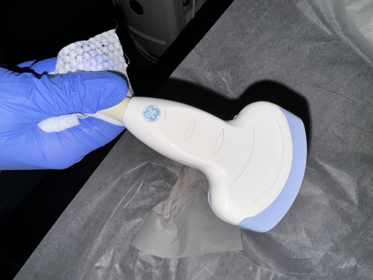
A handheld ultrasound probe. A wireless transvaginal probe packs the same kind of scanner into a slim endocavity body, chosen on the specifications this guide works through. The internal-use head has a different, slimmer shape from the probe shown here.
A wireless transvaginal probe is a slim endocavity transducer with its electronics, its battery and its wireless link built into the one handheld body. There is no cart and no cable to a console; the probe sends its picture to a phone or a tablet running the clinic’s app. The whole machine is the part held in the hand, which is what makes the choice of probe the choice of the whole system. Nothing else is bought alongside it. A wireless probe holds the scanner, the array and the picture’s source in one body, so the specification on the box is the specification of the system. Reading that specification well is the whole of choosing the machine.
That packing of everything into the probe raises the stakes of the choice. The frequency, the field, the seal, the wireless link, the battery and the software all live in the one device, and a clinic buys them together. Weighing each specification against the work the probe will do is how the right one is found. The table below sets out the specs to read. The sections that follow take them one at a time. The specifications pull on each other, which is why they are weighed together. A high frequency that drains the battery, a wide field on a slow link, a fine app on a probe that will not seal: each is a mismatch a buyer reads for. The aim is a probe whose specifications fit one another and fit the work, a set that holds together in use.
What to check on a wireless transvaginal probe
Frequency
around 5 to 9 MHz for the transvaginal route
Field of view
wide enough to hold the uterus and an ovary
Endocavity head
slim, with an angled tip that fits comfortably
Sealing
IPX7, so it takes high-level disinfection
Wireless link
low latency to a phone or tablet, steady in the room
Battery
a working session on one charge
App and display
OB and gynecology presets, clear calipers
Frequency, the first thing to weigh
Frequency is the specification that decides how fine a picture the probe draws. It is the first one to read on a transvaginal probe. A higher frequency resolves finer detail over a short reach, the kind of reach the inside route works at. The probe sits an inch or two from the uterus and the ovaries, close enough that a high frequency reaches them with detail to spare. That is the whole reason the inside route runs at a higher frequency than the belly probe. Distance is what forces a probe lower. The transvaginal probe has almost none to cross. Freed from the long reach, it spends its frequency on detail, the strength the route is chosen for.
How high a frequency suits the route is a settled question. A clinical reference for transvaginal sonography, the StatPearls review by Nahlawi and Gari, puts the working range at about five to seven and a half megahertz, the band that the short internal path is suited to. The closeness of the probe to the organs lets that higher frequency do its work, drawing the fine detail the pelvis is read for. A probe whose frequency sits in or around that range is built for the transvaginal job, and many modern probes reach a little higher still.
The work the clinic does sets where in the range to land. A practice reading early pregnancies, follicles and the lining of the uterus leans on the higher end, where the millimeter detail those measurements need is drawn at its cleanest. A probe that offers a span of frequencies, tuned in the app for the task at hand, covers more of the work than one fixed at a single figure. Reading the frequency on the specification is the first check a buyer makes. The higher figure sharpens the near detail the pelvic measurements turn on. A probe that reaches it serves the finest work a clinic does, and a buyer reading for early pregnancy and fertility looks for the upper end of the band.
A single probe rarely runs at just one frequency. A modern transvaginal probe spans a band, set in the app for each task, reaching the higher end where the finest detail is needed. A buyer reads the span the probe offers, since a probe with room to move covers more of a clinic’s work than one fixed at a point. The frequency is best read as a range, the span the probe can work across.
The field and the depth it reads
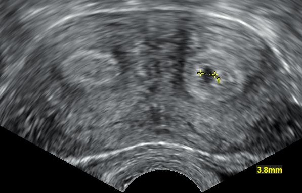
A transvaginal scan holding the uterus in one pelvic field, here a bicornuate uterus with an early pregnancy measured. The width of a probe’s field of view decides how much of the pelvis sits in a single picture like this. The markers are the scanner’s own.
The field of view is how much of the pelvis the probe holds in one picture. A transvaginal probe reads a sector that fans out from its tip. The width of that fan sets how much sits on the screen at once. A field wide enough to hold the uterus and an ovary together saves the sonographer sweeping piece by piece. It is a figure to read on the specification alongside the frequency. A field too narrow makes for a slower scan. A sonographer working through a narrow window reads the pelvis in pieces, angling from one structure to the next. A wider field holds more of them together in one view, which speeds the scan and eases the read. The width of the field is a comfort the buyer reads for.
Depth matters less on the inside route than it does from the belly, since the organs sit close to the tip. A transvaginal probe needs only to read a few centimeters ahead of itself to reach the uterus and the ovaries. A probe built for the route focuses its detail in that near field, where the work is, and leaves the far depth the route never reaches for.
The shape of the field is set by the array in the tip. The makers describe it by an angle and a depth. A buyer reads those figures against the scans the clinic does, looking for a field that holds the structures it measures without strain. The field and the frequency together decide the picture. They are read as a pair. Neither figure tells the whole story alone. The two set the detail and the view together, and a buyer reads them against each other for the balance the clinic’s scans call for. A probe strong on one and short on the other suits a narrower run of work than one balanced across both, which is why the pair is weighed as one.
The shape that has to fit
The endocavity head is the part that goes inside, and its shape decides how it sits and how it is tolerated. A slim shaft with a smoothly angled tip reads the uterus ahead and the ovaries to each side with a gentle turn. It is comfortable for the patient through the scan. A buyer handles the probe, or reads its dimensions, to judge that the head is the slim, well-shaped kind the route needs, since a probe that is awkward to angle or uncomfortable to use is one a clinic will fight with every day. The fit is read with the patient in mind as much as the sonographer. A head shaped to sit and angle easily makes the scan quicker and the few minutes inside more comfortable, which matters in a route already more intimate than a scan on the belly. A buyer who handles the probe, feeling its weight and the balance of its head, judges the fit better than a dimension on a sheet can show. The shape that fits is the one a clinic reaches for without a second thought.
Wireless, connection and power
The wireless link is what makes a handheld probe handheld, and its quality decides how the probe feels to use. The probe sends its picture to the screen over a wireless connection, and that connection has to carry a live image with little delay. A lag between moving the probe and seeing the picture move would make a guided scan hard, so a low latency is the thing to look for in the link. Latency is the delay between the probe moving and the screen catching up. A probe built for live work keeps that delay small enough to go unfelt, the picture tracking the hand as it moves. A lag long enough to notice would throw off any scan that follows a moving structure or a needle. The smaller the delay, the closer the live picture is to the hand that makes it.
The link also has to hold steady in a working room. Other devices, walls and bodies can all interfere with a wireless signal, and a probe that drops its picture mid-scan is no use. A probe built for clinical work keeps a reliable connection across the room it is used in, on a band that holds up in a busy clinic. Reading how the probe connects, and how far it holds, is part of weighing it.
Power is the other side of going wireless. The probe carries its own battery, and that battery sets how long it works between charges. A battery that lasts a working session, a clinic list or a procedure, without a stop to recharge, is what the work needs. A probe that runs flat in the middle of a list is a probe that holds the clinic up. Battery life is read in the work the probe does. A figure measured in still standby tells little; what counts is the hours of live scanning the probe gives. A clinic reads the battery against its busiest day, looking for a charge that carries the list without a stop. The honest measure is scanning time, the work the battery does on a live picture.
How the probe charges weighs into the choice as well. A spare battery that swaps in, or a charge fast enough to top up between patients, keeps a single probe working through a long day. A clinic running back-to-back scans reads the battery life and the charging method together, sizing them to the list it runs. The power has to match the pace of the work.
The connection a probe uses also touches on the data it carries. A scan holds patient information. The link between the probe and the screen, and onward to where the images are stored, is built to keep that information secure. A buyer checks that the system handles the images and the patient data to the standard the clinic works to. The wireless convenience is matched to the care the data needs. The security travels with the picture from end to end. The link from the probe to the tablet, and the path onward to where the scans are kept, each holds patient data, and each is built to keep it safe. A clinic checks that the system meets the data rules it works under before the probe joins the work. Convenience that loosened the hold on patient data would cost more than it saved.
The seal that lets it be cleaned
An internal probe has to be cleaned to a high level between patients, and the seal is what lets it take that cleaning. A probe rated IPX7 is watertight enough to be soaked or run through a disinfection cabinet without harm, which is the reason the rating is read before a probe is bought for internal use. A probe without it cannot be cleaned to the standard the work demands.
The cleaning a probe is built for is a check the buyer makes alongside the seal. The maker lists the disinfectants and the methods the probe is cleared for, and a clinic matches its disinfection to that list so the cleaning does not harm the probe over time. Confirming the seal and the cleaning together is part of choosing a probe that will last the work it meets.
The app and the picture
A wireless probe runs from an app on a phone or a tablet, and that app is as much a part of the probe as the array. It carries the presets for the work, the obstetric and gynecologic settings that tune the picture for each scan, and the tools the sonographer reads the image with. A buyer tries the app, since the probe is used through it, and an app that is clear and quick makes the probe quick to use.
The measurement tools are where the app earns its place in this work. So much of a pelvic scan is a number, a sac, a follicle, a lining, a cervix, and the app has to lay a caliper cleanly and read a length to the millimeter. A clinic checks that the app measures the things its scans measure, with the calipers and the reports the work needs. The picture is only as useful as the tools that read it. The reports the app builds matter as much as the calipers. A scan ends in a record, a set of measurements written into a report the clinic keeps and shares. An app that gathers the numbers into a clean report, ready to file, saves the time a hand-built one would take. A buyer reads how the app reports as closely as how it measures.
The screen the app runs on is the last link in the picture. A phone or a tablet bright and sharp enough to read the fine detail of a pelvic scan shows the probe’s picture at its best, and a clinic chooses a device that does the probe justice. The probe, the app and the screen are read as one system, since the picture passes through all three on its way to the eye.
Putting the choice together
A wireless transvaginal probe is chosen by reading its specifications against the work it will do, one at a time and then as a whole. The frequency for the detail, the field for the view, the head for the fit, the seal for the cleaning, the link and the battery for the wireless work, the app for the reading: each is checked, and the probe that meets them together is the one to buy. The order of the checks follows the work. The frequency and the field come first, since they set the picture every scan depends on. The seal and the cleaning come close behind, since a probe that cannot be cleaned cannot be used at all. The link, the battery and the app round out the read, each weighed for the way the clinic works.
The right probe then disappears into the work, a slim tool that reads the pelvis closely, cleans to standard, connects without fuss and runs a full list on a charge. A clinic that chooses well buys a probe it can carry from room to room and trust through the day, the whole of its pelvic imaging running from a device that fits in a pocket. The choice made with care is the one a clinic stops thinking about, because it works.
Common questions about choosing a wireless transvaginal probe
What frequency should a transvaginal probe be?
Higher than a belly probe, because the close internal path suits a higher frequency. A clinical reference for transvaginal sonography puts the working range at about five to seven and a half megahertz, and many modern probes reach a little higher. A practice reading early pregnancies, follicles and the endometrium leans on the higher end, where the millimeter detail those measurements need is drawn at its cleanest.
Does a wireless probe need an IPX7 rating?
Yes, for an internal probe. IPX7 means the probe is watertight enough to be soaked or run through a disinfection cabinet for high-level disinfection without fluid reaching its electronics. A transvaginal probe is disinfected to a high level between patients, so it has to carry a seal that survives the cleaning. The rating is a first check before such a probe is bought.
What to look for in the wireless link?
A low latency and a steady connection. The probe sends a live picture to a phone or a tablet, and a lag between moving the probe and seeing the picture move would make a guided scan hard. The link also has to hold across a working room without dropping the image. A probe built for clinical work keeps a reliable, low-delay connection in a busy clinic.
How long should the battery last?
Long enough for a working session without a stop to recharge, such as a clinic list or a procedure. A probe that runs flat in the middle of a list holds the clinic up. Many probes pair a working battery life with a spare that swaps in or a quick charge between patients, so a single probe keeps going through a long day. The battery is sized to the pace of the work.
Why does the app matter when choosing the probe?
Because a wireless probe is used entirely through its app. The app carries the obstetric and gynecologic presets that tune the picture, and the calipers and reports the sonographer measures with. So much of a pelvic scan is a precise number, so the app has to lay a caliper cleanly and read a length to the millimeter. A clear, quick app makes the whole probe quick to use.
Can one wireless probe cover a clinic’s obstetric and gynecologic work?
A dual-head system can, pairing the transvaginal head with a curved abdominal one on a single wireless handle. For the internal work alone, a well-chosen transvaginal probe reads early pregnancy, follicles, the cervix and the gynecologic pelvis from close range. Matching the frequency, the field, the seal, the link and the app to that work is how one probe is made to cover it.
About the Author
Julien Mercier
Senior R&D Engineer · Medical Ultrasound Transducer Development
Senior R&D Engineer with an M.S. in Applied Physics and over 15 years of experience in medical ultrasound transducer development, specializing in the design verification and performance testing of high-frequency imaging transducers. Currently leading the development and verification of the company’s next-generation high-frequency linear-array transducer, responsible for imaging performance evaluation and reliability analysis in preclinical testing. Brings extensive hands-on experience in piezoelectric element tuning, beamforming parameter optimization, and system-level performance testing.