Transvaginal Versus Transabdominal Ultrasound for OB GYN Comparison
Transvaginal and transabdominal ultrasound are the two routes to the same pelvic organs, one with the probe inside the vagina and one with it on the lower belly. Each reads the same uterus and ovaries by a different path, with its own set of strengths.
The two routes set side by side
The transabdominal route places the probe on the lower belly, a curved transducer pressed to the skin above the pubic bone. A full bladder lies beneath it, the fluid carrying the sound down to the pelvic organs and lifting the loops of bowel up out of the path. The probe runs at a lower frequency, in the range of three to six megahertz, the frequency that reaches the depth of the pelvis from the surface. From this position the scan looks down across the whole pelvis at once. It is the route a pelvic scan opens with for almost every patient. The curved shape of the probe spreads its beam into a fan, wide at the depth of the pelvis. That fan is what lets a single placement take in the whole region, the uterus in the midline and an ovary out to either side. The sonographer slides and angles the probe over the lower abdomen, building the picture from a series of sweeps. The full bladder underneath holds the view open throughout.
The transvaginal route places a slim probe a short way inside the vagina, its tip resting an inch or two from the uterus and the ovaries. No bowel lies in the path. The bladder is left empty so the organs fall close to the probe. The probe runs at a higher frequency, in the range of six to twelve megahertz, the frequency that draws fine detail over a short reach. From this position the scan reads the pelvic organs from close range. It is the route a scan turns to when the question depends on detail. The probe suits the path, long and slender, with the array set in its tip. The sonographer guides it gently into the vagina and angles the tip toward each structure in turn, the uterus ahead, an ovary to each side. A fresh single-use cover goes on before it enters, removed and replaced for every patient. This short path, kept free of bowel, is what the whole route is built on.
Both routes end at the same organs. The uterus, the ovaries, the lining of the womb, the spaces around them: each route reaches these and returns a picture of them. The difference lies in the path taken and in what that path does to the picture that comes back. The rest of this comparison works through those differences one at a time, from the detail each route resolves to the comfort each one costs.
What each one is like for the patient
The belly scan asks little of the patient beyond a full bladder. The patient drinks water and waits for the bladder to fill, then lies down for the scan, the probe moving over the lower abdomen through a layer of gel. Nothing enters the body. The full bladder is the one discomfort, a pressure held through the scan. It empties the moment the scan ends. For a patient who finds an internal scan difficult, or who is young, or for whom an internal scan is unsuitable, the belly route is the gentler way in. The preparation is the main demand it makes. A patient drinks several glasses of water an hour before and holds the bladder full until the scan, a wait that can stretch when a clinic runs late. That wait buys a clear acoustic window and an exam that touches only the skin. For a great many patients the belly scan is the whole of the visit, the bladder emptied and the appointment done.
The inside scan asks the patient to accept a probe within the vagina. The probe is slim, covered with a fresh single-use sheath and gel, and a sonographer explains the step and offers a chaperone before it begins. An empty bladder makes it comfortable, with no waiting beforehand. The scan is more intimate than a probe on the belly. A patient’s consent and comfort with it sit inside every decision to use it. A patient may decline. The scan then stays on the abdomen. The report says what that view could answer.
Resolution and reach
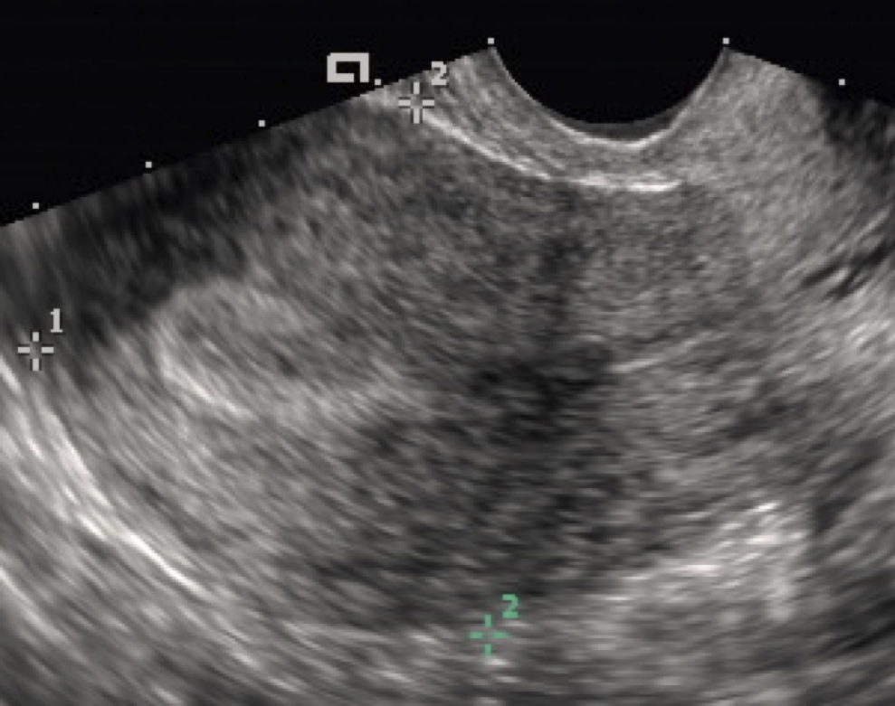
A transvaginal scan of the uterus, the endometrial lining resolved as a bright stripe through the middle. The close route reads fine structures like this lining to the millimeter. The markers are the scanner’s own.
Resolution is the heart of the comparison. It turns on two things, the frequency of the probe and the distance to the target. A higher frequency draws finer detail. A shorter distance lets a probe use that higher frequency, since high-frequency sound fades quickly as it travels through tissue. The two factors work together. The closer the probe sits to an organ, the higher the frequency it can use. The higher the frequency at close range, the finer the picture of that organ. This single rule sits under the whole comparison. It explains the softness of the far picture and the sharpness of the close one in one stroke. Every difference that follows, in detail, in depth, in the measurements each route can trust, traces back to this trade between frequency and distance. Holding the rule in mind makes the rest read as one idea.
The transvaginal route has both factors in its favor. Its probe sits an inch or two from the uterus and the ovaries, a short enough reach to run at six to twelve megahertz. At that frequency and that range it resolves structures a millimeter or two across, an early sac, a single follicle, the separate layers of the endometrial lining. The fine detail is the route’s defining strength, the reason a sonographer reaches for it whenever a small structure has to be measured to the millimeter.
The transabdominal route reaches the same organs from much farther off. Its probe sits five to fifteen centimeters from the pelvic organs, a distance that calls for a lower frequency of three to six megahertz to arrive at the depth at all. The sound also crosses the abdominal wall and any bowel and fat along the way, each layer softening it a little further. The picture it returns holds the large and the medium structures clearly, across a wide field. Reading something a millimeter or two across from that distance asks more of the picture than the long path can give. The wall and the bowel add their own toll. Sound that has crossed skin, fat, muscle and a layer of bowel gas arrives at the uterus already scattered. What returns has crossed all of it again. The thinner the wall and the quieter the bowel, the better the belly route reads. A heavy wall or a gassy bowel sets the limit of what it can show.
Depth shifts the comparison further. The transabdominal route loses ground when the target sits deeper or behind a thicker abdominal wall, the sound weaker by the time it returns from so far down. The transvaginal route is barely troubled by depth in the pelvis, since it begins its short path already close to the organs. For a deep ovary, or a pelvis behind a heavy abdominal wall, the close route keeps its full detail even at that depth, working from where it already sits.
The resolution gap is the single biggest difference between the two routes. A measurement that has to be exact, of an endometrium, a follicle, an early pregnancy, comes from the close route as a number a decision can rest on. The closer the probe sits, the more a fine measurement holds up. Wherever the answer depends on fine detail, the inside route is the one that supplies it. This single difference drives a large share of the choices a clinic makes between the two. The gap shows plainly on the screen. A follicle measured from within shows a crisp edge a caliper sits on exactly, sharp enough that two readers land on the same millimeter. Where a treatment turns on that millimeter, the close edge is the one a clinic works from. The fine measurements of fertility and gynecology are built on it.
The field of view
Field of view is the transabdominal route’s chief advantage. The belly probe, looking from a distance, takes in the whole pelvis in one picture, a large fibroid uterus or an ovarian mass reaching up out of the pelvis held whole on the screen. The wide single sweep locates everything large or high in one look. A close probe gives up that wide field for its detail, reading a window only an inch or two wide, one structure at a time. Sitting an inch from the uterus, it sees that uterus clearly and little beyond it, so the sonographer reads the pelvis piece by piece, angling the tip from one structure to the next. A large mass that spills past the window is read in parts, or read whole from the belly route. The wide single picture is the belly route’s to give.
Where each route is stronger
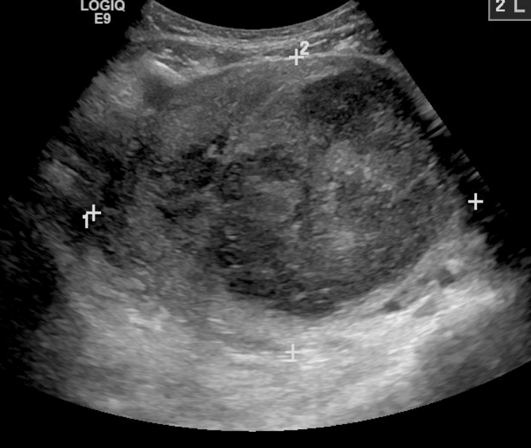
A large fibroid, around nine centimeters, held whole on one screen with calipers across it. A structure this size is read in a single wide sweep, the strength the wide field brings. The calipers and label are the scanner’s own.
The transabdominal route is the stronger choice wherever size or reach matters. A large structure that overflows the close window, a fibroid uterus, a big ovarian cyst, an organ sitting high in the pelvis or up into the abdomen, reads whole from the belly. The wide survey at the start of an exam belongs here, the single sweep that locates everything before any of it is looked at closely. So does the scan of a patient for whom the inside route is unsuitable or unwanted. The belly route covers the broad, the large and the high. Screening and follow-up sit here too. A known fibroid watched for growth over years needs the same wide measure of its size each visit. A check of the pelvis for free fluid, or of the bladder for what it holds after voiding, reads from the belly in a moment. These are jobs of size and presence. A single wide sweep does them quickly and well.
The transvaginal route earns its place wherever detail or depth matters. A small structure that the wide field cannot resolve, a precise measurement that a decision rests on, a deep organ behind a heavy wall, an early pregnancy whose location has to be settled: each of these reads from close range in the detail the question needs. The close route covers the small, the deep and the precise. Guidance belongs here as well. When a needle has to reach a follicle for egg collection, or a small target for sampling, the close view watches the tip the whole way in. The detail that resolves the target also resolves the needle against it. Any procedure that turns on seeing a fine structure and a needle together leans on the inside route. The same closeness that measures well also guides well.
The split follows from the two differences already drawn, the reach of one route and the resolution of the other. A clinician reading a case knows from the question which kind of answer it needs. The route follows from that. The choice is read off the case as it comes.
Comfort weighs in the choice too, on the side of the belly route. For a patient who finds the inside scan difficult, or for whom it is unsuitable, the abdominal route answers what it can without going inside. A clinic opens with the belly view for this reason as much as for its wide field. It goes inside for the questions that only the close view can settle, with the patient’s consent.
Using the two together
The routine pelvic exam uses both routes in turn. The scan opens on the belly for the wide survey, reading the whole pelvis and anything large in one pass. It then moves inside for the close detail of whatever the survey flags, an ovary that needs its texture read, a lining that needs measuring to the millimeter. The two readings stack into one exam, the broad layout and the fine detail recorded together. Neither route alone gives the full picture a pelvis often needs. The order of the two is settled by what each does best. The wide survey comes first, locating everything and flagging what needs a closer look. The close read comes second, answering the specific question the survey raised. Broad then close is the order that suits the work. The reverse would mean reading detail before knowing where to look.
The handheld dual-head probe makes that pairing easy. One wireless handle carries a curved head for the belly and an endocavity head for the inside scan, switched in the app. A sonographer runs the wide survey, changes the head, and reads the close detail without leaving the room or reaching for a second machine. The two routes that a comparison sets apart are, on a dual-head probe, two heads on the one handle.
The comparison in everyday scanning
In daily practice the comparison rarely comes down to picking one route over the other for good. It comes down to reading each case for what it needs and reaching for the route, or the sequence of routes, that answers it. Each case carries its own answer: a known large fibroid stays on the belly, an early-pregnancy question goes inside, a general pelvic pain takes the survey first and the close look after. The comparison is a tool for that reading. No single rule replaces that case-by-case read. A clinic could not serve its patients well by running everything from the belly, nor by taking every patient inside. The skill is in matching the route to the question, sometimes one route, sometimes both in sequence. A sonographer who holds the comparison clearly makes that match quickly, often before the probe touches the patient.
The patient’s comfort sits inside the decision throughout. The belly route, asking nothing internal, is the default opening and the fallback for anyone the inside scan does not suit. The inside route is offered with consent and a chaperone, for the questions that need it. A scan that the belly view answers on its own often stays there. The inside route is held for the questions it alone can settle.
Equipment shapes how freely a clinic moves between the two. A dual-head handheld puts both routes on one device, so switching costs nothing but a change of head. Carrying both heads lets a sonographer follow each case to the route it needs without friction. A single belly probe leaves the close questions to be sent elsewhere.
Set side by side, the two routes are less rivals than partners, each bringing what the other lacks: reach and a wide field and an easy approach from the belly, resolution and depth and a close detailed read from within. A clinic that understands what each one offers reads the pelvis more completely than either could alone. It chooses the route by the question. More often than not, it uses both in the one sitting. The comparison, in the end, is a guide to using the two well together.
Common questions comparing the two routes
What is the difference between transvaginal and transabdominal ultrasound?
The path to the organs. The transabdominal scan looks at the pelvis from the lower belly, through a full bladder, at a lower frequency over a long reach, giving a wide view. The transvaginal scan reads the pelvis from a probe inside the vagina, an inch or two away, at a higher frequency over a short reach, giving fine detail. Both reach the same uterus and ovaries by their own path.
Which gives a clearer picture of the pelvic organs?
The transvaginal route, for the small structures. Its closeness lets it run a higher frequency. That higher frequency over the short reach resolves detail finer than the belly route can draw from across the abdomen. A measurement of an endometrium, a follicle, or an early sac comes from the inside route as a number sharp enough to act on. The detail is its defining strength.
Which has the wider view?
The transabdominal route. Looking from a distance, the belly probe takes in the whole pelvis in one picture and holds a large structure, a fibroid uterus or a big ovarian mass, whole on the screen. The transvaginal probe sees a narrow window an inch or two wide. Anything too large or sitting too high for that window is read from the belly.
Is one route more comfortable than the other?
The transabdominal scan is the less intrusive of the two, since nothing enters the body. Its one demand is a full bladder, held through the scan. The transvaginal scan is more intimate and is offered with consent and a chaperone, on an empty bladder with no waiting. A patient who finds the inside scan difficult can keep the scan on the abdomen.
Do pelvic scans usually use both?
Many do. The exam opens on the belly for the wide survey and goes inside for the close detail of whatever the survey flags. The two readings stack into one picture, the broad layout and the fine detail together. Used in sequence, the two routes answer more than either does alone.
Can a handheld probe do both routes?
Yes, a dual-head one. A single wireless handle carries a curved array for the belly and an endocavity array for the inside scan, switched in the app. A sonographer runs the wide survey, changes the head, and reads the close detail on the same device. One handheld covers both routes a clinic needs.
About the Author
Julien Mercier
Senior R&D Engineer · Medical Ultrasound Transducer Development
Senior R&D Engineer with an M.S. in Applied Physics and over 15 years of experience in medical ultrasound transducer development, specializing in the design verification and performance testing of high-frequency imaging transducers. Currently leading the development and verification of the company’s next-generation high-frequency linear-array transducer, responsible for imaging performance evaluation and reliability analysis in preclinical testing. Brings extensive hands-on experience in piezoelectric element tuning, beamforming parameter optimization, and system-level performance testing.