When Transvaginal Ultrasound TVUS Is Required Versus Transabdominal
Transvaginal ultrasound becomes the required route when the view from the lower belly cannot answer the question a pelvic scan is asking. A pelvic scan begins on the abdomen. The probe moves inside in the cases where the structures sit too early, too deep, or too small for the belly view to read clearly. The situations that force that move, and the reasoning behind each, are the subject here.
The scan starts on the belly
A pelvic ultrasound opens with the transabdominal view for almost every patient. The probe rests on the lower belly, a full bladder beneath it acting as a window that carries the sound through to the uterus and the ovaries and pushes the gas-filled bowel up out of the way. From there the scan takes in the whole pelvis in a few sweeps, the uterus in the midline, an ovary to each side, the spaces around them. The approach asks nothing of the patient beyond a full bladder and a few minutes on the couch. It gives the broad layout that the rest of the exam builds on. For a large share of referrals the transabdominal scan answers the question on its own, and the exam ends there. The full bladder is the price of that wide view. A patient drinks a measured amount and waits for the bladder to fill, half an hour or more, before the scan can begin. The wait is the cost the belly route carries, paid in time and in the discomfort of a full bladder held through the exam. For the wide questions it answers, the cost is a fair one.
The wide view suits the things that are large or that sit high in the pelvis. A fibroid uterus enlarged to the size of a fist, an ovarian mass reaching up out of the pelvis, a pregnancy past the first trimester, a full bladder being checked for residual volume: each of these reads well from the abdomen, where the field is wide enough to hold the whole structure in one picture. The belly scan also reaches a uterus or ovary that sits too high for an internal probe to reach from below. The transabdominal view is the starting point and, for these wider questions, the finishing point. The move to the inside route is the step a sonographer takes when the belly view leaves the question unanswered. That step is the whole of what follows here. A scan the abdomen answers needs no probe inside; the patient is spared the inside exam. The cases that send the probe in are a defined set, each with its own reason. The skill lies in reading which case is in front of you.
When the belly view runs out
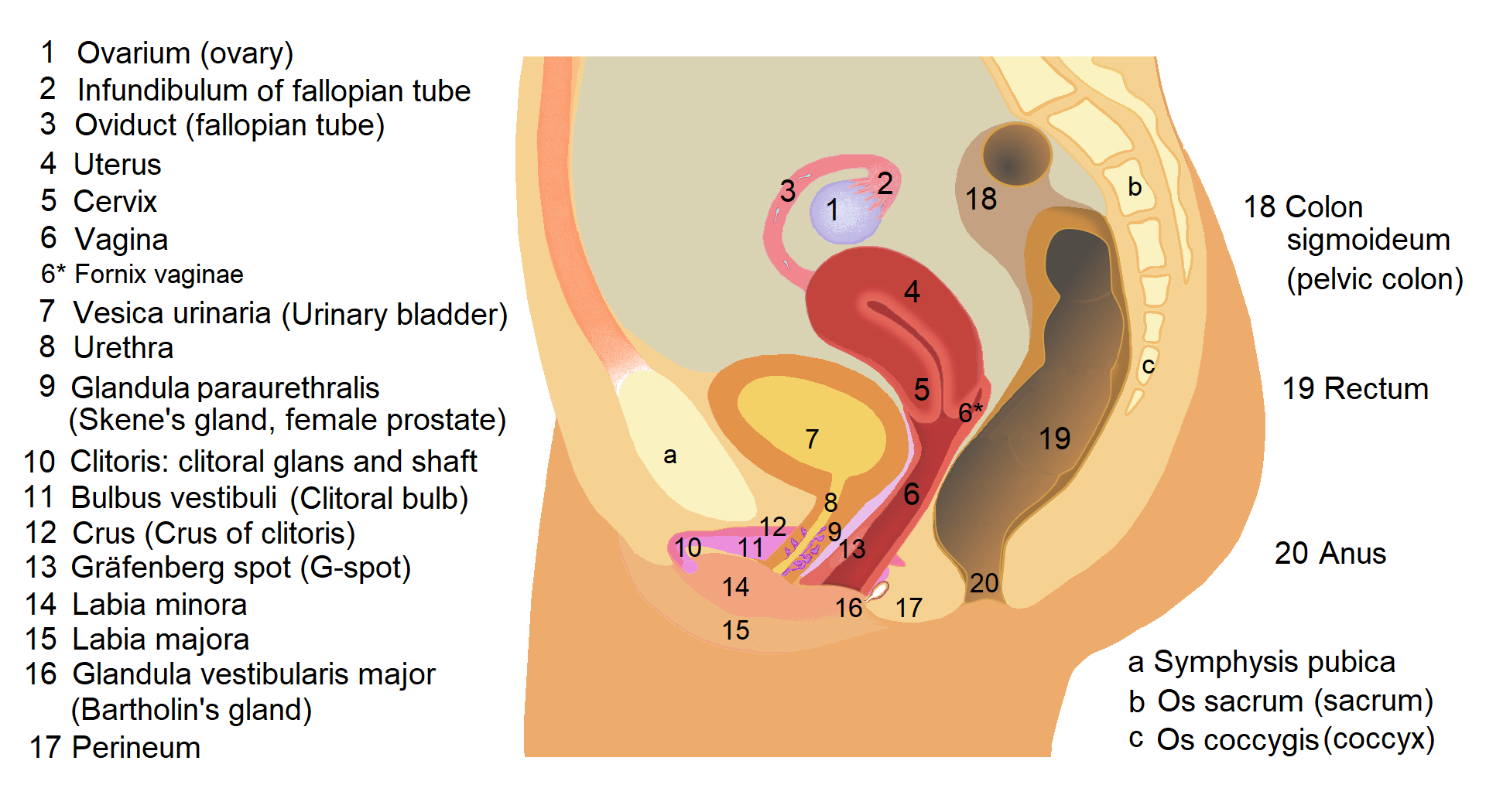
The female pelvis in side view: the uterus and an ovary sit deep behind the bladder, reached from the belly through the abdominal wall or from close range through the vagina. The numbered labels are the diagram’s own. This is an anatomical drawing, not an ultrasound image.
The earliest weeks of pregnancy are the clearest case for going inside. A gestational sac begins at two to three millimeters around five weeks from the last period, a structure far below what the transabdominal view resolves at that stage. The inside scan reads it from close range, confirms that the pregnancy sits inside the uterus, and finds the yolk sac and then the fetal heartbeat in the days that follow. When a woman presents in early pregnancy with pain or bleeding, the question of where the pregnancy sits is urgent. The transvaginal scan is the one that answers it. The belly view at six weeks often shows a uterus that looks empty, a picture that settles nothing. The required route in early pregnancy is the inside one. The dates make the point concrete. At four to five weeks the belly scan shows nothing definite in the early weeks. By the time it can see a sac with confidence, often past seven weeks, the inside scan has already read the heartbeat and dated the pregnancy. Those two or three weeks are the window in which a question of location or viability presses hardest. They belong to the transvaginal route.
A uterus that tilts backward makes the second case. A retroverted or retroflexed uterus folds away from the abdominal probe, its lining and its cavity turned toward the patient’s spine and away from the sound coming in from the front. The belly view of such a uterus is foreshortened and soft, the endometrium hard to measure and the cavity hard to read. The transvaginal probe sits behind the uterus from below, looking at it from the side it has turned toward, and reads the lining and the walls in full. A retroverted uterus on the belly scan is a standing reason to finish the exam from within. The angle is the heart of it. Sound reflects best off a surface it strikes head-on. A uterus tipped back presents its lining edge-on to a probe coming from the front. The picture softens with it. The same lining, seen from below by the transvaginal probe, turns its face to the sound. The retroverted uterus is read in full from the one direction that meets it square.
Body habitus makes the third. The transabdominal probe has to send its sound through the full thickness of the abdominal wall before it reaches the pelvis. A thick wall absorbs and scatters that sound on the way down, so the deep pelvis arrives faint and blurred. The transvaginal probe begins its path already past that wall, an inch or two from the organs, with nothing but a thin layer of tissue in between. For a patient whose abdominal wall defeats the belly scan, the inside route is the one that reaches the pelvis at all. The decision here is a practical one read off the first few belly images, the moment they come back too soft to use. The depth is the whole of the problem. Sound weakens as it travels. A wall several centimeters thick takes a heavy toll before the pelvis is even reached. Lowering the frequency to push through costs the detail the pelvis needs. The transvaginal probe sidesteps the wall entirely, starting its short path on the far side of it. It reads the pelvis at a frequency the belly route could never use at that depth.
Small structures make the fourth. A measurement of the endometrium to the tenth of a millimeter, a count of follicles a few millimeters across, the texture of an ovary, a small early sac: each of these asks for detail that lives only in the close view. The transabdominal scan, looking across a long path, softens these fine edges into their surroundings and returns a figure too rough to act on. The transvaginal scan resolves them. Whenever the question depends on a precise measurement of something small, the inside route is the one that supplies a number solid enough to act on. These small measurements drive real decisions. An endometrium read at a few millimeters sends a postmenopausal patient one way or another on the question of a biopsy. A follicle count guides the drug dose in a fertility cycle. A figure softened by the long belly path can mislead the decision it feeds. The close view gives the measurement the weight a decision can lean on.
The bladder question
The state of the bladder sets the two routes against opposite needs. It often decides which one a scan can use on the day. The transabdominal view depends on a full bladder, the fluid forming the acoustic window and lifting the bowel clear of the pelvis. A patient who arrives having just emptied her bladder, or who cannot hold it full through the wait, leaves the belly scan without its window, the pelvis hidden behind bowel gas. The transvaginal scan asks for the opposite, an empty bladder, since a full one pushes the organs up and away from the probe sitting below. An empty bladder is quicker to arrange than a full one, with no waiting and no discomfort, which is why an urgent scan or a scan on a patient who cannot fill the bladder so often runs from within. The bladder, full or empty, is one of the first things that points a scan toward one route or the other. In an emergency the difference is starkest. A woman in pain in early pregnancy cannot wait an hour for a bladder to fill. The empty-bladder transvaginal scan reads her pelvis at once. The bladder that the belly route waits on is the bladder the inside route needs out of the way.
The situations that demand the inside view
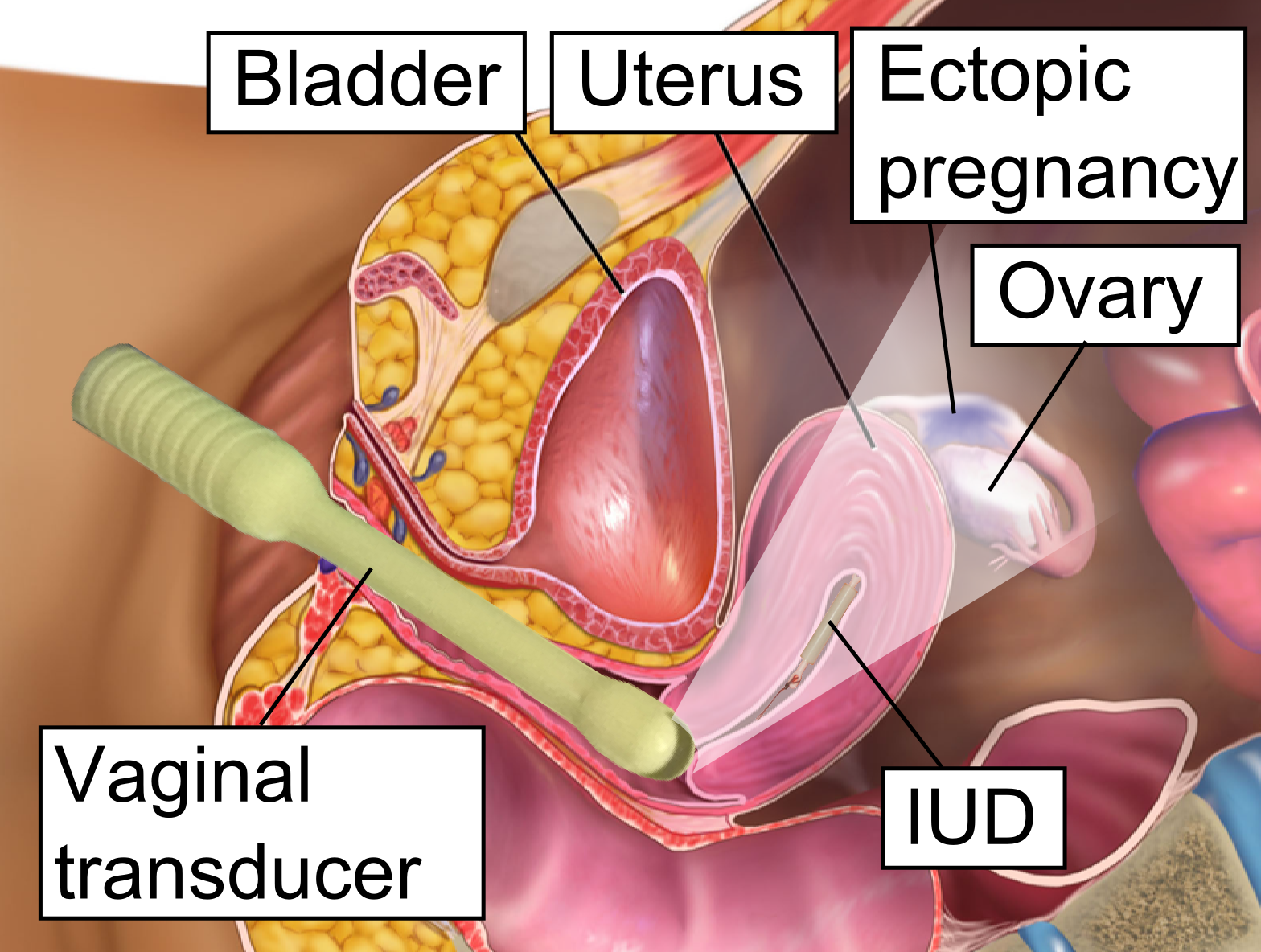
A schematic of the transvaginal scan: the vaginal transducer reads the uterus from close range and the spaces beside it, where an ectopic pregnancy can sit. Reading this from within is the standard when an early pregnancy’s location is in doubt. This is a diagram, not an ultrasound image.
Suspected ectopic pregnancy is the situation that demands the inside view first. A woman with a positive pregnancy test and pain or bleeding needs the location of that pregnancy established quickly, because a pregnancy growing outside the uterus is an emergency. The transvaginal scan reads the uterus for a sac that should be there by a known level of pregnancy hormone, and it reads the tubes and the spaces beside the uterus for a sac or a mass that should not. The detail it gives at close range is what lets a clinician say whether the pregnancy is where it belongs. The belly view in this setting rarely carries enough detail to settle the question, and the time spent on it is time the situation cannot spare. The inside route is the standard of care for early pregnancy of uncertain location. The reading pairs with the blood test. A level of pregnancy hormone above a known threshold means a normal pregnancy should be visible inside the uterus. An empty uterus at that level points toward a pregnancy sitting elsewhere. The transvaginal scan is the eye that reads the uterus and the tubes against that number. Together they locate the pregnancy when the test alone cannot.
Bleeding after menopause makes a second demanding case. The question there is the thickness of the endometrium, the lining of the uterus, because a thin lining points away from cancer and a thicker one calls for a sample. That measurement has to be exact, read at the right plane through the middle of the uterus, and it has to be repeatable from one visit to the next. The transvaginal scan gives a clean millimeter figure that a management decision can rest on. A belly-scan estimate of the same lining, softened over the long path and thrown off by a retroverted uterus or a thick wall, cannot carry that weight. Postmenopausal bleeding is an indication that takes the scan inside almost every time.
Acute pelvic pain in a woman of reproductive age makes a third. The causes that have to be ruled in or out, a torsion of the ovary, a cyst that has burst or bled, a tubo-ovarian abscess, an ectopic, all turn on fine detail of the ovaries and the tubes and the fluid around them. The close view reads the size and the blood flow of an ovary, the wall and the contents of a cyst, the collection in the pouch behind the uterus. These are the readings that separate a problem that can wait from one that needs the operating room within the hour. An acute pelvis is a frequent reason the scan finishes from within.
An inconclusive transabdominal scan is the fourth, and the commonest in plain numbers. Bowel gas across the pelvis, a deep ovary, a habitus that blunts the picture, a structure the wide view cannot fully resolve: any of these leaves the belly scan with a question still open. The transvaginal scan is the step that completes the exam, reaching from close range what the abdomen could not. A great many transvaginal scans are done for exactly this reason, as the second half of an exam that the first half could not finish. The decision is made the moment the belly images come back short of an answer. The pattern is the same across them. The belly scan opens the exam and carries it as far as it goes. The inside scan then takes over where it stops. A report that records both makes clear what each view contributed, the wide survey and the close detail set down in order. The two halves of the one exam answer between them what neither could alone.
Fertility treatment makes the fifth, a demand that recurs on a schedule through a treatment cycle. A stimulated cycle has to be tracked with a count and a measurement of the follicles in each ovary, done every few days, each reading exact enough to time the trigger and the egg collection. Follicles a few millimeters apart have to be told from one another and measured one by one, work that asks for the close view at every visit. The whole of monitored fertility treatment runs on the transvaginal scan, the belly view never having the detail the counting needs. A fertility unit reaches for the inside route as the routine tool of the cycle.
Making the call
The decision follows a simple order in practice. The sonographer starts on the belly for the wide survey, reads what it gives, and moves inside the moment the question outruns the picture. Some referrals carry the answer in advance, an early pregnancy with pain, a postmenopausal bleed, a fertility cycle, each one a known trip inside before the probe touches the skin. Others declare themselves on the first few belly images, the retroverted uterus that foreshortens, the wall that blurs the pelvis, the lining that will not resolve. A sonographer reads the indication and the first images together and picks the route that the case needs, often using both in the one sitting. The order rarely reverses. A scan that starts inside and then moves to the belly is the exception, used when a high or large structure outruns the close probe’s short reach. The usual path runs outward to inward, the belly survey first and the transvaginal detail after it. A sonographer holds that order as the default and departs from it only for a clear reason.
The inside route asks more of the patient, and that shapes the call too. A transvaginal scan is more intimate than a probe on the belly, and it needs the patient’s understanding and consent, a chaperone offered, the reason for going inside explained. A patient who declines, or for whom the route is unsuitable, keeps the scan on the abdomen, and the report says plainly what the belly view could and could not answer. The choice weighs the information the inside view would add against the patient’s comfort with it, a balance the sonographer strikes case by case.
The set of triggers, once learned, is short and steady. Early pregnancy and its emergencies, a uterus turned away from the belly probe, a body habitus that swallows the pelvis, a measurement of something small and precise, a bladder that cannot be filled, a belly scan that came back inconclusive: each of these is a standing reason to read the pelvis from within. A clinician who carries that list reaches for the right route without hesitation, starting broad and going close when the question asks for it. Knowing when the inside view is required is the judgment that gets the answer on the first visit.
Common questions about choosing the transvaginal route
Is a transvaginal scan always better than a transabdominal one?
No. Each route suits a different question. The transabdominal scan gives the wide view of the whole pelvis and reaches structures that sit high or that are large, and for many referrals it answers the question on its own. The transvaginal scan is required when the structures sit too early, too deep, or too small for the belly view, where its close range supplies detail the wide view cannot. The right scan is the one that fits the question in front of it.
When must a scan use the transvaginal route?
In a defined set of situations. Early pregnancy and its emergencies, including suspected ectopic, take the inside route for the detail and the speed it gives. A retroverted uterus, a body habitus that blurs the belly view, and any precise measurement of a small structure such as the endometrium or the follicles also call for it. So does an inconclusive transabdominal scan. In each, the close view answers a question the abdomen left open.
Why can the belly scan miss an early pregnancy?
Because the structures are too small for it at that stage. A gestational sac starts at two to three millimeters around five weeks, sitting below what the transabdominal view resolves across its long path. The belly scan at six weeks often shows a uterus that looks empty, a picture that settles nothing. The transvaginal scan reads the sac from close range, confirms the pregnancy sits inside the uterus, and finds the heartbeat in the days that follow.
Does the bladder need to be full or empty?
It depends on the route. The transabdominal scan needs a full bladder, which forms the acoustic window and lifts the bowel out of the pelvis. The transvaginal scan needs an empty one, since a full bladder pushes the organs away from the probe below. An empty bladder is quicker to arrange, with no waiting, which is part of why an urgent scan often runs from within.
Is the transvaginal scan used in pregnancy?
Yes, mainly in the early weeks and for the cervix. In the first trimester it confirms the location of the pregnancy, finds the heartbeat, and dates it from the crown-rump length. Later in pregnancy it measures the cervix when there is a risk of early delivery. For the routine mid-pregnancy anatomy scan, when the baby is large enough to read from the abdomen, the belly view does the bulk of the work.
Can a patient decline the transvaginal scan?
Yes. The route is more intimate than a scan on the belly, and it needs the patient’s consent, with a chaperone offered and the reason explained. A patient who declines keeps the scan transabdominal, and the report states plainly what that view could and could not answer. The choice weighs the added information against the patient’s comfort, struck case by case.
About the Author
Julien Mercier
Senior R&D Engineer · Medical Ultrasound Transducer Development
Senior R&D Engineer with an M.S. in Applied Physics and over 15 years of experience in medical ultrasound transducer development, specializing in the design verification and performance testing of high-frequency imaging transducers. Currently leading the development and verification of the company’s next-generation high-frequency linear-array transducer, responsible for imaging performance evaluation and reliability analysis in preclinical testing. Brings extensive hands-on experience in piezoelectric element tuning, beamforming parameter optimization, and system-level performance testing.