Cervical length assessment is a transvaginal scan that measures the cervix during pregnancy to judge the risk of an early birth. The cervix is the lower neck of the uterus, closed and firm through a healthy pregnancy, holding the baby in until term. A cervix that shortens too soon raises the chance of a preterm delivery. The scan is how that shortening is caught early.
What the cervix does in pregnancy
The cervix is the lower end of the uterus, the narrow channel that opens into the top of the vagina. Through nearly all of a pregnancy it stays long, firm and tightly closed, a plug of muscle and tissue that keeps the growing baby and the fluid around it held inside. The strength of that closed channel is part of what carries a pregnancy to term. The work it does is quiet and constant, holding shut against a weight that grows month by month. The strength of the cervix is part muscle and part the dense tissue around it. Through the early and middle months it holds a length of three to four centimeters, the long closed channel the scan later measures against. A cervix doing its job gives no sign of the effort, sitting quietly closed under a growing pregnancy. The length and the firmness together keep the pregnancy where it belongs.
Near the end of pregnancy the cervix is meant to change. In the weeks before labor it softens, shortens and begins to open, the ripening that lets the baby pass through when the time comes. This change belongs at the end of pregnancy, after the baby has grown enough to be born safely. The timing of it is the whole point: the right change at the wrong time is what cervical assessment watches for. The ripening is a normal process driven by the body’s signals near term. The fibers of the cervix loosen, water moves into the tissue, and the firm channel softens into one that can open for birth. Happening in the last weeks, this prepares the way for a baby ready to be born. The assessment looks for the same softening and shortening arriving far ahead of that point.
A cervix that begins to shorten and open weeks or months too early sits on the path toward a preterm birth. The channel gives way under the growing weight, shortening from the inside, sometimes with no pain and no warning the woman can feel. By the time it would declare itself through symptoms, the process is often well along. The silence of early shortening is the reason it has to be looked for on a scan.
Preterm birth carries real weight for the baby, and that is what gives the cervix its importance. A baby born too soon faces the problems of organs not yet ready for the world. The earlier the birth, the steeper those problems climb. Catching a cervix shortening early opens a window to act before the birth comes. The measurement of the cervix is, in the end, a way of reading the risk of a birth that arrives before its time. The numbers behind preterm birth give the measurement its weight. A birth before thirty-seven weeks counts as preterm. The births that come much earlier carry the heaviest risks for the baby. Lungs, brain and gut that need their last weeks in the womb meet the world before they are ready. A scan that flags a cervix heading toward an early birth points to where that risk is gathering, in time to work against it.
Why the length is measured
The length of the cervix is a number that forecasts the risk. A long, closed cervix in mid-pregnancy points toward a pregnancy likely to reach term. A measurement is taken because that single figure, read at the right weeks, sorts the pregnancies carrying a raised risk of early birth from those that are not, in time for something to be done. The number turns a hidden process into a reading a clinician can act on. The forecast is read from a measurement taken at the right weeks. A cervix read in the middle of pregnancy, when it should still be at its full length, gives the clearest signal: a short reading then stands out plainly against the long cervix expected. The figure is held against the length a healthy cervix keeps at that stage, the gap between them marking the risk. One careful measurement, taken in the window, carries the forecast.
The measurement is offered where the risk runs higher. A woman who has had a preterm birth before, who is carrying twins, who has had surgery on the cervix, or whose history points that way, is watched with cervical scans through the middle weeks of pregnancy. Some services measure the cervix in every woman at the mid-pregnancy scan, reading the length once as a screen. The scan finds the short cervix in time only when it is done in the window before the birth would come. The history a woman carries shapes how closely she is watched. A previous birth that came early is the strongest pointer, raising the chance of another and earning a close watch on the cervix from early in the next pregnancy. Surgery that removed part of the cervix, a cervix injured before, a pregnancy carrying twins: each adds to the case for measuring. The clinician reads the history first and sets the watching to fit it.
Measuring the cervix on the scan
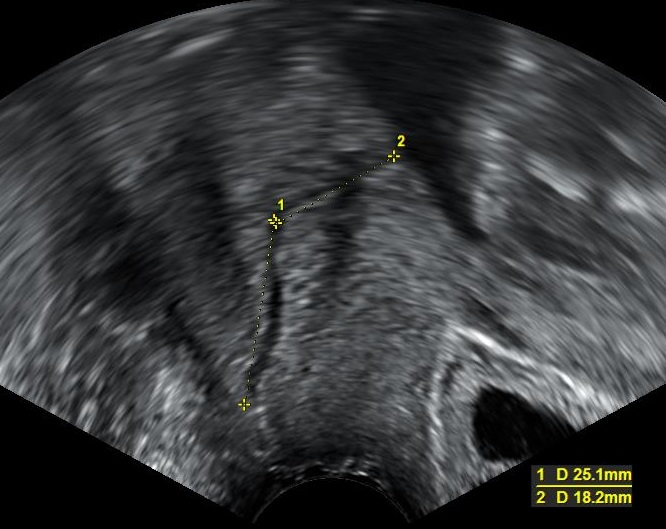
The cervix measured on a transvaginal scan, the calipers laid along the closed canal between its inner and outer openings. The readings, in millimeters, are the lengths this scan recorded. A measurement near or under twenty-five millimeters in mid-pregnancy marks a cervix to watch. The figures and markers are the scanner’s own.
The cervix is measured on a transvaginal scan, the route that reads it clearly. The probe sits in the upper vagina, an inch from the cervix. The screen shows the cervix in long section, its inner opening at the uterus, its outer opening at the vagina, and the closed canal running between them. From this close view the whole length of the cervix lies open to measurement. The long section is the view the measurement needs. The sonographer turns the probe until the cervix lies along the screen as a single straight channel, both its openings in view at once. A cervix caught at an angle reads shorter or longer than its truth, so the sonographer works to bring it fully into line before measuring. The clean long view is the starting point every cervical measurement is built on.
The bladder is emptied before the scan. A full bladder presses on the cervix and can stretch it longer than it truly is, hiding a shortening the scan is there to find. With the bladder empty and the probe held with a light touch, the cervix keeps its true shape on the screen. The sonographer takes care not to press, since pressure on the probe can lengthen the cervix in the picture and mask the reading.
The measurement runs from the inner opening to the outer along the closed canal. The sonographer marks the internal os, where the cervix meets the body of the uterus, and the external os, where it meets the vagina, and reads the length of the closed channel between them. Several measurements are taken across a few minutes. The shortest good one is recorded, since the cervix can shorten and lengthen a little as the scan watches. The shortest reliable reading is the one that counts. A cervix can tighten and ease over the minutes of a scan, giving a spread of lengths over the minutes. Taking several and recording the shortest good one captures the cervix at its shortest, the reading that matters for the risk. The method guards against a single relaxed measurement reading falsely long.
The scan reads the shape of the inner opening as well as the length. An internal os giving way opens from the top, drawing the membranes down into it in a wedge. This funnel marks the cervix yielding from the inside before its full length has gone. Reading the funnel adds to what the length alone tells, a sign the change has started at the os.
The close view is what makes the measurement trustworthy. A cervix of twenty or thirty millimeters has to be read to the millimeter, because a few millimeters move it across the line that calls for action. The transvaginal probe, an inch from the cervix and running at a high frequency, draws the inner os, the outer os and the canal between as clean points a caliper can sit on. The reading the cervix is judged by comes from the close route that resolves it this sharply. A scan from the lower belly cannot draw the cervix this way. It sits deep and low in the pelvis, behind the bladder, too far for the wide abdominal view to give its true length sharply. The close transvaginal route is the standard for cervical length for that reason. A cervical length that calls for action is a transvaginal measurement.
The length that raises concern
A cervix is called short when it measures under about twenty-five millimeters in the middle weeks of pregnancy, and that figure marks a raised risk of an early birth. The shorter the reading falls below that line, the steeper the risk climbs, a cervix of fifteen millimeters carrying more concern than one of twenty-four. The single number, set against the threshold, is what turns the scan into a decision, a short reading calling for a closer look and a plan. The threshold is a line drawn from large studies of how cervical length tracks with early birth. It is read as a guide, weighed with the woman’s history and the look of the internal os, never as a verdict on its own. A reading near the line on a woman with a worrying history is taken more seriously than the same reading in a woman with none. The number opens the conversation about what to do, set in the fuller picture of the pregnancy.
Watching an at-risk pregnancy
A pregnancy at raised risk is watched with cervical scans repeated across the middle weeks. The scans run from around sixteen weeks toward twenty-four, the window in which a shortening cervix can be caught with time to act. A woman with a worrying history comes in every week or two for the cervix to be read, each scan added to the last to show how the length is holding. The schedule is set to the level of risk, the strongest histories read weekly and the rest a little less often, the interval chosen short enough to meet a shortening early, before much of the length has gone. A clinician reads the cervix each visit against the readings before it. The watching is built to see a change begin, to catch it underway.
The trend across the scans tells as much as any single reading. A cervix that has begun to slip a few millimeters at each visit shows the change underway in time to step in, the slippage caught at its start. The run of measurements, plotted across the weeks, lets a clinician see the shortening start, catching it underway.
The watching has a window it has to work inside. The value of catching a short cervix lies in catching it with weeks of pregnancy still to protect, when a treatment can add days and weeks that matter for the baby. A scan that finds the shortening early gives those weeks. The repeated cervical scan is built around reaching the short cervix inside the window where acting on it still changes the outcome. Missing that window costs the chance to act. A short cervix found late, with the birth already near, leaves little a treatment can do to hold it. The whole design of the repeated scan is to reach the cervix before that point, when a stitch or a course of progesterone still has weeks to work across. Reaching the short cervix in time is the entire value of the watching.
What a short cervix calls for
A short cervix found in time opens a set of treatments meant to hold the pregnancy longer. The aim of each is the same: to keep the cervix doing its job for more weeks, buying the baby growing time toward a safer birth. The scan that finds the short cervix is what puts these options on the table, early enough for one to help. The choice among them rests on the whole picture. The cervical length, the look of the internal os, the woman’s history and how far along she is all feed the decision of which treatment fits. A clinician reads these together to choose between a course of progesterone, a stitch, or a closer watch alone. The scan that found the short cervix is the center of that reading.
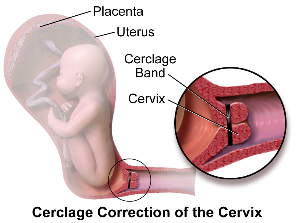
A cervical cerclage, a stitch placed around the cervix to hold it closed, shown in a medical illustration. A short cervix found on the scan is one of the findings that can lead to a stitch like this, placed to reinforce the cervix through the weeks ahead. This is a diagram, not an ultrasound image.
Vaginal progesterone is one of the treatments a short cervix calls for. A nightly dose of the hormone, placed in the vagina, helps quiet the uterus and support the cervix through the weeks ahead, lowering the chance of an early birth in a woman whose cervix has been found short. The treatment follows directly from the scan, started on the strength of a length under the line. The cervix is read again across the weeks that follow to watch how it holds.
A cervical stitch is the other main answer, for selected pregnancies. A cerclage is a strong suture placed around the cervix to hold it closed, an operation offered when the history and the scan together point to a cervix likely to give way. The stitch reinforces the closed channel through the weeks it would otherwise be losing. It is removed near term to let labor take its course. The decision to place one rests heavily on the cervical length the scan reads. The stitch is placed under anaesthetic and watched on the scans that follow. After a cerclage, the cervix is read again across the weeks to confirm it is holding above the stitch and to catch any change early. The cervical scan that pointed to the stitch goes on watching the cervix it helped protect. The measurement runs through the whole story, from the reading that called for the stitch to the readings that follow it.
The handheld probe suits the repeated reading a watched cervix needs. A woman at risk comes in often for a quick cervical scan, and a portable probe gives that scan at the bedside, run from a phone or a tablet, without a booking into a scanning suite each time. A clinic following many at-risk pregnancies reaches for probes it can keep close. The scan that has to happen again and again fits a tool that stays within reach.
Across a pregnancy at risk of coming early, the cervical scan is the thread the management hangs on. It reads the length that forecasts the risk, follows the shortening as it starts, and times the treatment that holds the pregnancy longer. On a handheld probe at the bedside, repeated through the middle weeks, the measurement of the cervix turns a hidden risk into a reading a clinician can act on, in the window where acting still adds the weeks a baby needs.
Common questions about cervical length scans
What is cervical length assessment?
A transvaginal ultrasound measurement of the cervix during pregnancy, taken to judge the risk of an early birth. The scan reads the length of the closed cervical canal, from its inner opening at the uterus to its outer opening at the vagina. A cervix that measures short for the weeks of pregnancy marks a raised risk of preterm delivery. The reading is taken in the middle weeks, when there is still time to act on a short result.
What cervical length is considered short?
Under about twenty-five millimeters in the middle weeks of pregnancy is taken as short, marking a raised risk of an early birth. The shorter the reading falls below that line, the higher the risk climbs. A length well above the line points toward a pregnancy likely to reach term. The figure is read against the threshold along with the woman’s history to decide what comes next.
Why is the cervix measured transvaginally?
Because the close route reads the cervix clearly and to the millimeter. The transvaginal probe sits an inch from the cervix and shows the whole canal, the inner opening, the outer opening, and the closed channel between, as clean points a caliper can sit on. The bladder is emptied first, since a full bladder can press the cervix longer than it truly is. The reading the cervix is judged by depends on this close, careful view.
When are cervical scans done in pregnancy?
Mainly between about sixteen and twenty-four weeks, the window in which a shortening cervix can be caught with time to act. A woman at raised risk, such as one who has had a preterm birth before, is scanned every week or two through this window. Some services also read the cervix once at the mid-pregnancy scan as a screen. The timing matters, since the value of the scan is in catching the shortening early.
What is funneling of the cervix?
The opening of the inner end of the cervix before its full length has gone. On the scan the membranes draw down into the internal os in a wedge, a funnel that marks the cervix giving way from the inside. Reading the funnel adds to what the length alone shows, a sign the change has started at the top of the canal. It is read alongside the cervical length as part of the picture.
What is done if the cervix is found short?
The scan opens a set of treatments meant to hold the pregnancy longer. Vaginal progesterone, a nightly dose placed in the vagina, helps lower the chance of an early birth. A cervical stitch, a suture placed around the cervix to hold it closed, is offered for selected pregnancies where the history and the scan point to a cervix likely to give way. The cervix is then read again across the following weeks to watch how it holds.
About the Author
Julien Mercier
Senior R&D Engineer · Medical Ultrasound Transducer Development
Senior R&D Engineer with an M.S. in Applied Physics and over 15 years of experience in medical ultrasound transducer development, specializing in the design verification and performance testing of high-frequency imaging transducers. Currently leading the development and verification of the company’s next-generation high-frequency linear-array transducer, responsible for imaging performance evaluation and reliability analysis in preclinical testing. Brings extensive hands-on experience in piezoelectric element tuning, beamforming parameter optimization, and system-level performance testing.