Thyroid ultrasound decides whether a lump in the neck needs a needle. A high-frequency linear probe reads the gland in fine detail, sizes any nodule inside it, and judges from the look of that nodule whether it can be watched or has to be sampled. That judgement, made on every nodule a neck turns up, is the whole work of the scan. Almost every nodule proves harmless.
What the gland looks like
The thyroid sits low in the front of the neck, a butterfly of tissue wrapped around the windpipe, each wing about the size of a thumb. On ultrasound a healthy gland reads as an even, fairly bright gray, finer in its grain than the muscle around it, with the dark round windpipe at its centre and the large neck vessels running down each side. The whole thing sits barely a centimetre under the skin, square in the sharpest range of a linear probe. Around the gland sit the landmarks a reader uses to keep their place. The carotid artery and jugular vein run as large channels at the outer edge of each lobe. The strap muscles lie as thin dark bands across the front. Behind the windpipe on the left hides the gullet, a target that can mimic a nodule until a swallow sets it moving and gives it away.
A reader scans it with the patient’s neck tipped gently back. The probe sweeps up and down each lobe and across it, building the whole gland in two planes, then crosses the midline to the strip of tissue joining the lobes. The lobes are measured in three dimensions and the gland’s volume noted, the two need not match, one often sitting a little larger than the other without alarm. The study takes a few minutes and asks nothing of the patient but to lie still and swallow once or twice, a swallow that lifts the gland and brings a part hidden behind the collarbone into view.
The even gray of the normal gland is the reference everything is read against. A nodule shows because it breaks that evenness. A diffuse disease shows because it spoils the whole texture. The first skill of thyroid ultrasound is knowing the healthy gland well enough that any change stands out from it. The normal gland carries a gentle, even flow on color Doppler, a soft scattering of vessels through the tissue. Its size depends on the person, larger in a big adult, smaller in a child, swollen a little in pregnancy. A reader keeps that range in mind, since a gland at the top of normal in one patient would be plainly enlarged in another. The volume, taken from the three measurements of each lobe, puts a number on a goiter, a measure the eye alone could never give. A simple goiter, a gland grown large, is watched on size alone, the numbers from each scan charting whether it presses on the windpipe or drifts down behind the breastbone.
Finding a nodule
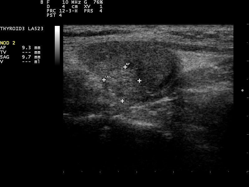
A thyroid nodule on a 10 MHz linear probe, measured with on-screen calipers. The crosses mark the nodule’s edges and the readout at the left gives its dimensions, here about nine millimetres across, the routine sizing every nodule gets. Image: Nevit Dilmen, CC BY-SA 3.0.
The bulk of thyroid ultrasound comes down to the nodule. A nodule is a lump within the gland, a spot that reads differently from the even tissue around it. The great majority are found by chance on a scan run for something else. They are remarkably common: over half of adults carry at least one, more of them in older necks. Almost all of them are harmless. Whether a nodule even reaches a report depends on its size. The tiniest, a few millimetres across, are passed over, too small to matter, too common to chase. A nodule earns a full description and a score once it grows to the size the guidelines care about, a centimetre give or take.
Screening the gland for nodules is a careful sweep through both lobes, every focal change measured in three planes and its position noted, the same nodule found again on a later scan and read against its old size. The reader counts them, sizes them, and marks the one or two that stand out from the rest. A nodule’s position is logged as carefully as its size, the upper, middle, or lower third of a named lobe. The next reader lands on the same one. A nodule that has grown between scans earns a closer look, as does one that stands apart from its neighbours in the way it reads.
A nodule comes in a few basic forms. A simple cyst is a clear bag of fluid, smooth-walled and clean inside, almost never anything to fear. A solid nodule is a lump of tissue that reads as gray as the gland or darker. Many are a mix, part fluid and part solid. The form is the first thing a reader notes. A solid nodule carries the question the rest of the scan is built to answer. A few looks reassure on sight. A spongiform nodule, riddled with tiny fluid spaces like a sponge, is benign almost without exception. A cyst with a bright comet-tail trailing from a fleck inside it is a colloid nodule, harmless. Many necks hold a dozen nodules at once, a multinodular goiter. The work there is to find the one in the crowd that reads differently from its neighbours, since that is the one carrying any risk. The sheer commonness of nodules is a trap of its own. Scan enough necks and almost everyone has one. Treating every speck as a threat would bury a clinic in needless needles and frightened patients.
The art is restraint.
A reader reports the nodule big enough to matter, scores it honestly, and passes over the tiny and the plainly benign. A solid nodule can also break down in its centre, a cystic degeneration that is benign far more often than not.
Reading the risk
The look of a solid nodule carries its risk. A handful of features mark the one that needs attention. It stands taller than it is wide, grown against the natural lie of the gland. Its edge is ragged or ill-defined. Its body reads darker than the thyroid around it. It carries flecks of bright calcium, the tiny specks that often sit inside a cancer. The features that mark a thyroid cancer are read together, since no single one decides it, the judgement built from the whole set.
Each feature earns its weight from how often it travels with a cancer. The shape comes first: a nodule taller than it is wide has grown across the grain of the gland, the way a cancer pushes. The margin comes next: a ragged, lobulated, or ill-defined edge raises concern. Brightness follows: the darker a solid nodule reads against the gland and the muscle, the more it worries. The bright specks come last. The fine ones, the microcalcifications, are the dots that often sit inside a papillary cancer. Color Doppler adds a little, the tangle of vessels in a nodule’s core one more soft sign, weighed against the shape and the calcium that carry the real weight. Newer probes add elastography, a read of how stiff a nodule is, a hard one worrying more than a soft one. The plain gray picture and its few scored features stay the heart of the scan all the same.
To keep that judgement consistent, the field scores it. TI-RADS, the thyroid imaging reporting and data system, rates a nodule on each of those features, adds the points, and sorts the total into a category from clearly benign to highly suspicious. Each category carries a rule tied to the nodule’s size: which one needs a needle, which needs only a repeat scan in a year or two, which needs nothing at all. The score gives two readers the same language for the same nodule, and gives a patient a number that follows them from one visit to the next. The categories carry rough numbers a clinic can hold in mind. A clearly benign nodule needs nothing. A mildly suspicious one is sampled once it passes a couple of centimetres, a moderately suspicious one at around a centimetre and a half, a highly suspicious one at a centimetre. The thresholds fall as the score climbs. The more a nodule worries, the smaller it has to be before a needle is reached for.
Size alone is not the danger it once seemed. A small nodule with suspicious features earns a needle on its look, its size beside the point. The scoring is what untangles the two, weighing the look of a nodule ahead of its bare measurement. It spares the many harmless lumps a needle they never needed. A nodule left unsampled is not forgotten. It is watched on a repeat scan in a year or two, measured the same way, and reconsidered if it grows or its look changes. Growth alone is rarely cancer. A benign nodule can grow larger and stay harmless. So a reader watches the look of a nodule more closely than its size. A few friendly looks are as useful to know as the worrying ones. A thin dark halo around a nodule is the mark of a slow, benign growth easing the tissue aside. A bright patch standing out pale against a dark Hashimoto gland, the so-called white knight, is usually spared normal tissue, no nodule at all. Knowing the benign saves as many needles as spotting the malignant does.
Guiding the needle
A suspicious nodule ends in a needle. An ultrasound-guided fine-needle aspiration draws a few cells from the nodule through a thin needle, the probe watching the tip the whole way in to land it in the worrying part of the nodule. The cells go to a pathologist, who reads them under a microscope and sorts them into one of six grades, from plainly benign through suspicious to malignant. A fair share come back indeterminate, neither clearly safe nor clearly cancer, a result that calls for a watchful eye, a repeat needle, or a gene test on the cells to settle the odds. The grade sets the path from there, the scan’s part already played. A needle that comes back empty of useful cells is repeated, since a nodule that earns a needle earns a careful one. When the cells read indeterminate, a panel of gene tests can weigh the chance of cancer and point a patient toward surgery or toward watching. Many a nodule is spared an operation it did not need.
Ultrasound is what makes the sample reliable. With the probe guiding it, the needle reaches a centimetre-wide target a finger could never find, draws cells from the exact spot the scan flagged, and stays clear of the vessels and the windpipe close by. A blind needle, working by feel, cannot reach a small or deep nodule at all. The same scan that raised the concern places the needle that settles it, in one sitting. The aspiration is a brief office procedure under local anaesthetic, a fine needle passed two or three times, the soreness gone within a day. Bleeding is slight and serious harm rare, the vessels and the windpipe held clear under the probe’s eye the whole time. The whole sequence runs in one room. A clinician scans the nodule, numbs the skin, guides the needle, and smears the cells on a slide, often with a pathologist next door to say at once whether the sample holds enough to read.
When the whole gland is the problem
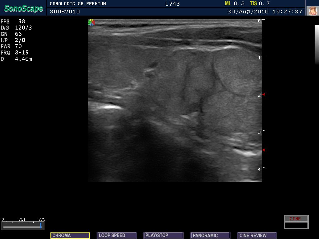
A thyroid in Hashimoto’s thyroiditis on a high-frequency linear probe. The even gray of a healthy gland has broken into a coarse, mottled texture across the whole organ, the mark of a diffuse disease. The red arrowheads at the right are the machine’s depth markers. Image: Schomynv, public domain.
Not every thyroid trouble is a lump. Sometimes the whole gland is diseased, and ultrasound reads that too. The look of Hashimoto’s thyroiditis, the common cause of an underactive thyroid, is a gland turned dark, the texture gone coarse. The even gray breaks into a mottled, patchy field, the gland often shrunken and criss-crossed by bright bands of scarring, the change spread through the whole organ.
An overactive gland reads its own way. It runs hot with blood. Color Doppler lights it up in a blaze the field has long called a thyroid inferno, the flow so heavy the gland glows almost solid color. The pattern spread across the whole gland tells the reader the disease is diffuse. The management is set by that: blood tests and medication. The needle is held back here, with no focal lump to chase. Graves’ disease is the overactive gland’s commonest cause, an enlarged thyroid carrying that inferno of flow on Doppler. A painful gland of sudden onset, dark, its texture broken for a few weeks, points to a viral thyroiditis that settles on its own. The scattered dark patches of a diffuse disease can throw up false nodules. A reader reads a pseudonodule as one more patch of the diffuse pattern.
The labs and the look go together. A blood test names the gland overactive or underactive. The scan that follows shows the cause. When treatment takes hold, the scan follows the gland back toward normal, a Graves gland shrinking and settling on Doppler over the months. Hashimoto carries one quiet long-term risk, a small rise in the odds of thyroid lymphoma, that a new dominant mass in a known Hashimoto gland is read against.
The glands behind the gland
Behind the thyroid sit four tiny parathyroid glands, each about the size of a grain of rice, governing the calcium in the blood. A normal one is too small to pick out. Scanning for an enlarged parathyroid looks for a small dark oval behind or below the thyroid, in the spot a normal gland hides unseen. A patient with too much calcium in the blood often has one parathyroid gone overactive and swollen, and ultrasound is the first look for it. Found and marked on the scan, the enlarged gland gives the surgeon a target before an operation that may take only minutes. The same linear probe that reads the thyroid reads its small neighbours with no change of setting, the parathyroid hunt folded into the end of a thyroid scan. The clue that sends a reader looking is in the blood: a high calcium beside a high parathyroid hormone, the signature of a gland gone rogue. The culprit is usually one swollen gland, a parathyroid adenoma, and finding it turns a long neck exploration into a quick targeted operation. A small artery feeding the gland at one pole helps mark it out. Color Doppler picks up that polar vessel. A gland that has slipped down behind the breastbone sits beyond the probe’s reach, the one place a surgeon may need a scan of another kind. A few parathyroids hide elsewhere too, low in the neck, behind the gullet, or down within the chest, the ectopic glands a surface scan can miss. Found by ultrasound or by a nuclear scan, a single adenoma is removed through a small cut, the blood calcium falling toward normal within hours and the cure checked by a hormone test on the table. Hyperparathyroidism turns up more often than it once did, caught by a routine blood test in a patient with no symptoms at all. A scan that finds the one guilty gland turns a chance finding into a quick, targeted cure. The old route, a blind exploration of all four glands, is needed no more.
The nodes of the neck
A thyroid cancer spreads first to the lymph nodes of the neck. The scan that finds the nodule sweeps the nodes too. A normal node is a flat oval with a bright fatty streak running down its middle, a shape a reader passes over without a second thought. The sweep through the neck is part of any scan of a worrying nodule.
A node taken over by cancer changes its look. It rounds out and fills, losing the bright fatty streak, and can carry the same bright specks or tiny cysts as the nodule that fed it. Color Doppler shows a chaotic, scattered flow run through the node. A node found this way before an operation changes the surgery a patient needs, since the surgeon must clear the involved nodes along with the gland. The scan maps those nodes out beforehand. The neck is mapped in numbered levels a surgeon and a radiologist both use. A node is reported by the exact zone it sits in. After a thyroid cancer is treated, those same zones are swept on every follow-up scan, a returning node often found on ultrasound before any blood test stirs. A node already removed leaves a scar bed a reader learns to tell apart from a true regrowth, the surgical change fading over the months that follow. A node in doubt can take a needle of its own. The same fine-needle aspiration that samples a thyroid nodule samples a suspicious node. The fluid drawn is tested for thyroid proteins that betray a spread even when the cells are few.
On a handheld
The thyroid is the ideal organ for a handheld. It sits a centimetre under the skin, square in the linear probe’s sharpest range. The whole exam is a surface scan a pocket probe runs as well as a cart. A clinician feels a lump in a neck and reads it on the spot, the patient’s question answered in the same visit, the patient not sent away to wait.
The image saves to the phone, sizes a nodule on the screen with a fingertip, and travels to a specialist or a surgeon for a second opinion in seconds. A thyroid clinic far from a radiology department gets the whole nodule workup in one hand, from the first scan through the risk score to the needle that takes the sample. The cost of the hardware has fallen far enough that a single doctor can carry the tool that once filled a room.
The handheld suits the long watch a thyroid asks for. A nodule scored low needs a look again in a year or two, then again after that, a string of quick scans across a patient’s life. A pocket probe makes each one a two-minute job in the clinic, the old images on the phone to measure against, with no appointment at a distant department to wait for. The reach of the tool matters above all where care is thin: a district clinic with a pocket probe screens necks, scores nodules, and guides needles on the spot, with no journey to a city hospital.
Thyroid ultrasound is operator-dependent in a way an X-ray is not, the same gland telling two stories in two pairs of hands. A trained reader matters more than a costly machine. The spread of pocket probes has put scanning into the hands of the clinician who knows the patient. The thyroid was among the first organs the handheld proved itself on. It stays the clearest case for one. The thyroid is a shallow gland, the complaint common, the scan quick to a clear plan. A pocket linear probe gives exactly that, a shallow target met by a sharp shallow picture.
Common questions about thyroid ultrasound
What does a thyroid nodule look like on ultrasound?
A spot inside the gland that reads differently from the tissue around it. It may be a dark solid lump, a clear fluid-filled cyst, or some of each. A reader judges its risk by its shape, its edges, its brightness, and any flecks of calcium in it, and sizes it in three planes for the record.
What is TI-RADS?
A scoring system for thyroid nodules. It rates a nodule on a handful of ultrasound features and adds the points into a category, from benign to highly suspicious. The category sets what happens next: a needle, a follow-up scan, or nothing. It gives every reader the same rule for the same nodule.
Can a handheld ultrasound find thyroid cancer?
It can find the nodules that need testing. A handheld linear probe reads the suspicious features of a nodule to the same standard as a cart, and guides the needle that gives the diagnosis. The diagnosis itself comes from the needle, since only the cells under a microscope make a cancer certain.
What does Hashimoto’s thyroiditis look like on ultrasound?
A whole gland turned dark, its texture gone coarse. The mottling runs patchy across the organ, the thyroid often shrunken and crossed by bright bands of scar tissue. The change runs across the whole gland, the mark of a diffuse disease handled with blood tests and medication.
Can a handheld probe scan the parathyroid glands?
Yes, with the same linear probe. The parathyroids are tiny and hide behind the thyroid, seen on ultrasound only when one enlarges. A swollen parathyroid shows as a small dark nodule below or behind the gland. A handheld probe finds it and marks it for surgery as well as a cart does.
About the Author
Julien Mercier
Senior R&D Engineer · Medical Ultrasound Transducer Development
Senior R&D Engineer with an M.S. in Applied Physics and over 15 years of experience in medical ultrasound transducer development, specializing in the design verification and performance testing of high-frequency imaging transducers. Currently leading the development and verification of the company’s next-generation high-frequency linear-array transducer, responsible for imaging performance evaluation and reliability analysis in preclinical testing. Brings extensive hands-on experience in piezoelectric element tuning, beamforming parameter optimization, and system-level performance testing.