Thyroid Cancer Ultrasound Characteristic Features Handheld Imaging
Thyroid cancer takes more than one form, and the form changes what the scan looks for. Papillary, the commonest, tends to calcify into tiny bright flecks; follicular can pass for a benign nodule on the screen; medullary and anaplastic each carry their own marks. The type a scan suspects steers what it looks for, with a metastatic neck node often the finding that settles the question.
Why the cancer type matters on the scan
The four types do not appear in equal numbers. Papillary carcinoma is the large majority, around 80 to 85 percent of thyroid cancers. Follicular carcinoma follows at 10 to 15 percent. Medullary carcinoma sits near 1 to 2 percent. Anaplastic carcinoma is rare, under 2 percent.
The four also run far apart in outlook. Papillary cancer is slow, with a ten-year survival above 95 percent. Follicular cancer carries a fair outlook once it is removed. Medullary cancer is tracked by its calcitonin level for years. Anaplastic cancer is fatal within months for the large majority. The scan often finds it late, as a mass that has already crossed the gland.
Age and sex change the odds. Papillary cancer favors women by about three to one, often in the thirties or forties. Anaplastic cancer appears late, past sixty, in a gland that may have held a quiet cancer for years. A new hard nodule in an older man carries more concern than the same nodule in a young woman.
The look on the screen tracks the type. A cancer that calcifies suggests papillary disease. A bland solid nodule fits a follicular or benign picture. The scan cannot name the type by itself, since the cell line comes from the needle or from surgery. The features narrow the field and flag the nodule that needs a sample. They also separate a cancer that stays put from one that has reached the neck nodes.
Papillary cancer, the calcifying type
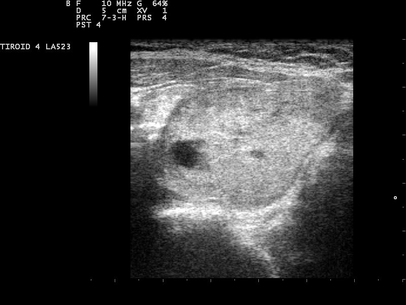
A hypoechoic thyroid nodule with an unclear margin. A dark nodule that breaks from the smooth gland around it raises concern for papillary cancer. Image: Nevit Dilmen, CC BY-SA 3.0, Wikimedia Commons.
Papillary thyroid carcinoma is the commonest type a handheld scan meets. Its hallmark is the microcalcification, a bright echogenic focus one millimeter or smaller, usually with no acoustic shadow behind it. These specks are psammoma bodies, laminated calcium laid down inside the tumor. A nodule carrying many of them holds a high suspicion of papillary cancer.
Papillary cancer also reads hypoechoic, darker than the surrounding gland. It often grows taller than it is wide, a shape that crosses the normal tissue planes. Its margin runs irregular or lobulated. No one of these features belongs to papillary cancer alone. Together, in a dark solid nodule with microcalcifications, they make the call.
The classic papillary cancer is the one that calcifies. The follicular variant can lose the specks and read softer, closer to a follicular lesion. The tall cell and columnar variants run more aggressive, with higher rates of spread past the capsule. The variant is a pathology label. A soft-looking papillary cancer stays a known trap on the scan.
A papillary cancer under one centimeter is a papillary microcarcinoma. Many turn up by chance on a scan done for another reason. A microcarcinoma with no suspicious node and no spread past the capsule is sometimes placed under active surveillance, watched with serial scans when surgery is not pressed.
Molecular tests add to the picture after the needle. A BRAF mutation, found in many papillary cancers, confirms the type and flags a slightly higher risk of spread. For a small low-risk papillary cancer, some centers now offer ultrasound-guided ablation, heating the nodule through a fine probe without an operation.
Follicular cancer, the one ultrasound misses
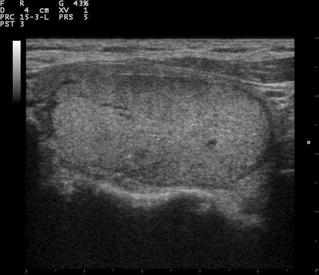
A bland, evenly solid nodule with a smooth border. This calm look is the one a follicular cancer can hide behind, since it lacks the dark echo and microcalcifications of papillary disease. Image: Nevit Dilmen, CC BY-SA 3.0, Wikimedia Commons.
Follicular thyroid carcinoma is the type ultrasound struggles with. It tends to look bland. It is usually solid, iso- to hyperechoic, smooth-bordered and free of microcalcifications. On the screen it can pass for a benign follicular adenoma.
The reason sits in the pathology. Follicular cancer is defined by capsular or vascular invasion, a feature the microscope reads and the probe cannot. A follicular lesion goes to surgery for that answer, since even a needle sample cannot separate a follicular cancer from a benign follicular tumor. The cytology returns as a follicular neoplasm, a Bethesda category that names the uncertainty without settling it.
Follicular cancer also takes a separate route when it spreads. It spreads through the blood to the lungs and bone. Papillary cancer spreads through the lymph to the neck. A solitary lung or bone lesion in a patient with a follicular nodule carries weight for that reason. The Hurthle cell type, an oncocytic variant, runs a more aggressive course.
So the scan flags a follicular lesion by what it lacks. A solid nodule with no benign sign, no spongiform change and no clean cyst warrants a closer look, even when it reads calm. A thick irregular halo, or a break in the capsule line, raises the concern.
A follicular neoplasm on cytology usually goes to a diagnostic lobectomy, the removal of one lobe for a full pathology read. The single lobe answers the question for the majority of patients. A second operation follows only when the lobe holds a wide-invasive cancer that calls for more.
Medullary and anaplastic cancer
Medullary thyroid carcinoma comes from the calcitonin-producing C cells. The other three types arise from follicular cells. On ultrasound it resembles papillary cancer: solid, hypoechoic, marked with calcifications. Its calcifications tend to run coarser than the fine specks of papillary disease. It often sits in the upper third of a lobe, where the C cells cluster. A raised blood calcitonin level points to it before any needle.
Medullary cancer can come alone or as part of MEN 2A or MEN 2B, the inherited syndromes that pair it with adrenal and parathyroid tumors. A family history calls for genetic testing and a calcitonin check. The cancer is often bilateral when it runs in a family, so both lobes get a careful sweep.
Medullary cancer also raises a second blood marker, CEA, which tracks the disease alongside calcitonin. A new diagnosis prompts a check for a pheochromocytoma, the adrenal tumor of MEN 2, before any neck surgery.
Anaplastic thyroid carcinoma is the aggressive extreme. It appears as a large, ill-defined, hypoechoic mass that grows over weeks. Necrosis shows in around 82 percent of cases, as ragged fluid-like areas inside the mass. Extrathyroidal extension reaches roughly 91 percent, with invasion of the trachea in over half of cases and the esophagus close behind. The carotid sheath is encased in more than 40 percent.
The mass can press on the airway within days. A hard, fixed neck swelling in an older patient points to it on contact. The scan maps the airway and the great vessels for the team planning an urgent biopsy or a tracheostomy.
Treatment moves fast. Surgery comes where it can be done, then radiation and targeted drugs for tumors with a BRAF mutation.
Anaplastic cancer needs urgent care.
What microcalcifications mean
The microcalcification is worth a closer look, since it is the strongest single sign of thyroid cancer on ultrasound. A microcalcification is an echogenic focus one millimeter or smaller, with little or no acoustic shadow behind it. A macrocalcification measures larger than a millimeter and shows a shadow. The two carry unequal weight. Microcalcifications track with papillary and medullary cancer, where they mark psammoma bodies or tumor calcium. Macrocalcifications are less specific, since they turn up in old benign nodules as well as in cancers. A macrocalcification at the rim of a nodule, the eggshell pattern, is usually benign. A broken eggshell rim raises concern. The trap is the colloid speck. A bright dot in a cystic or spongiform nodule is often inspissated colloid. It shows a comet-tail artifact behind it. That comet tail marks the dot as benign. Reading colloid as a microcalcification turns a calm nodule into a false alarm. The pattern within the nodule adds detail. Microcalcifications clustered at the center of a solid nodule carry weight. A single speck means little on its own. Three or more in one nodule raise the suspicion. The specks hold their brightness with depth, since they reflect strongly for their size, which sets them apart from the speckle of normal tissue. The same dot reads two ways by setting. In a purely cystic nodule, bright dots are almost always colloid. In a solid hypoechoic nodule, the same dots are read as calcium until proven otherwise. Microcalcifications carry high specificity for cancer, around 85 to 95 percent in reported series. Their sensitivity runs lower, since not every cancer calcifies. A follicular cancer rarely shows them at all. So a nodule without microcalcifications is not cleared. A nodule with them is not condemned on the speck alone. Under the microscope, a microcalcification is a psammoma body, a ring of calcium laid down around dead tumor cells. The rings build up as the cancer grows, which is why a fast papillary cancer can be dense with them. Color Doppler shows the flow. A papillary cancer often shows chaotic internal flow, with vessels in odd directions. A benign nodule keeps a neat rim of flow, or none at all. The flow pattern alone does not diagnose, since some benign nodules carry brisk flow. It counts toward a nodule that already looks suspicious on grayscale. The microcalcification is one feature among several, weighed alongside echogenicity, shape and margin.
The look of a cancerous lymph node
Thyroid cancer travels first to the lymph nodes of the neck. A normal node is oval, with a bright fatty hilum running down its center. A node that has taken up cancer changes in ways the probe can read.
The earliest change is loss of the fatty hilum. The node turns round as the tumor replaces its core. Microcalcifications can appear inside it, the same psammoma specks of papillary cancer. They are the strongest sign of a metastatic node. Cystic areas can open up within it. Blood flow moves from the hilum out to the rim, supplying the node around its edge.
Size alone does not make a node benign.
The neck is mapped by compartment. The central compartment, level six, sits beside the gland and drains it first. The lateral compartments, levels two through four, take spread that has moved on. A node high in the lateral neck carries weight, since reactive nodes are common lower down. A round node with a lost hilum, inner microcalcifications and peripheral flow is suspicious enough to sample. The needle rinse is tested for thyroglobulin, a protein that should not sit in a lymph node. A positive wash confirms spread even when the cells are sparse.
Node size matters less than shape and content. A small round node with microcalcifications counts for more than a large oval one with a clear hilum. After surgery, the same features flag a recurrence in the neck, which is why a treated patient keeps returning for scans.
Spread past the capsule and multiple foci
Cancer that breaks past the thyroid capsule changes the surgery. Extrathyroidal extension shows on ultrasound as a break in the bright capsule line where the tumor reaches the edge, or as tumor bulging into the strap muscle or against the trachea. Anaplastic cancer shows this in the large majority of cases. Papillary cancer shows it in a minority, where finding it shifts a small nodule toward surgery.
The line between a nodule that abuts the trachea and one that invades it can be subtle, so the operator watches the nodule move against the airway as the patient swallows. A cancer fixed to the airway does not slide. A benign nodule moves freely.
Multiple foci are common in papillary cancer, reported in a fifth to a third of cases. The disease is bilateral in many of these, in both lobes at once. A scan that finds one papillary nodule checks both lobes for another, since multifocal disease moves the surgery from a partial removal to a total one. The handheld probe sweeps the whole gland for a second or third focus before the report is closed.
Reading cancer features on a handheld unit
A handheld probe shows the same cancer features as a cart system, given a high-frequency linear transducer. Microcalcifications need that high frequency to stand out, since they are so small. The operator sweeps the gland and both sides of the neck, then records any suspicious nodule or node for a sample.
Common questions about thyroid cancer on ultrasound
What does thyroid cancer look like on ultrasound?
It depends on the type. Papillary cancer is a dark solid nodule with microcalcifications. Follicular cancer is a bland solid nodule that can look benign. Medullary cancer resembles papillary cancer with coarser calcium. Anaplastic cancer is a large invasive mass with necrosis.
Which thyroid cancer shows microcalcifications?
Papillary cancer shows them most often, where they represent psammoma bodies. Medullary cancer shows calcifications too, usually coarser. Follicular cancer rarely shows microcalcifications, which is part of why it is hard to spot.
Why is follicular cancer hard to see on ultrasound?
It looks bland, solid and free of microcalcifications, much like a benign follicular adenoma. The diagnosis rests on capsular or vascular invasion, which only the microscope reads. Such nodules go to surgery for the answer.
What does a cancerous lymph node look like?
It loses its fatty hilum and turns round. It can hold microcalcifications, cystic areas, or a bright echogenic core. Blood flow runs around the rim, away from the center.
Are microcalcifications always cancer?
No. They carry high specificity, around 85 to 95 percent, so they raise concern. A bright dot with a comet-tail artifact is usually benign colloid. The number and pattern of specks matter.
Can a handheld ultrasound find thyroid cancer?
Yes, with a high-frequency linear probe. It shows the same nodule and node features as a cart system. Microcalcifications need the high frequency to stand out, since they are small.
About the Author
Julien Mercier
Senior R&D Engineer · Medical Ultrasound Transducer Development
Senior R&D Engineer with an M.S. in Applied Physics and over 15 years of experience in medical ultrasound transducer development, specializing in the design verification and performance testing of high-frequency imaging transducers. Currently leading the development and verification of the company’s next-generation high-frequency linear-array transducer, responsible for imaging performance evaluation and reliability analysis in preclinical testing. Brings extensive hands-on experience in piezoelectric element tuning, beamforming parameter optimization, and system-level performance testing.