Thyroid Fine Needle Aspiration FNA Ultrasound Guided Handheld
Fine needle aspiration only works on a small thyroid nodule if the needle tip stays inside it the entire time, which is what ultrasound provides. The probe keeps the tip in view while the needle draws cells, so the sample comes from the nodule itself, never the normal gland around it. Those cells go to a pathologist, who reads them and reports a category that carries a known cancer risk.
What guidance adds to the biopsy
A thyroid nodule can measure under a centimeter, or sit deep against the trachea. A needle placed by feel misses a target that size. The patient lies with the neck extended over a pillow, which brings the gland forward under the skin. Ultrasound puts the nodule on the screen and holds the needle tip in the picture. The operator watches the tip reach the nodule, then samples the spot that looked suspicious on the scan.
Before ultrasound, a biopsy went in by palpation, so only a nodule large enough to feel could be sampled. A small nodule went unchecked. A deep one was reached half by guesswork. Guidance closed that gap. A 5 mm nodule flagged on a scan can now be biopsied under the same probe that found it.
Guidance also raises the yield. A biopsy placed by feel returns a non-diagnostic slide more often than a guided one, since touch does not confirm the needle reached the nodule. Watching the tip enter the target cuts the wasted passes.
Guidance avoids trouble as well. Color Doppler shows the vessels along the planned path. The carotid artery and the jugular vein sit close to the gland, so the operator routes the needle clear of both. A vessel that crosses the path shifts the entry point by a few millimeters.
Setting up the needle
The needle is thin. A thyroid aspiration usually runs a 25 to 27 gauge needle, finer than the needle used for a core biopsy. The fine bore draws a cell sample with little bleeding into the specimen. A thinner needle also parts the tissue without cutting a core, so the procedure stays close to painless. A syringe holder, a pistol-grip frame, lets one hand drive the suction and the needle together, which frees the other hand for the probe.
The exact gauge is a small judgment call. A 25 gauge needle pulls a richer cell sample. A 27 gauge needle bleeds less into that sample. The operator reads the nodule and chooses, a finer needle for a vascular one, a wider needle for a fibrous one that gives up few cells.
The operator then picks a path to the nodule. A trans-isthmic route crosses the isthmus in the midline. It shortens the distance to a deep nodule and pins the nodule against firmer tissue, which steadies the target for the pass. A trans-isthmic path also keeps the needle away from the large vessels at the side of the neck. A lateral route comes in from the side, a closer reach for a nodule near the surface.
A guide attachment can clamp to the probe to set a fixed needle angle. Many operators work freehand, which trades the fixed angle for a wider reach and a faster change of plane.
Keeping the tip in view
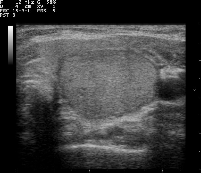
A thyroid nodule on ultrasound. Guidance keeps a nodule like this on screen so the needle tip can be driven into it and held in view. Image: Nevit Dilmen, CC BY-SA 3.0, Wikimedia Commons.
The single rule of guided biopsy is to see the tip before it moves. A needle shaft looks the same on screen whether the tip sits one millimeter ahead or one centimeter ahead. The operator confirms the tip, never the shaft alone. Two approaches put the tip on screen. Each one shows the needle in its own way. In the in-plane approach, the needle travels along the long axis of the probe. The whole shaft and the tip appear as one bright line. The operator follows the needle from skin to nodule without losing it. This approach gives close control over depth, since the tip stays in the picture for the entire pass. It asks for a steady hand to hold the needle inside the thin scan plane. In the out-of-plane approach, the needle crosses the probe at an angle. It shows up as a single bright dot where it passes through the scan plane. That dot can be the tip, or it can be a point along the shaft. The operator rocks the probe to find the true tip, then advances in small steps. This approach reaches some nodules the in-plane path cannot. It carries a cost in certainty, since a dot tells less than a full line. A steep needle adds a second problem. A needle driven at a sharp angle reflects less sound back to the probe, so the line dims on screen. Dropping to a shallower angle brightens it again. A needle with an etched, echogenic tip reflects more sound, so it holds a brighter line even at a steep angle. That tip costs a little more, so a clinic keeps it for the deep, awkward nodules. Probe pressure plays a part too. Light, steady pressure flattens the path to a shallow nodule, which lowers the needle angle and brightens the line on its own. The depth setting matters as well. Zooming the image down to the nodule enlarges the tip on screen, so a one-millimeter advance reads as a clear move. Some machines layer in needle-enhancing software that thickens the line on the display. A small jiggle of the needle marks the tip when the line fades, since motion stands out against still tissue. The choice of plane follows the anatomy. A wide, shallow nodule takes the in-plane line well. A nodule tucked behind the trachea may open only to an out-of-plane dot. The operator picks the plane the neck allows, then holds it for every pass.
Taking the sample
A hypoechoic thyroid nodule. The operator places the needle tip in the suspicious part of a nodule like this, then works it in short strokes. Image: Nevit Dilmen, CC BY-SA 3.0, Wikimedia Commons.
With the tip in the nodule, the operator works the needle to load it with cells. The needle moves in short strokes, a few millimeters in and out, ten to twenty times over a couple of seconds. Each stroke shears loose a few cells that ride up the bore by capillary action.
Two methods fill the needle. The capillary method, also called fine-needle non-aspiration, leans on that capillary draw alone. No suction is applied, so the sample stays clean. Less blood dilutes the cells. The aspiration method adds suction from a syringe, which pulls harder on a fibrous or sparsely cellular nodule. Suction also draws more blood, so the operator releases it before the needle comes out.
The suction stays gentle. A pull of one or two milliliters on the syringe is enough, since hard suction fills the sample with blood. The operator drops the pull the moment cells or fluid show in the hub.
A full study runs three to five passes, each into a slightly different part of the nodule.
Preparing the slide
The cells reach the pathologist as a slide. Speed matters here. Cells dry and distort within seconds off the needle, so the smear and the fixative wait ready before the first pass. The operator expels the aspirate onto glass and spreads it into a thin film. Part of the film is air-dried for one stain. Part is fixed in alcohol at once for another. The two fixations bring out their own features, so a lab often asks for both. A rushed smear lies too thick, which presses the cells together and costs a clean read. Some clinics rinse the needle into a liquid medium, which collects the cells in a vial for processing. Leftover material can form a cell block, a small core that holds the special stains a hard case needs.
When the nodule holds fluid
A part-cystic nodule needs a plan. The fluid carries few cells, so a needle parked in the cystic part draws a non-diagnostic sample. The operator aims at the solid wall or the solid nodule within the cyst, where the cells live. Draining the fluid first can help, since it collapses the cyst and brings the solid part within reach. The drained fluid still goes to the lab, since a rare cystic cancer sheds cells into it. A nodule that fills straight back after drainage gets a note for follow-up. A purely cystic nodule with no solid part rarely hides cancer, so a clear fluid tap often ends the workup.
Knowing the sample is enough
Not every pass yields a readable sample. Even under ultrasound, some aspirates come back non-diagnostic, with too few cells to call. Reported non-diagnostic rates run around 8 to 10 percent. A cystic nodule is the common culprit, since fluid carries few cells. Blood, ultrasound gel or a thick smear can also spoil a slide. Gel on the skin can ride the needle in, so the operator wipes the puncture site clean before the pass.
Rapid on-site evaluation catches a thin sample before the needle leaves. A technician stains one slide and reads it under a microscope in the room. A poor pass means another pass, taken while the patient stays positioned on the table. On-site evaluation lowers the non-diagnostic rate, since a weak sample is repeated on the spot.
A non-diagnostic result is not a dead end. The nodule is re-aspirated after a few months, often with on-site evaluation the second time around. A solid nodule that reads non-diagnostic twice may go to a core biopsy, a slightly thicker needle that takes a tissue fragment.
Reading the result with the Bethesda system
A pathologist sorts the cells into one of six Bethesda categories, each tied to a measured cancer risk. The category drives the next step.
Category one is non-diagnostic, the slide that could not be read. Category two is benign, with a risk near 4 percent. It returns the patient to routine surveillance. Category three is atypia of undetermined significance, a gray zone near 22 percent. Category four is a follicular neoplasm, around 30 percent, a result that often needs a diagnostic lobectomy. Category five is suspicious for malignancy, near 74 percent. Category six is malignant, at 97 percent and above, a result that goes to surgery.
Categories three and four sit in the middle on purpose. A repeat aspiration clears some category three nodules into a benign read. Molecular testing on the original sample sorts many of the rest, which reads the genes in the cells to refine the risk before any surgery is planned.
The risks rise in order, so the number carries real weight. A benign read spares surgery on a nodule that looked alarming on the scan. A suspicious read moves a quiet-looking nodule toward the operating room. A handful of benign reads still return for biopsy, since one sample can miss a small cancer inside a large nodule.
Staying out of trouble
Thyroid aspiration is a low-risk procedure. The needle is thin, so serious harm is rare. The common aftereffect is mild soreness, reported in up to roughly 9 percent of biopsies. A small bruise can show where the needle crossed a vessel.
Bleeding is the event to plan for, reported in under 3 percent of cases. Color Doppler before the pass maps the vessels, so the needle passes between them. Pressure on the neck for a few minutes after withdrawal settles a small bleed. A patient on a blood thinner needs that pressure held longer.
Blood thinners take a word before the visit. Many centers keep aspirin running through a thyroid FNA, since the needle is so fine. A stronger anticoagulant may be paused on the prescribing doctor’s advice, then restarted the same day.
Rarer events have been recorded. The list runs to recurrent laryngeal nerve irritation, a tracheal nick with a brief cough and Horner syndrome from a needle near the sympathetic chain. These events stay unusual. They resolve on their own in nearly every report. Lasting harm from a thyroid FNA is rare enough that large series report none.
FNA on a handheld unit
A handheld probe runs the same biopsy. One hand holds the probe over the nodule. The other steers the needle along the probe face for an in-plane view. A wireless probe frees the operator from a cart, a help at the bedside or in a clinic with no fixed procedure room.
Common questions about ultrasound guided thyroid FNA
How big a needle is used for a thyroid FNA?
A thyroid aspiration uses a fine needle, usually 25 to 27 gauge. It is finer than a core biopsy needle. The thin bore draws a cell sample with little bleeding and modest discomfort.
Why is ultrasound used during the biopsy?
Ultrasound keeps the needle tip in view inside the nodule. It lets the operator sample a small or deep target. Color Doppler shows the vessels to route around, which keeps the needle clear of the carotid artery and the jugular vein.
How many passes does an FNA take?
A full study takes three to five passes. Each pass samples a slightly different part of the nodule. More passes raise the chance of a readable sample.
What is the Bethesda system?
The Bethesda system sorts the cells into six categories, each with a measured cancer risk. The risks run from about 4 percent for a benign result to 97 percent and above for a malignant one. The category decides whether the nodule is watched, re-sampled, or removed.
Does a thyroid FNA hurt?
Patients usually feel mild soreness for a short time. Reported pain reaches up to about 9 percent of biopsies. Serious complications are rare.
What does a non-diagnostic result mean?
It means the slide had too few cells to read. It happens in roughly 8 to 10 percent of aspirations, often from cystic nodules. The nodule is re-aspirated after a few months, usually with on-site evaluation.
About the Author
Julien Mercier
Senior R&D Engineer · Medical Ultrasound Transducer Development
Senior R&D Engineer with an M.S. in Applied Physics and over 15 years of experience in medical ultrasound transducer development, specializing in the design verification and performance testing of high-frequency imaging transducers. Currently leading the development and verification of the company’s next-generation high-frequency linear-array transducer, responsible for imaging performance evaluation and reliability analysis in preclinical testing. Brings extensive hands-on experience in piezoelectric element tuning, beamforming parameter optimization, and system-level performance testing.