





































Thyroid scanning
How to Screen Thyroid Nodules with Handheld Ultrasound
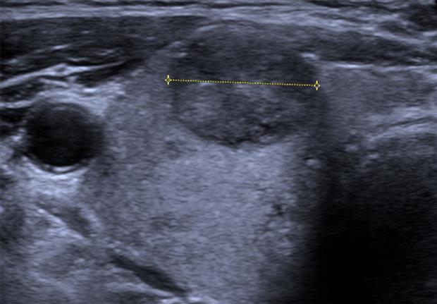
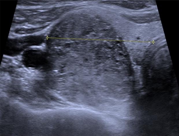
Thyroid nodules are common, usually harmless, and turn up by accident more often than not, which is why a screen is mainly a sorting job, separating the few nodules worth a closer look from the many that are not. A handheld probe does it at the bedside in minutes, its high-frequency face reading the gland a centimeter or two under the skin. The scan covers the gland so no nodule is missed, then weighs each one on the features that flag the ones worth a needle.
Covering the whole gland
The patient lies back with the neck extended over a pillow, the chin lifted to open the field below the jaw. The linear probe runs at the top of its band, around 12 megahertz, with the depth set shallow so the gland fills the screen. The scan starts in the transverse plane, the probe laid across the neck and swept from the top of one lobe down to the bottom, then across the isthmus and down the other lobe, so every part of the gland passes under the beam. The same ground is covered again in the longitudinal plane, the probe turned along the length of each lobe, since a nodule that hides between two transverse sweeps shows on a length-wise pass. The gland reads as an even mid-grey tissue, brighter than the muscle around it, and a nodule stands out as a patch that breaks that even grey. Each lobe is measured in three planes, its length, its width, and its depth, and the isthmus is measured front to back, the figures written down so a later scan can tell a stable gland from a growing one. The machine settings carry their own weight in the sweep. The focus is dropped onto the depth of the gland a centimetre or two down, where the beam is narrowest and the detail sharpest. The gain is set so the gland reads an even mid-grey, a nodule standing out cleanly against it on a picture set neither too bright nor too dark. The field is widened to hold a whole lobe across the screen where the neck allows, and narrowed onto a single nodule when it comes to be measured. Colour Doppler is laid over a nodule to read its blood supply, the flow inside it and around its rim, one more feature the same probe draws without reaching for a second tool. A whole-gland sweep takes only a minute or two, but it is the part that decides whether a nodule is found at all, and a hurried scan that skips the deep edge of a lobe or the low pole behind the collarbone misses the one nodule the screen is for. The discipline of the sweep, top to bottom and side to side in both planes, is what makes the screen worth running. A second pass over a doubtful patch settles it, the probe returning to read a shadow again before it is logged as a nodule or set aside as gland.
Finding and measuring a nodule
A nodule is caught as a break in the even gland, a rounded patch darker or brighter or lumpier than the tissue around it. Its position is noted, the lobe and the level within it, so the same nodule is found again on a follow-up scan. A gland often holds more than one, and each is logged in turn, the largest and the highest-risk followed closely whether or not they are the biggest.
Each nodule is measured in three planes, the same length, width, and depth taken for the lobe. The largest diameter is the figure that drives much of what follows, since the threshold for sampling a nodule is set in centimetres and a nodule is watched for growth against its own earlier size. The measurement is taken at the nodule’s edge, the calipers set where the nodule meets the gland, and a nodule with a fuzzy border is measured where the bulk of it sits. The three diameters also give a volume by the ellipsoid formula, the figure a growing nodule is tracked by from one year to the next.
Reading what a nodule is made of
The first thing read off a nodule is what fills it. A cyst is a clear dark pocket of fluid, the easiest call on the screen and almost always harmless, its back wall bright where the sound passes cleanly through. A spongiform nodule, a honeycomb of tiny cysts, is read the same reassuring way. A solid nodule, grey tissue filling the whole of it, is the one that carries the bulk of the risk, and a nodule part solid and part fluid is read on the solid part. A bright comet-tail flare inside a cystic nodule, the ring-down a fleck of colloid throws, reads as a reassuring sign of the harmless colloid a benign nodule holds.
The brightness of a solid nodule against the gland is the next feature. A nodule the same brightness as the gland, or brighter, sits at the low-risk end. A nodule darker than the gland, the low echogenicity that comes from packed cells, climbs the scale, and a nodule darker still than the strap muscle in front of it, the markedly low echo, climbs higher again. The reader sets the nodule’s grey against the gland and the muscle in the same frame, since the call is a comparison made on the one screen.
The texture inside a solid nodule is read alongside its brightness. An even texture reassures, and a patchy one is logged as a step up the scale. A nodule with a bright spongy core set inside a darker rim is noted for that pattern. The reader takes the make-up of the nodule as the foundation of the score, the feature the rest is read on top of.
The shape, the edge, and the bright specks
The shape of a nodule carries a clear sign. A nodule taller than it is wide, measured on the transverse view, grows against the natural lie of the gland, and that taller-than-wide shape is one of the strongest marks of a nodule worth sampling. A nodule wider than it is tall, spreading along the gland, sits low on the scale for shape.
The edge of the nodule is read next. A smooth well-defined border is reassuring, the nodule sitting cleanly in the gland. An irregular, lobulated, or spiky border, the nodule’s edge blurring into the gland around it, raises the concern, and a nodule that reaches through the gland’s capsule toward the strap muscle or the windpipe raises it further. The reader follows the whole rim, since a single ragged stretch counts.
The bright specks inside a nodule are the last feature and one of the clearest. A scatter of tiny bright dots, the punctate microcalcifications that mark the deposits a papillary cancer leaves, is a strong sign, each dot smaller than a millimetre and often without the shadow a larger stone casts. A single coarse calcification, or a bright rim of calcium around the nodule, is read on its own terms, since a broken rim can hide a growth pushing through it.
Each of these is read against the others, the score weighed from all of them at once, never resting on a single feature.
Turning the features into a decision
The features feed a structured score. A reporting system weighs each one on a scale and adds them into a category, every feature read so far contributing its points, and the category points to whether a nodule is watched or sampled. The handheld draws the picture every one of those features is read from, in the minutes a bedside scan allows.
The size of the nodule meets the score to set the threshold for a needle. A highly suspicious nodule is sampled at a smaller size than a low-suspicion one, the cut-off falling as the features climb, so a small nodule with microcalcifications and a taller-than-wide shape is sampled where a larger bland nodule is left alone. The screen sorts the gland’s nodules into the ones that call for a fine-needle aspiration and the ones that are watched.
A watched nodule is followed on its size and its features. A later handheld scan measures it against its logged figures, and a nodule that grows or sharpens a suspicious feature is brought back for a needle it did not need before. The bedside probe that found the nodule is the same one that follows it, scan after scan, the record built from one visit to the next.
The score has its blind spots, and a careful reader keeps them in mind. A follicular cancer can read as a bland nodule, smooth and no darker than the gland, so a nodule that grows is watched even when its features read low. The patient’s own history shifts the threshold too, a course of neck radiation in childhood or a family pattern of thyroid cancer lowering the size at which a needle is taken. The screen grades the picture, and the clinic reads the patient, the needle decision resting on both.
The aim of the whole screen sits in this sorting. The great majority of nodules are harmless and stay watched or ignored, a few carry the features that call for a needle, and the screen exists to tell the two apart without sending every neck for a biopsy.
Five features, one read
The five features build the score together, and no single one carries the call.
The diffuse gland and the bedside screen
Not every thyroid question is a nodule. A gland that reads coarse and dark all through, its texture broken into patches with raised flow, points to an inflammatory process across the whole organ rather than a single lump, and the handheld reads that diffuse change in the texture and the colour the same scan draws. A gland enlarged smoothly without a nodule is measured for its size and watched.
The probe reads more thyroid questions than nodules alone. A goitre, the whole gland swollen, is sized and searched for any nodule hiding in the bulk. A cyst grown tense and sore is drained under the same probe, the needle guided into the fluid pocket and the cyst emptied at the bedside. Each is a question the screen answers in the one sitting, the gland and its trouble read on the spot.
The handheld puts this screen where the patient is. A neck lump felt in a clinic room is scanned on the spot, a thyroid checked at a bedside or in a community clinic far from a cart-based machine, the gland and its nodules read in one hand against the front of the neck. The screen that once meant a referral and a wait now runs in the same visit, the picture and the measurements taken before the patient leaves the room.
The bedside scan does not replace the formal study where one is needed, and a suspicious nodule still goes on to a full department scan and a needle. What the handheld does is sort early, finding the nodule that needs that path and reassuring the many necks that do not, the first read taken where the question is first asked, the answer often in hand before the patient stands to leave the room.
Common questions about screening thyroid nodules
What probe is used to screen the thyroid?
A high-frequency linear probe, run around 10 to 14 megahertz, since the gland sits a centimetre or two under the skin and rewards the fine detail of the high-frequency band. The depth is set shallow so the gland fills the screen.
How is the whole gland covered?
The neck is extended over a pillow and the probe is swept across both lobes and the isthmus in the transverse plane, top to bottom, then turned and swept again along the length of each lobe, so no part of the gland is missed. Each lobe is measured in three planes.
What features mark a nodule worth sampling?
A solid make-up, a low echogenicity darker than the gland, a taller-than-wide shape, an irregular or invasive margin, and punctate microcalcifications. Each feeds a structured score, and no single feature decides the nodule alone.
What size of nodule is sampled?
It depends on the score. A highly suspicious nodule is sampled at a smaller size, often around a centimetre, while a low-suspicion nodule is left alone until it is larger. Size meets the feature score to set the threshold for a needle.
Can a handheld probe screen the thyroid reliably?
Yes, for finding nodules and reading the features a score is built on, at the bedside in a few minutes. A suspicious nodule still goes on to a formal department scan and a fine-needle aspiration; the handheld sorts early and reassures the many necks that need no further step.
What does a thyroid cyst look like?
A clear dark pocket of fluid with a bright back wall where the sound passes cleanly through. A pure cyst, and a spongiform nodule of tiny cysts, are almost always harmless and sit at the low-risk end of the screen.

About the Author
Julien Mercier
Senior R&D Engineer · Medical Ultrasound Transducer Development
Senior R&D Engineer with an M.S. in Applied Physics and over 15 years of experience in medical ultrasound transducer development, specializing in the design verification and performance testing of high-frequency imaging transducers. Currently leading the development and verification of the company’s next-generation high-frequency linear-array transducer, responsible for imaging performance evaluation and reliability analysis in preclinical testing. Brings extensive hands-on experience in piezoelectric element tuning, beamforming parameter optimization, and system-level performance testing.