





































Ophthalmic Eye Ultrasound Handheld 103g
Ophthalmic ultrasound reads the eye with sound where light cannot reach. A small high-frequency probe rests on the closed lid and sends its echoes through the eye, drawing the front chamber, the lens, the gel-filled cavity, and the wall at the back onto the screen. A clinician turns to it when a cataract, a dense bleed, or a swollen lid shuts the view a light would need. A handheld probe of barely a hundred grams carries that read to the clinic, the ward, and the bedside.
What the eye probe shows
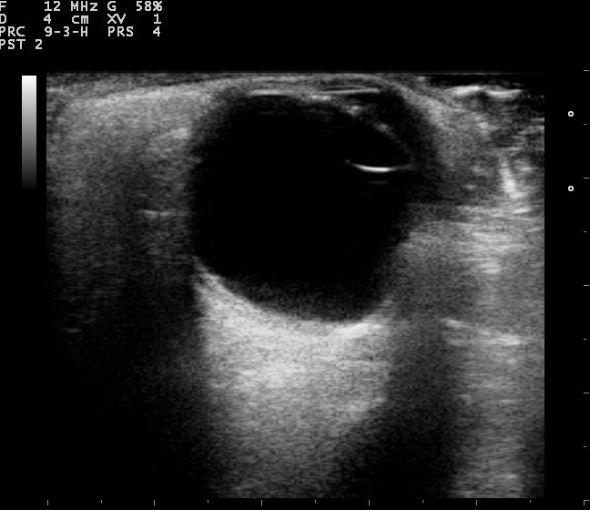
The eye sits close under the lid, so the probe runs at a high frequency to read fine detail. A clinician lays the probe on a bead of gel over the closed lid and sweeps the eye through its planes. The probe reads in two modes, one for a picture and one for a measurement. The grey slice of the picture mode shows the shape of the eye and anything adrift inside it. A clinician reads the wall at the back, the gel in the middle, and the lens up front in that one sweep. The picture comes up in seconds on a screen the clinician holds in one hand. A clinician marks the orientation of each sweep so the next reader knows which slice of the eye the picture shows. A clinician zooms in on the part of the eye in question to fill the screen with the wall or the lens. The closer view sharpens a small finding that would blur at a wider setting.
The eye answers ultrasound well because it is a ball of fluid close to the surface. Sound passes cleanly through the clear gel and bounces sharply off each layer it meets. A clinician reads the cornea, the iris, the lens, and the retina as a set of bright marks at known depths. The shallow path lets the probe read quickly in fine detail, so a small structure stands out against the dark of the fluid. A clinician builds the whole study from gentle sweeps across the closed lid. The fluid-filled eye is one of the kindest organs a probe ever reads. A clinician learns its layers quickly. The clean black of a healthy cavity makes any stray echo easy to catch. A clinician reads the depth of each layer against the known shape of the eye to place a finding exactly. A bright mark at the wrong depth gives itself away as an artefact.
A clinician keeps the study gentle on a tender eye. The probe rests on the lid over the gel and never presses on the globe, since a light hand keeps the picture true and the patient still. A clinician sweeps from side to side and up and down to bring every quadrant of the back of the eye into the beam. The patient turns the eye on command to carry a far corner under the probe. A clinician watches how a finding moves while the eye turns, since the way a membrane sways tells one kind apart from another. A careful study reads the whole eye with no touch on the eye itself and no drop of discomfort. A clinician steadies the probe on the bony rim around the eye to hold the view through a long sweep. The hand rests on the brow and the cheek so the beam stays where the clinician sets it.
A clinician learns the look of a normal eye before reading a troubled one. The healthy gel cavity reads as a clean black space, empty of echoes from front to back. The wall at the back curves as a single bright line. The optic nerve leaves it as a dark notch. The lens up front catches the beam at its edges. A clinician sets the gain low so the black cavity stays dark, since a gain pushed too high paints a haze that mimics a bleed. A clinician reads any haze in the black cavity or any line lifting off the smooth wall as the first sign of trouble. A clinician compares the troubled eye against its healthy fellow to judge what counts as out of place. The good eye sets the look a clinician measures the bad one against.
A clinician reaches for the probe the moment the eye stops giving its own answers. A scan at the bedside takes a few minutes and asks nothing of a lab down the hall. A clinician scans the eye where the patient lies, in the clinic chair or on the emergency trolley. The read comes back while the patient is still in the room. A clinician pairs the scan with the story of the eye, the blow it took or the sight it lost, that sent the patient in. A handheld study turns a long wait for an eye specialist into an answer in the first hour. A clinician records the study so the eye team reads the same picture later from the clip. The saved sweep carries a moving find a still frame cannot hold.
Seeing past a cloudy eye
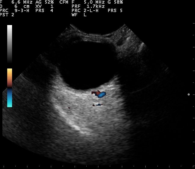
The probe earns its place the moment light stops getting through. A dense cataract, a pool of blood in the gel, or a scarred cornea hides the back of the eye from any lens or torch. A clinician sends sound past the cloud to read the retina and the cavity that light can no longer show. A clinician scans behind a white cataract to check the retina is flat before the surgeon removes the lens. The picture behind an opaque eye is what ophthalmic ultrasound does best. A clinician scans an eye that has lost its sight overnight to read the cause the clouded view hides. The answer often sits on the wall or in the gel a light cannot reach.
A bleed into the gel of the eye scatters the light and blots out the view. A clinician reads a vitreous hemorrhage as a haze of faint dots adrift in the cavity, settling and swirling when the eye stops moving. The scan grades how dense the bleed is and hunts the wall beneath it for a tear. A clinician turns the gain down to clear the faint dots of blood and leave only a true membrane on the screen. A clinician watches the dots stream on the moving clip to read the bleed apart from the layers behind it. The cause of a sudden loss of sight that comes without pain often shows on this one scan. A clinician follows a dense bleed across the weeks to watch it clear or to catch a tear it hides. A bleed that stays thick keeps the wall behind it out of view until it settles.
The wall at the back can peel away from its bed, a sight-threatening turn that an opaque eye hides from a light. A clinician reads a retinal detachment as a bright line that folds as it lifts off the back of the eye, staying anchored where the retina holds at the disc and the front edge. The scan tells a detachment apart from a lesser membrane by where it tethers and how stiffly it sways with the eye. A total detachment draws a funnel toward the optic nerve, a shape a clinician reads at a glance. A clinician who finds a peeled retina behind a bleed sends the eye on for urgent repair. A clinician grades how high and how wide the retina has lifted to tell the surgeon what the eye needs. A detachment near the centre of sight calls for the fastest repair.
A clinician carries the bedside read straight into the decision the eye needs. A fresh detachment goes to the operating room within days to save the sight it threatens. A clinician dates the find and measures how far the retina has lifted for the surgeon who takes it on. A clinician notes whether the centre of vision still lies flat, since that detail shapes how fast the eye must be fixed. The scan turns a blinded eye into a picture a surgeon can act on, read through the cloud before the sight is lost for good. A clinician marks the day the sight went, since a retina lifted only days ago saves better than one down for weeks. The clock on a detachment runs from the moment the centre of sight goes. A clinician reads the freshness of the lift to tell the team how fast the eye must reach the table.
Measuring the eye
The eye is one of the few organs a clinician measures to a tenth of a millimetre. The measuring beam times its echoes from the front of the eye to the back and reads a precise length. A clinician keeps the beam square to the eye, since a beam off the straight line reads the eye short. A clinician carries that figure into the plan for surgery and the watch on a disease. The number the eye gives is as much the point of the scan as the picture is. A clinician repeats a measurement that reads odd, since a single off reading can mislead the whole plan. A figure checked twice carries the weight a surgical plan rests on.
Before a cataract is removed, the eye needs a new lens of the right power. A clinician reads the axial length, the distance from the front of the eye to the retina, as the figure that sets that power. A length off by a fraction of a millimetre throws the result of the surgery off by a margin the patient sees. A clinician floats the probe just clear of the cornea to keep from pressing the eye shorter, and averages a run of readings. The averaged length, paired with the curve of the cornea, feeds the formula the surgeon picks the lens from. A clinician measures both eyes and compares them, since a wide gap between them flags a reading to take again. Two eyes of one person usually fall close in length.
The depth of the front chamber tells a clinician how much room the eye has at its drainage angle. A clinician reads the anterior chamber depth as a guide to the risk of angle-closure glaucoma, where a crowded angle can shut and drive the pressure up. A chamber that reads shallow marks an eye prone to a sudden painful rise in pressure. A clinician folds that depth into the watch on an eye at risk and the plan to open its angle before it shuts. A clinician reads a shallow chamber as a reason to watch the pressure across the seasons. A narrow angle can shut in the dark, when the pupil widens and crowds it further. A clinician reads the depth in both eyes, since a shallow angle in one warns of the same in the other.
A window on the pressure in the skull
The nerve that carries sight runs in a sheath that widens when the pressure inside the skull climbs. A clinician reads the optic nerve sheath diameter about three millimetres behind the globe, a width that grows when the pressure in the head rises. A sheath past about five millimetres points to a pressure raised inside the skull. A clinician reads it at the bedside, where no brain scan is at hand. A clinician uses that width in a patient who cannot be moved or who lies far from a scanner, as a fast read of a danger inside the head. The eye gives a window on the brain through the same probe that reads the retina. A clinician scans both eyes to be sure of the find. A clinician reads the width at a set distance behind the eye each time, so one study compares cleanly with the next. A sheath that settles once treatment lowers the pressure tells the clinician the head is easing. A clinician scans the nerve again over the hours to follow a pressure on the move.
Finding what does not belong
The clear path through the eye lets ultrasound spot a thing that should not be there. A clinician reads a bright fleck with a long shadow or a tail of repeating echoes behind it as a sign of metal or glass lodged in the eye. The scan places an intraocular foreign body in the eye and measures its depth for the surgeon who must reach it. A clinician maps the fragment against the lens and the wall so the team plans the safest way in. The same scan checks the wall for a bleed or a detachment the fragment has caused. The scan finds a fleck a torch would miss behind a swollen eye full of blood. A clinician reads the shadow a dense fleck throws to judge what the fragment is made of. Metal and glass throw a hard shadow a softer speck never makes.
A growth on the wall of the eye reads on ultrasound by its shape, its height, and the way it takes the sound. A clinician reads an intraocular tumour by these features, telling a solid mass apart from a clot or a fold of detached retina. A melanoma often rises as a dome or a collar-button mound with a hollow look on the beam, a clue to what the growth is made of. A clinician measures the height and the base of a tumour to size it and to follow it across the months. The scan turns a shadow at the back of the eye into a mass a clinician can name and track over time. A clinician sends a suspected tumour to the eye specialist for the full work-up the bedside read opens. The bedside scan starts the trail the specialist follows to a diagnosis. A clinician watches a quiet tumour across the visits to catch the day it starts to grow.
The probe and the rules
The eye’s probe is a small head that weighs almost nothing, suited to the delicate surface of the eye. A clinician holds a probe of about a hundred and three grams against the lid with the ease of a pen, steady through a long study. The high frequency it runs at suits the shallow eye and the fine detail the eye asks for. A light probe stays steady on a moving eye and tires no hand across a clinic full of patients. A wireless head that charges on a bench and pairs with a tablet reads the eye anywhere a patient sits. A clinician reads the whole eye with a tool that slips into a coat pocket. A clinician charges the probe between clinics and scans a full list of eyes on one charge. A wireless head leaves no cart to wheel from room to room.
The eye scan runs to a set of rules that keep it safe and its readings sound. The ophthalmology ultrasound guidelines set out how to scan the eye, how much sound power is safe on it, and how to read what comes back. A clinician keeps the power low on the eye, since the eye takes more care than the thicker tissue elsewhere in the body. A clinician follows the standard sweep so every quadrant of the eye comes under the beam, and labels each view the same way every time. The rules turn a scan of a precious organ into one a clinician runs the same safe way on every patient. A clinician keeps the power low and the study brief, since a gentle scan reads the eye safely. The eye needs less sound than the deeper organs to give a clear picture. A clinician logs the settings used so the same safe scan repeats on the next visit.
Putting the eye study together
A clinician reads the eye in one gentle study that answers several questions at once. The picture shows the gel, the wall, and anything adrift inside the eye. The measurements set the lens power, flag a crowded angle, and read the pressure behind the eye. A clinician carries the whole read into the plan for the patient in the few minutes the study takes, and writes each figure down with the view it came from. A clinician lays the picture and the figures side by side in the note so the eye team reads the whole study at once. The single record holds the look of the eye and its measurements together.
A clinician runs the part of the study the question in front of them needs. An eye that has suddenly gone blind calls for the picture behind the cloud. An eye headed for cataract surgery calls for the length that sets its new lens. A sick patient with a headache calls for the width of the nerve sheath. A clinician runs the part that answers the question and saves the rest of the eye for the record. A clinician notes the answer plainly so the next reader sees what the scan settled. The record names what was found and what the eye still needs.
The eye scan reaches places a clinic without a machine could never read. A handheld probe brings the study to the ward, the emergency room, and the field where a full eye lab sits far away. A clinician reads a peeled retina, a metal fleck, or a swollen nerve sheath wherever the patient is met. A clinician on a remote round carries the eye specialist’s first read in a pocket. A handheld scan travels in the hand to places a darkened room and a heavy console never reached. A clinician on a ship, a remote clinic, or a home visit reads the eye that once needed a trip to the city. The reach of the eye lab now follows the patient.
A clinician treats the bedside scan as the first read, with the eye lab behind it for the harder case. The handheld answers the urgent question in the first minutes a sight is at risk. A clinician passes the eye to the specialist for the fine work the bedside read points to. A clinician keeps a clip and the figures so the specialist starts from the bedside find. The eye gives up its secrets to a probe that weighs nothing in the hand, carried to wherever a patient with a troubled eye is found.
| Measure | Figure | What it tells |
|---|---|---|
| Probe frequency | about 10–20 MHz | high frequency for the shallow eye |
| Axial length, normal eye | about 23–24 mm | sets the power of a cataract lens |
| Anterior chamber depth | about 3.0–3.5 mm | a shallow chamber flags angle-closure risk |
| Optic nerve sheath diameter | above about 5 mm | points to raised pressure in the skull |
| Bedside scan for retinal detachment | about 90% and up | a fast read behind an opaque eye |
Common questions about ophthalmic eye ultrasound
What does ophthalmic ultrasound show that a light cannot?
It shows the back of the eye when a cataract, a bleed in the gel, or a scarred cornea blocks the view. Sound passes through the cloud and reads the retina, the gel cavity, and the wall behind it. A clinician finds a retinal detachment or a vitreous hemorrhage on a scan where a light shows nothing. The picture behind an opaque eye is the main reason to reach for the probe.
What are the two modes of an eye scan?
One mode paints a grey slice of the eye for a clinician to read at a glance. The other sends a single beam straight back and times its echoes into a precise length. A clinician reads the picture and measures off the line of spikes. The two modes read the same eye for two kinds of answer.
Is an eye ultrasound safe?
Yes, with a light hand and a low power. The probe rests on the closed lid over a bead of gel and never presses on the globe. A clinician keeps the sound power low, since the eye takes more care than thicker tissue. The guidelines set the safe limits a clinician scans within.
Why measure the length of the eye?
The length of the eye sets the power of the lens implanted at cataract surgery. A clinician reads it to a tenth of a millimetre, since a small error throws the result off by a margin the patient sees. The measuring beam times its echoes from the front of the eye to the retina for that figure. A clinician averages a run of readings for a length the surgeon can trust.
Can a handheld probe read the eye as well as a cart?
A handheld probe runs the high frequency the eye needs and brings the study to the bedside. A clinician reads a detachment, a foreign body, or a swollen nerve sheath on a probe that weighs about a hundred grams. The deeper work-up and the specialist scan stay with the eye lab. A clinician runs the bedside read where a full machine sits far away.

About the Author
Julien Mercier
Senior R&D Engineer · Medical Ultrasound Transducer Development
Senior R&D Engineer with an M.S. in Applied Physics and over 15 years of experience in medical ultrasound transducer development, specializing in the design verification and performance testing of high-frequency imaging transducers. Currently leading the development and verification of the company’s next-generation high-frequency linear-array transducer, responsible for imaging performance evaluation and reliability analysis in preclinical testing. Brings extensive hands-on experience in piezoelectric element tuning, beamforming parameter optimization, and system-level performance testing.