





































Ophthalmic Ultrasound A Scan and B Scan Handheld Probe
Ophthalmic ultrasound works in two scan modes, the B-scan and the A-scan. Both run from the same small probe resting on a closed lid over a drop of gel. Between them they answer the two questions a whole eye study is built on: the look of the eye and the measure of it. These two modes come first in any training, well before a single condition of the eye.
What the two modes are
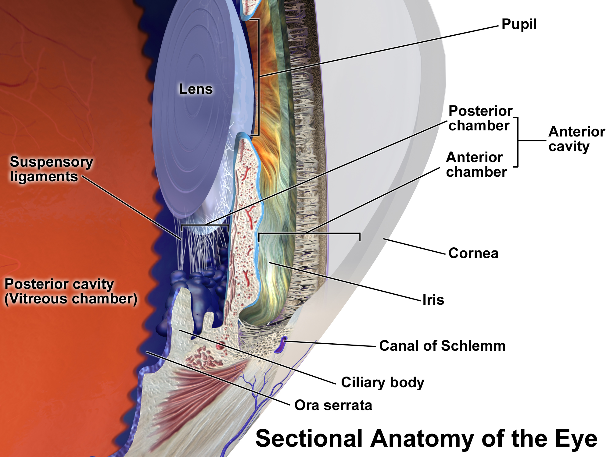
The B-scan draws a grey picture of a slice through the eye. The probe sweeps a thin beam back and forth across the closed lid, and the machine stacks the returning echoes into a two-dimensional image. That slice looks like any other ultrasound picture: bright lines mark each surface the sound struck. The dark round space of the vitreous fills the centre, with the curved wall of the back of the eye bounding it behind. Sweeping the beam through the whole globe builds the eye plane by plane. The picture shows the shape of the globe and anything floating or lifting inside it, taken in at a glance. Turning the probe catches the eye in three directions, across, along, and straight through, so a finding shows up in more than one plane. The mark on the probe tells which way each slice runs. The cornea, the lens, and the back wall stand out as the bright landmarks the picture is built around, sharp against the dark of the vitreous. The probe runs at a high frequency, around ten megahertz, suited to the shallow eye and the fine detail it needs.
The A-scan works along a single line, with the echoes drawn as a row of spikes. The probe sends one beam straight into the eye and times what comes back, each surface throwing up a spike at its own depth. The spacing between the spikes gives the distances inside the eye, and the height of each spike marks the strength of its echo. The front of the cornea, the two faces of the lens, and the retina each raise a spike of their own along the line. The beam has to be lined up with care, since the spikes only ring true when it runs straight through the eye. This mode turns the eye into a row of marks to be measured and judged for the make-up of what the beam passed through. The corneal spike, the two lens spikes, and the retinal spike serve as the landmarks along that line. To turn timing into length, the machine assumes a set speed of sound through the eye.
These two modes are the whole of ophthalmic ultrasound. Every later study of the eye is one of the two aimed at a single job, a detachment to find or a lens power to set. Whoever can handle a grey slice and a row of spikes can handle any eye scan built on them. One probe often carries both modes, ready at the press of a key. The picture and the line of spikes come first in any training, the ground every later study stands on. In practice the picture finds a thing, and the line of spikes then measures or characterises it. Both modes share one probe and one screen, switched at a key.
How the eye is scanned
Both modes run from a small probe resting on the closed lid. The clinician lays a drop of gel on the lid and sets the probe down with a light touch, never pressing on the globe. The high frequency the probe runs at suits the shallow eye and brings out its fine detail. A few simple instructions, hold still or turn the eye on command, bring each part of the globe under the beam. The closed lid and the drop of gel are the whole of the preparation. Rolling the eye under the lid through its full range carries the beam out to the far edges of the retina. A gentle hand and a generous bead of gel carry the sound in with no press on the globe. When one eye looks wrong, both are scanned, the healthy fellow giving the baseline the troubled one is set against. For a scan that touches the eye directly, a drop of topical anaesthetic goes in first; the over-the-lid scan needs none.
The machine is set for the eye before the first sweep. Depth runs only a few centimetres, since the whole eye sits close under the lid. The sound power stays low, gentler than on the thicker tissue elsewhere in the body. Gain is set so the vitreous looks dark, with a true echo still standing clear of it. Saving the settings with the study lets the next scan repeat them, and the same gain and depth keep two studies of one eye comparable. Each view is marked the same way every time, so the next clinician knows the plane a picture or a line came from. A turn of the gain brings out a faint membrane or clears the soft dots of a bleed. Settings noted with the study let the next person work the picture the same way. An ophthalmic preset holds the thermal and mechanical indices to the low limits set for the eye, the lowest of any scan. The eye carries no flow of its own to wash heat away, so the gentle output matters more here than elsewhere.
The B-scan, the picture
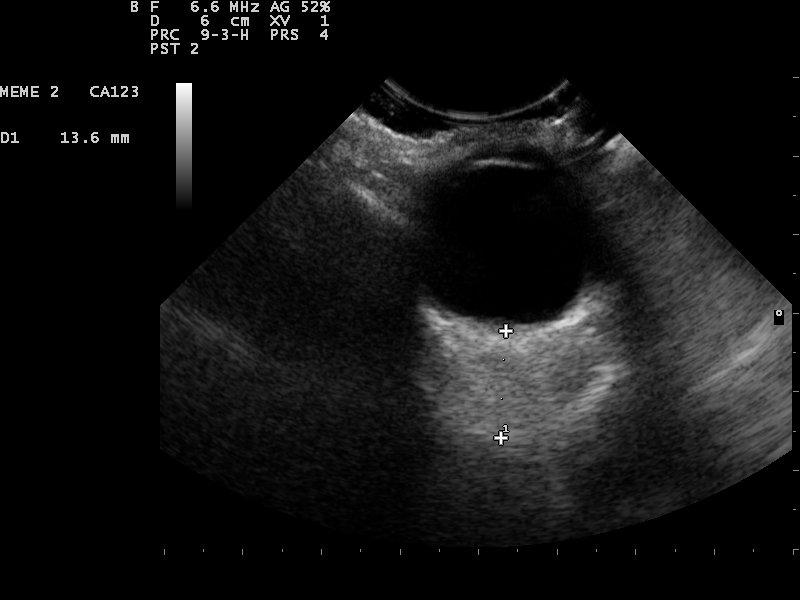
The B-scan is the mode for looking inside the eye. A sweep of the beam across the lid builds the grey slice on the screen. The dark anechoic space of the vitreous makes any echo inside it easy to catch. The smooth bright curve of the back wall stands clear, with a dark notch where the optic nerve leaves it. Point-of-care ocular ultrasound shows the back of the eye on this picture when an opaque view hides it from a light. A blinded eye becomes a slice to study. The lens sits as a bright curve near the front of the picture, and the optic nerve leaves a dark wedge at the back. The depth of each bright line, set against the known build of the eye, places it. Every finding lands at its true distance from the front. A colour overlay on the same picture shows blood moving in a vessel behind the eye, a help in a mass or in the vessels of the optic nerve.
The picture shows the danger behind a cloudy eye. A retinal detachment appears as a bright folded line lifting off the wall, anchored where the retina still holds. A vitreous hemorrhage shows as a haze of faint dots adrift in the cavity. A tumour rises as a solid mound from the wall. A foreign body sits as a bright fleck with a shadow behind it. Each of these comes up on the picture where a light shows nothing, behind a cataract or a bleed. A total detachment takes the shape of a funnel running back to the optic nerve, its two leaves meeting at the disc. The shape alone names it on the screen.
The moving picture carries more than a frozen frame. On a live clip, with the patient moving the eye from side to side, the way a finding shifts tells its own story. A detachment stays anchored at the disc and sways only a little, which sets it apart from a loose strand drifting free in the cavity. The dynamic sweep is a clue the moving B-scan alone offers, lost in any single still frame. A loose vitreous membrane undulates after the eye stops, drifting on for a moment, the lazy after-movement marking it as a loose strand. The eye has to move for the picture to tell one finding from another, so the patient is asked for a slow look up and down.
The B-scan measures as much as it shows. Calipers dropped on the picture size a tumour, a detachment, or the height of a mound on the wall. A measurement on the slice sets a baseline to follow across the visits. A mound that climbs in height over the months is one on the move. The picture holds both the look of a finding and a number for its size. The base and the height of a mound, marked with the calipers, both go down on the record. Measured again at the next visit, the same mound shows whether it has grown.
The B-scan is worked slice by slice through the whole eye. A small finding hides in all but one plane, so a slow sweep returns to the plane that showed it best. Each view is labelled and the clip saved for the next clinician. A careful sweep of the grey picture finds the danger an opaque eye hides, the first look behind a clouded eye. The probe travels through every clock hour of the eye, so no quadrant of the retina goes unseen. A finding caught in one plane earns a second look from a fresh angle, to be sure.
The A-scan, the measurement
The A-scan works the eye as a row of spikes along one beam. A single line of sound goes straight into the eye, and the echoes it sends back draw the row. Each surface the beam crosses raises a spike, set at the depth where the sound struck it. The spacing between the spikes is the distance between the surfaces inside the eye. Those gaps give the lengths the eye is built from. In a healthy eye the spikes stand steep and clean, the mark of smooth surfaces. A weak or doubled spike sends the clinician back to line the beam up again. The machine works from set speeds of sound, one for the watery aqueous and vitreous, a higher one for the dense lens.
The A-scan measures the length of the eye to a tenth of a millimetre. The distance from the front of the cornea to the retina, the axial length, comes off the spacing of the spikes. That length sets the power of the lens a surgeon implants at cataract surgery. The probe floats just clear of the cornea, since a touch that presses the eye shortens the figure. Paired with the curve of the cornea, the measured length feeds the formula the surgeon picks the lens from. Floating the probe in a small bath of fluid that bridges to the cornea keeps the eye from being pressed at all. An error of a single millimetre in the length shifts the lens power by a few dioptres, a miss the patient sees. Of the whole study, this length earns the closest care.
The height of each spike carries its own message. A taller spike means a denser surface, so the strength of a spike hints at the tissue the beam crossed. The pattern of spike heights through a mass tells one kind of growth from another. A melanoma sends back a run of low spikes a clinician comes to know. The standardized A-scan turns the make-up of a lesion into a row to study off the screen. Taken at a fixed gain, the row of spike heights lets two clinicians grade the same mass the same way. A standardized setting turns reflectivity into a number to compare. The reflectivity of a mass weighs against the patterns learned from known growths. The row of spikes carries a fingerprint of the tissue the beam crossed. The standardized method rests on three habits: placing a finding, watching how it moves, and measuring the height of its echoes.
The A-scan does the precise work the picture cannot. For the length that sets a lens, the figure no grey slice gives to a tenth of a millimetre, this is the mode. A run of readings, averaged, yields a length the surgeon can trust. Run with care along a straight beam, the A-scan gives the eye its hardest numbers. Those numbers carry into the plan for the surgery the eye is headed for. The speed of sound the machine used goes in the note too, since a different setting makes the same eye a touch longer or shorter. The hardest numbers of the eye come from a beam held dead straight. The final length is the figure the whole surgery is planned around. A number taken with care spares the patient a lens of the wrong power.
| Item | Figure | Note |
|---|---|---|
| Probe frequency | about 10 MHz | the standard for the eye picture |
| Axial length, normal eye | about 23–24 mm | the A-scan figure that sets a lens |
| A-scan precision | to about 0.1 mm | the length for cataract surgery |
| B-scan depth | a few centimetres | the whole globe in one sweep |
| Front-of-eye detail | up to 35–50 MHz | the ultra-high-frequency look kept for the lab |
Using the two together
Most eye studies run both modes on the one eye, each carrying part of the answer. A mass found on the grey picture invites a single beam dropped through it, the spikes giving its make-up. A detachment seen on the B-scan calls for the length of the eye on the A-scan, for the surgery that follows. The picture points to where to look; the spikes then say what sits there. A full study leans on the pair. Steering a single A-scan beam through a lesion the B-scan has found maps the make-up of exactly the spot in question. Taken together, the picture and the spikes name a finding the picture alone leaves open. A finding’s measure and its picture are two halves of one study. The eye gives up its place and its make-up to the one probe. A single probe holds both modes, so the switch from picture to spikes takes only a key, with no change of equipment.
What a handheld brings
A handheld probe carries both modes to the bedside. On a patient too sick to reach an eye clinic, the B-scan shows the back of a blind eye in minutes. Where a full biometry suite sits far away, the A-scan gives a quick length. The probe weighs about a hundred grams and charges on a bench, ready in a coat pocket. The eye exam comes to where the patient lies, on the ward or the emergency trolley. A fresh loss of sight gets its first look on the ward round, with no trip to the eye clinic. The probe pairs with a tablet and saves the study to the record on the spot. A swollen optic nerve or a peeled retina turns up at the bedside in the first minutes a sight is at risk. The handheld brings the eye exam to a patient who could never reach an eye clinic. The picture and the spikes travel over a wireless link to the screen, with no cart to wheel in.
A handheld answers the urgent question at the bedside. The finest work belongs to the lab. A screening length and a first look at the back of the eye come at the bedside. The finest biometry for a tricky eye, and the ultra-high-frequency look at the front of the eye, stay with the dedicated machine. A hard case passes to the eye lab for the study a handheld cannot match. The bedside result sets the direction the fuller study confirms. The lab takes over when a tricky eye needs an exact length or a close look at the drainage angle. The handheld names the urgent finding and points the patient toward the fuller study. That first pass is the one the lab study then confirms.
The eye across visits
Both modes serve the same eye across the visits as well. A B-scan from one visit lines up against the next to show a detachment that has spread or a mass that has grown. An A-scan length, taken the same way each time, tracks an eye that is changing. One method and one set of views keep the studies comparable. The trend across the scans tells more than any single visit. The old picture laid beside the new shows a detachment creeping toward the centre of sight. A length that climbs across the years marks an eye stretching, a clue in a short-sighted patient. The run of lengths over the years is the story of an eye that is growing or holding steady.
Both results go in one place for the eye team. The note carries the picture’s findings and the measured lengths, each tied to the view it came from. A clip of the moving B-scan and the figures from the A-scan are saved together. That record turns a moment at the bedside into a baseline the team builds on. A date on each study lets the next clinician compare like with like. The findings go down in plain words and the lengths in plain figures, each tied to its view. The record means the same to the next clinician who opens it.
The eye is taught on these two modes before any single condition. Whoever knows the grey picture and the row of spikes can handle any eye study built on them. The detachment scan, the biometry, the tumour study, each is one of the two modes turned to a job. With the foundation in hand, the rest comes without relearning the tool. The two modes are the alphabet the whole eye study is written in. A clinician who has seen a thousand grey slices and a thousand rows of spikes knows the eye in any study. The two modes carry a clinician through every condition the eye can raise.
Ophthalmic ultrasound rests on a picture and a measurement, taken from one small probe. Both modes run on the one eye, the look and the number a full study needs. A handheld carries both to the bedside, the first look where the patient is met. These two modes come first in any training, and every later study of the eye builds on them. The picture and the line of spikes are where the work on any eye begins. On every eye, no matter how the case opens, the study comes back to the picture and the measurement. Together the two modes hold the whole of what ultrasound finds in the eye.
Common questions about the A-scan and B-scan of the eye
What is the difference between an A-scan and a B-scan of the eye?
The B-scan draws a grey two-dimensional picture of a slice through the eye, used to look at the globe and its contents. The A-scan works a single beam as a row of spikes, used to measure lengths to a tenth of a millimetre. Both run from the same probe on the closed lid over the same drop of gel.
What does a B-scan of the eye show?
It shows a grey cross-section of the eye, the dark vitreous cavity at its centre, bounded by the curved wall behind. On it a retinal detachment appears as a bright folded line, a vitreous hemorrhage as a haze of dots, and a tumour as a mound on the wall. The B-scan shows the back of the eye when a cataract or a bleed blocks the view. A live clip shows how a finding sways as the eye moves.
What is the A-scan used for?
The A-scan measures the length of the eye to a tenth of a millimetre, the axial length that sets the power of a cataract lens. The spacing of the spikes gives that figure. The height of the spikes also tells the make-up of a mass, a clue to what a growth is made of. A run of readings, averaged, gives a length the surgeon can trust.
Is the eye scan safe and how is it done?
Yes, with a light hand and a low power. The probe rests on the closed lid over a drop of gel and never presses on the globe. The eye sits close under the lid, so the depth runs only a few centimetres. The sound power stays low, since the eye takes more care than the thicker tissue elsewhere.
Can a handheld probe run both modes?
A handheld probe carries both the B-scan and the A-scan to the bedside. It shows the back of a blind eye and takes a screening length on a probe that weighs about a hundred grams. The finest biometry and the ultra-high-frequency look at the front of the eye stay with the dedicated machine. The bedside scan runs where a full machine sits far away.

About the Author
Julien Mercier
Senior R&D Engineer · Medical Ultrasound Transducer Development
Senior R&D Engineer with an M.S. in Applied Physics and over 15 years of experience in medical ultrasound transducer development, specializing in the design verification and performance testing of high-frequency imaging transducers. Currently leading the development and verification of the company’s next-generation high-frequency linear-array transducer, responsible for imaging performance evaluation and reliability analysis in preclinical testing. Brings extensive hands-on experience in piezoelectric element tuning, beamforming parameter optimization, and system-level performance testing.