





































Intraocular Foreign Body Ultrasound Localization Handheld Ophthalmic
An intraocular foreign body is a fragment of metal, glass, stone, or wood driven into the eye, usually by an accident at work. A hammer striking metal throws off tiny shards at high speed. One can pierce the front of the eye and bury itself inside, in the fluid, the lens, or the wall at the back. Most are small, a millimetre or two of metal, and most land in young working men. The eye may look almost normal from the outside, with only a small entry wound to show for it. The pain and the loss of sight can be slight at first, far out of step with the danger inside. Inside, the fragment sits where no light can reach it once blood or a cloudy lens blocks the view. Finding that fragment, and pinning down exactly where it lies, is the work a handheld ultrasound does at the bedside. How well the eye does turns on three things: where the fragment lies, how big it is, and how soon it comes out. The scan reads the first two in a minute and helps start the third.
A fragment in the eye
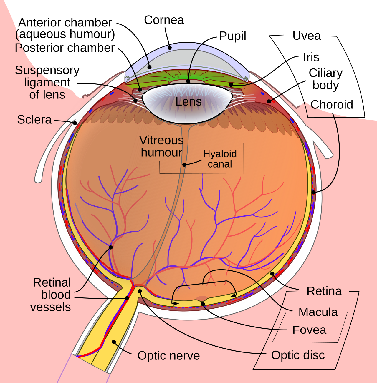
The classic story is a man striking steel on steel without eye protection. A metal chip flies off and goes through the cornea or the white of the eye faster than he can blink. Other fragments come from grinding wheels, explosions, gunshot, or a fall onto something sharp. Lawn mowers and strimmers fling stones and wire. A nail gun drives a fragment deep in a single shot. The fragment carries its own danger past the wound it makes. Iron rusts inside the eye over the following weeks and stains the retina, a slow poisoning called siderosis. Copper sets off its own steady inflammation. Any fragment can carry in the germs that cause a blinding infection within days of the injury. A piece of glass or stone may sit quietly for years. The same piece can tear the retina if it shifts. The fragment has to come out in most cases. The surgeon needs to know where it is before going in after it, and the deeper it lies, the more that knowing matters. The fragment enters through the cornea, the white of the eye, or the rim where the two meet. A high-velocity chip can pass right through the front and lodge in the wall at the back. Every penetrating wound also raises the question of tetanus and a course of antibiotics.
The trouble is seeing it. A fragment small enough to enter through a pinhole wound is small enough to hide. The entry can seal over within hours and look like a minor scratch. The eye can feel almost ordinary, the patient unaware anything went in. Blood from the torn vessels fills the inside of the eye and curtains off the view within minutes. The injured lens can go white and block the light as surely as a cataract. So the very eye most likely to hold a fragment is often the one a doctor cannot see into. The history does most of the work of suspicion: a hammer, a grinder, a blast, and a sudden sore red eye. A high-speed metal-on-metal injury is treated as a fragment in the eye until proven otherwise. From there the search turns to imaging. Ultrasound is one of the tools that finds the hidden piece. A few signs raise the alarm on exam: a drop in vision, a pupil that reacts poorly, a low pressure in a soft eye. None of them shows the fragment. Only imaging does that.
When the fragment can’t be seen
Ultrasound is at its most useful on the eye that light cannot enter. Sound crosses blood and a cloudy lens without trouble, reaching the back of the eye and returning a picture of everything inside. On that picture a fragment of metal or glass stands out sharply against the soft grey of the eye’s own tissues. The probe is the same high-frequency linear one used for the rest of the eye, run over a closed lid on a thick layer of gel. A few seconds of looking can settle whether a fragment is there. The scan does more than say a fragment is present. It shows where in the eye the fragment sits, how big it is, how many pieces there are, and what damage they have done to the retina and the lens around them. It reads all of this on an eye an ophthalmoscope cannot begin to examine. That map is what a surgeon plans an operation from. On the A-scan trace that runs alongside the picture, the fragment throws a single tall spike, taller than the echo from any tissue. Height and brightness together mark it out.
The first imaging for a suspected metal fragment in the eye is often a CT scan. A CT reads the orbit in thin slices and catches dense metal anywhere in or around the eye, with no instrument touching the wound. It also pins the fragment in three dimensions for the surgeon. One imaging test is kept off a metal injury entirely. An MRI uses a strong magnet. That magnet can drag a loose metal fragment through the eye and do fresh damage. So an MRI waits until metal is ruled out. Ultrasound works alongside the CT. The handheld reaches places a CT cannot: the bedside, an eye that cannot be moved safely, a clinic with no scanner at all. It reads the soft tissue around the fragment, showing a detachment or a bleed the metal has caused. The two build a fuller picture of the injured eye together. A CT cuts the orbit into slices a millimetre or two thick and reformats them in any plane the surgeon wants. Dense metal can flare on a CT and blur its own exact edge. The ultrasound adds a close read of the soft tissue around the fragment.
Time matters once a fragment is in the eye. The longer it stays, the higher the chance of an infection taking hold inside. An infection in the eye, called endophthalmitis, can blind it in a day or two. A fragment of iron or copper begins to dissolve and seed the retina with metal, a slow poisoning that steals sight over the following weeks. A small clean fragment is left in place and watched in a few cases. Most are taken out. The sooner that happens, the better, before infection or rust sets in. Locating the fragment fast lets the surgeon plan that removal. On an eye no one can see into, the scan is what starts the clock running toward the operating room. The first hours after a penetrating injury are the ones that decide the eye. Antibiotics go in early to hold off infection while the plan is made. The removal itself is usually a vitrectomy, the surgeon working inside the eye through tiny ports. The scan tells that surgeon what waits inside before the first port goes in. A patient with a fragment in one eye is asked about both eyes, since the same accident can throw chips into each. A magnetic fragment can sometimes be drawn out with a magnet, and the scan helps tell whether it sits free enough for that.
The scan is asked three plain questions. Is there a fragment in the eye at all. Where exactly does it lie. What has it damaged on its way in. The first is often answered in the first few seconds of looking, by a single point far brighter than anything around it. The second and third take a careful sweep of the whole eye and a steady reading of the picture. The examiner turns the gain down to keep the bright fragment from blooming over the tissue around it. Each question is read off the same grey picture, the fragment standing out on it like a spark in the dark. A clear answer to all three is what the surgeon needs before touching the eye. The examiner works through the eye in a set order, front to back, so the same ground is covered every time. A fragment found late in the sweep is as important as one found first.
The one rule on a hurt eye
One rule outranks everything else on a freshly injured eye: never press on it. An eye with a penetrating wound may be an open globe, its wall breached and its contents held in only by the pressure inside. Signs point to it: a soft eye, a teardrop-shaped pupil pulled toward the wound, a shallow front chamber, a trace of fluid weeping from the cut. The gentle weight of a probe on such an eye can push the inside out through the wound and lose the eye in an instant. So the scan over a suspected open globe is done with the lightest possible hand, the probe floated on a thick bed of gel, never bearing down. A shield goes over the eye between looks to keep any pressure off it. When the globe is clearly ruptured, many teams hold the ultrasound and let a CT do the looking, since a CT touches nothing at all. The rule states itself: a hurt eye is scanned feather-light, or not at all. The eye is shielded and the patient sent on to an ophthalmologist without delay. Nothing about the scan is allowed to make the injury worse. On a clearly open globe, the gentlest course is to image with a CT and save the ultrasound for later. The same gentle rule covers any badly bruised eye, even one with no clear cut, until an open globe is ruled out.
The bright spot in the dark
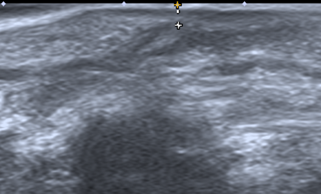
A foreign body is the brightest thing in the eye. Sound reflects off a hard fragment far more strongly than off any soft tissue, so the fragment returns a sharp, intense echo that the machine paints as a brilliant white point. The jump in density between soft tissue and metal or glass sends almost all the sound straight back. Against the black of a blood-filled or healthy vitreous, that point is unmistakable, brighter than the retina, brighter than any membrane or clot. A trained eye catches it in the first sweep. The fragment may be a fleck a millimetre across. It still shines out of the grey like a star, far out of proportion to its size. The brightness alone, in the right story, is enough to send a patient to surgery. A fragment that blooms too wide to measure at full gain shrinks to a clean dot when the gain comes down. The examiner drops the gain to size the piece and to keep its glare off the tissue around it.
Behind a solid fragment the scan often shows a shadow. A dense piece blocks the sound from passing through it, so the area directly behind the fragment goes dark, a clean band of shadow stretching away from the bright point toward the back of the eye. That shadow is a second confirmation that the bright point is a solid fragment. The deeper structures fall into that shadow and go unread, a small price for the certainty the shadow brings. The shadow also marks the line of the sound beam, pointing from the probe straight through the fragment, a clue to exactly where the piece sits along that line. Turning the probe to put the fragment in the centre of the beam sharpens both the point and its shadow. A bubble of air casts much the same kind of shadow. The bright point above the shadow is what separates a fragment from a bubble.
A metal or glass fragment can throw a third sign. Sound bounces back and forth inside a flat, hard surface, sending a train of fading echoes out behind the fragment, a string of bright dashes trailing into the dark. This reverberation, sometimes called a comet tail, is the mark of a smooth, hard, often metallic piece. The flatter and smoother the surface, the longer and more even the tail it throws. It tells the examiner that the fragment is dense and likely metal, the kind that corrodes and the kind a surgeon will want out soon. The brilliant point, the shadow, and the comet tail together name a fragment with little room for doubt. On the A-scan the same fragment throws a tall spike with a row of smaller spikes stepping down behind it, the trace’s own version of the comet tail. The two views say the same thing.
The kind of fragment shapes how it looks. Metal shows all three signs at their strongest, the brightest echo and the longest comet tail. Glass and stone come close, shining hard and shadowing well, often with no tail of their own. The trap is dry wood: it can trap air and read as a bright line, easy to mistake for a bubble of gas. Wood can also swell and rot and breed infection faster than metal, so a missed wooden fragment is its own emergency. Plastic may be nearly invisible, no brighter than the tissue around it. A fleck of calcium from an old scar can shine almost as bright as glass, so the story of a recent injury is what marks a fresh fragment out from an old deposit. The examiner reads the brightness, the shadow, and the tail together, and weighs them against the story of what struck the eye. An old fragment can wall itself off in a capsule of scar over months, its echo dimmed and its shadow softened by the tissue grown around it.
One fragment can hide another. A single blow can shower the eye with several pieces, so the examiner sweeps the whole globe slowly, corner to corner, never stopping at the first bright point. A blast or a shotgun can drive in a dozen at once. A tiny fragment can lodge in a blind spot, tucked against the wall or buried in the lens, and show only on one angle of the beam. Moving the probe through small arcs brings these out, each catching the light for a moment when the beam crosses it square. The count of fragments, and the place of each, goes into the plan for the operation. Missing a second piece means a second operation, on an eye that has already been opened once. The examiner notes each fragment’s depth and position the moment it is found, building a list the surgeon works down one by one. A scattered blast injury can take several minutes to map in full.
| Item | Figure | Note |
|---|---|---|
| Hammering metal as the cause | about 43% of cases | the classic mechanism |
| Probe frequency | about 10–15 MHz | linear probe, light on the lid |
| Typical fragment | about 1–3 mm | small enough to hide |
| Foreign-body echo | far brighter than retina | the spark in the dark |
| Behind a dense piece | an acoustic shadow | a clean dark band |
| Metal or glass clue | a comet-tail trail | reverberation behind the point |
| Open globe | no probe pressure | feather-light, or defer to CT |
What the scan pins down
Where the fragment sits decides how it comes out. The scan places it along several lines at once: how deep it lies from the front of the eye, how far it sits from the centre, whether it floats free in the vitreous or lies buried in the lens or the wall, whether it has passed clean through to the outside. The examiner measures its distance from the front of the eye and reads off the clock hour it sits at, the same coordinates the surgeon will use. A piece in the front chamber, a piece in the lens, a piece in the vitreous, a piece embedded in the retina each call for a different route in. The deeper and the more embedded the fragment, the bigger the operation to reach it. Each of those facts changes the way the surgeon goes after the piece. The handheld draws the map the operation follows. The map gives the surgeon a target before the eye is even opened, the depth and the clock hour fixing where to go in and how far to reach.
The same scan reads the damage the fragment did coming in. A fragment rarely travels alone. It tears a path going in. The scan shows the wreckage: blood filling the vitreous, the lens broken open, the retina lifted off the wall behind. A retinal detachment found alongside the fragment changes the whole operation, adding a repair to the removal. A heavy bleed shows how much blood will have to be cleared to reach the piece at all. The scan that finds the fragment finds these injuries in the same sweep, on the same blind eye, and hands the surgeon the full account before a single cut is made. The more the surgeon knows going in, the fewer the surprises on the table. If infection is feared, a drop of fluid is drawn from the eye and sent for culture in the same setting. The scan and the tap together start the treatment before the operating room is ready. A foreign body that has split the lens open can drag bits of lens into the vitreous, one more thing the scan counts for the surgeon. The fuller that count, the cleaner the operation that follows.
The scan has its blind spots. A fragment lodged in the very front, against the back of the cornea, can hide in the near edge of the picture where the beam is crowded. A speck of plastic or wood that gives no bright echo can pass unseen. A fragment resting in the bone of the orbit, outside the eye, sits beyond the reach of the probe. Small metal fragments still go to CT for a count and a precise map, the test that catches dense metal best. What the handheld gives is the early answer on the eye that cannot wait or cannot move: a fragment is in there, here is roughly where, here is what it has torn. From that the surgeon and the CT take over. The removal is planned in full. The scan that took a minute on a blind eye is what set the whole rescue in motion. A repeat scan after surgery checks that the fragment is truly gone and the retina lies flat. For a fragment caught in the very front, a higher-frequency probe maps the anterior segment in fine detail. A fragment that cannot be reached safely is sometimes left in place and watched with repeat scans for any sign of trouble. The handheld carries the eye from the first blind minute to the follow-up weeks later.
Common questions about ultrasound for an eye foreign body
How does a foreign body look on ultrasound?
As a single, very bright point, far brighter than anything around it in the eye. Behind a solid fragment the scan usually shows a dark shadow, where the dense piece has blocked the sound. A metal or glass fragment can also throw a comet tail, a train of fading echoes trailing back from the bright point. Those three signs together, the brilliant point, the shadow, and the tail, name a foreign body with little doubt.
Why use ultrasound when a CT can find a metal fragment?
Because the handheld reaches what a CT cannot. It works at the bedside, on an eye that cannot be moved safely, in a clinic with no scanner. It reads the soft tissue around the fragment, showing a retinal detachment or a bleed that the metal has caused. CT and ultrasound work together on a serious eye injury, each adding what the other leaves out.
Can ultrasound tell what the fragment is made of?
It gives strong clues. Metal shows the brightest echo and the longest comet tail. Glass and stone shine hard and cast a firm shadow. Dry wood can trap air and read as a bright line, easy to mistake for a gas bubble. Plastic may be nearly invisible against soft tissue. The scan reads these clues together with the story of what struck the eye. The final word on the material often waits for the fragment to come out.
Is it safe to scan an eye that may be cut open?
Only with great care. A penetrating wound may be an open globe, held together by the pressure inside. Pressing a probe on it can push the contents out through the wound. The scan over a suspected open globe is done feather-light, the probe floated on a thick layer of gel, never bearing down. When the globe is clearly ruptured, many teams hold the ultrasound and let a CT do the looking, since a CT touches nothing.
What does the scan tell the surgeon before the operation?
Where the fragment lies, how big it is, and how many pieces there are. It places the fragment in the front of the eye, in the lens, in the vitreous, or driven into the wall, each location calling for a different way in. It also shows the damage around the fragment: a torn retina, a broken lens, a bleed filling the eye. The surgeon plans the removal and any repair from that map, before making a single cut on an eye no light could enter.

About the Author
Julien Mercier
Senior R&D Engineer · Medical Ultrasound Transducer Development
Senior R&D Engineer with an M.S. in Applied Physics and over 15 years of experience in medical ultrasound transducer development, specializing in the design verification and performance testing of high-frequency imaging transducers. Currently leading the development and verification of the company’s next-generation high-frequency linear-array transducer, responsible for imaging performance evaluation and reliability analysis in preclinical testing. Brings extensive hands-on experience in piezoelectric element tuning, beamforming parameter optimization, and system-level performance testing.