





































Retinal Detachment Ultrasound Diagnosis Handheld Ophthalmic Probe
A retinal detachment is the sensory retina peeling away from the wall it normally lines, the layer that turns light into sight coming loose at the back of the eye. The warning signs are familiar to any emergency clinician. The patient describes a curtain or a dark shadow drawn across one part of the vision, almost always without pain. A shower of new floaters or a burst of flashing lights often comes first, in the days before the curtain falls. Sight in the covered field goes. It stays gone until the retina is laid back down on the wall. Detachment runs higher in the very short-sighted eye, in the eye that has had cataract surgery, and after a blow to the head or orbit. The job at the bedside is to find the detachment quickly, often long before an eye specialist can get to the patient. A handheld ultrasound is what finds it.
When the retina lifts off the wall
The reason ultrasound belongs here is the same one that brings it to the cloudy cataract eye. Light has to reach the retina for anyone to see it directly through the pupil. Many of the eyes most likely to hold a detachment are exactly the ones light cannot get into. A bleed in the vitreous turns the inside to fog. A dense cataract, a scarred cornea, a swollen lid that will not open, a pupil that will not dilate: each of these shuts the door on the ophthalmoscope. A high-frequency probe laid on the closed lid reads the eye all the same, sending pulses through the lid, the front of the eye, and the vitreous, then timing the echoes that come back. From those echoes the machine draws the whole back of the eye on its screen, the retina and any detachment laid out in grey and white. The view costs nothing but a smear of gel and a minute of the clinician’s time. In many emergency departments the same handheld probe that scanned a belly or a lung an hour earlier is the one that now reads the eye.
Speed matters because the clock is running on the retina from the moment it lifts. A detached retina is cut off from its main blood supply in the wall behind it. It survives only on the dwindling oxygen it can draw from the fluid around it. The longer the retina stays off the wall, the fewer of its light-sensing cells come back to work once it is laid down again. A detachment caught before it reaches the central macula is an emergency where hours count. Even after the macula goes, the outer edges keep their best chance of useful recovery when the repair is not left to drift. Finding the detachment fast, even on an eye no one can see into, is the whole point of putting a probe on the lid. A scan that takes a minute can move a patient up an operating list by a day.
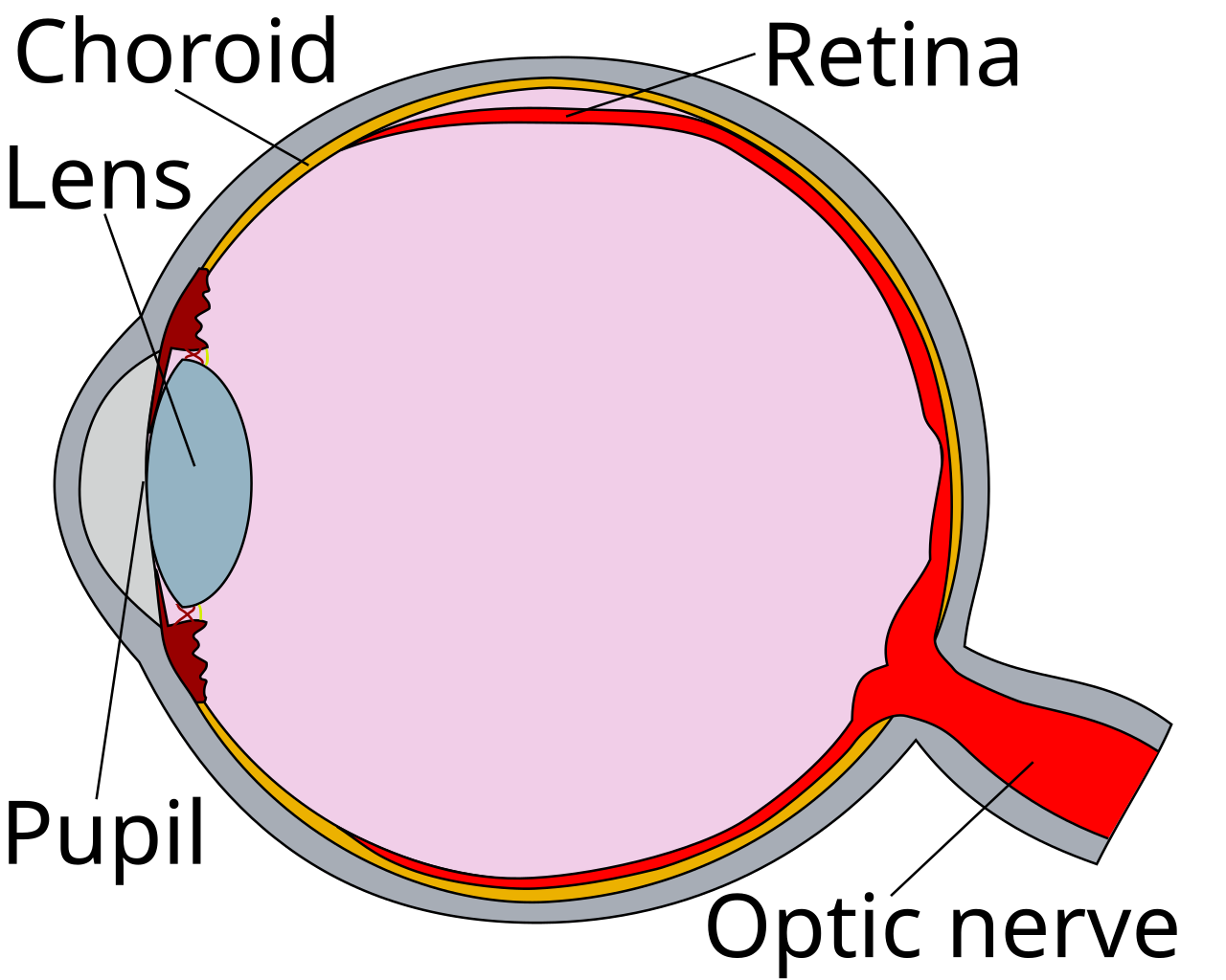
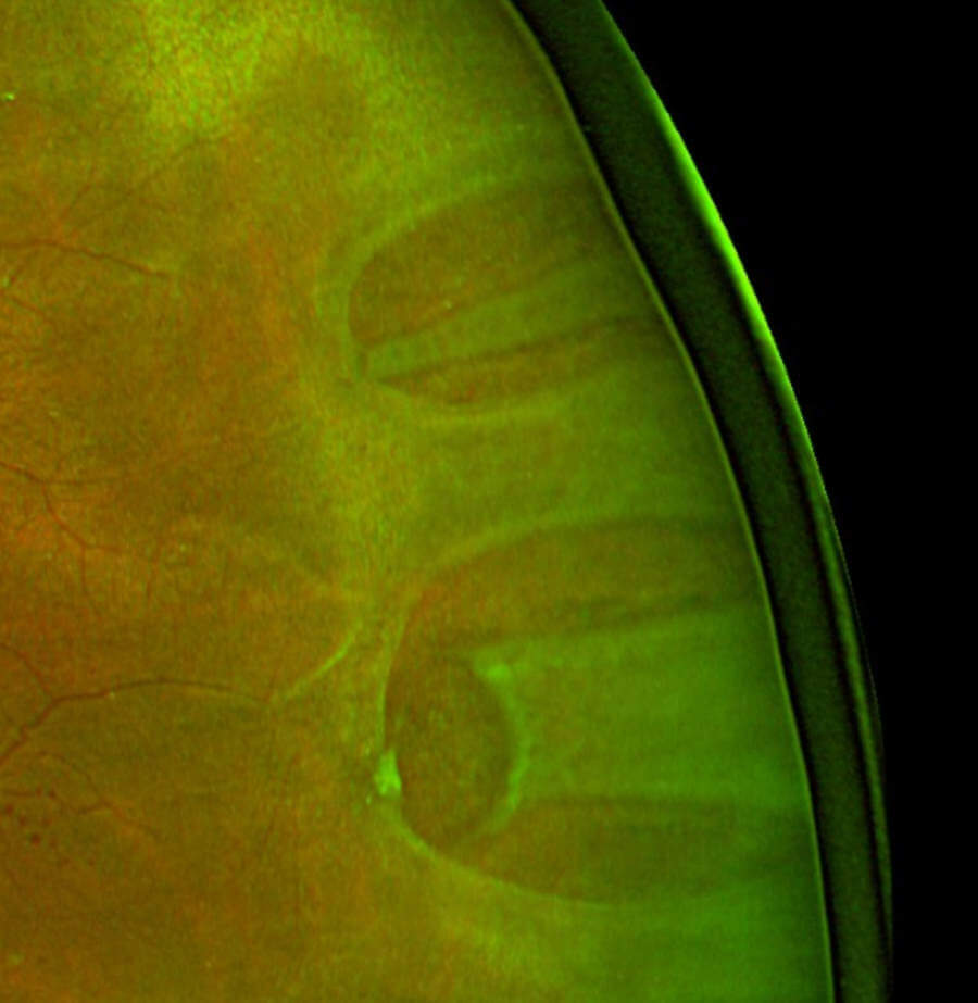
The retina is the innermost of three coats at the back of the eye. The white sclera makes the tough outer shell. The choroid, a dense bed of blood vessels, lines it from within. The retina rests against the choroid, held there mostly by the gentle suction of a single cell layer that pumps fluid out of the space behind it. A tear in the retina breaks that seal. Vitreous fluid passes through the hole and tracks underneath, floating the retina off its bed. This is the common rhegmatogenous detachment, the kind that starts with a tear. Two rarer kinds exist: one where scar tissue pulls the retina off, and one where fluid leaks under it from disease in the wall. Once any flap of retina lifts, the detachment tends to spread, fluid working its way around the curve of the globe. A small lifted edge can spread into a total detachment over hours to days. That spreading window is what the bedside scan is racing.
The folded bright line in the dark
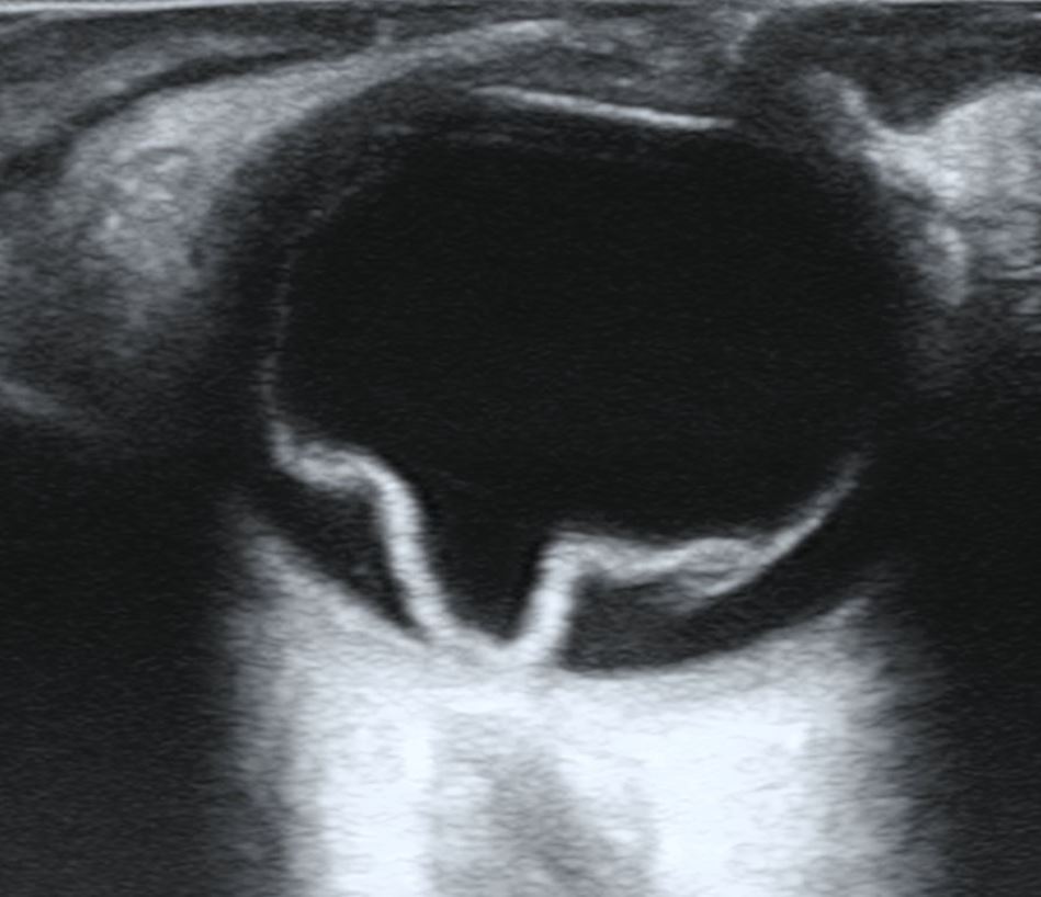
On the screen a healthy vitreous is a black, echo-free space filling most of the globe. The gel inside a normal eye is clear, with nothing in it to bounce sound back. The machine draws that silence as pure dark. A retinal detachment breaks the dark with a bright line. The detached retina, lifted off the wall, floats in the vitreous as a thin, sound-reflecting membrane. Where the beam strikes it, the echo comes back strong. The scan paints it as a pale streak laid across the black space, often with a curve to it. That contrast, a bright structure standing inside an otherwise empty dark globe, is what draws the eye to a detachment in the first second of looking.
The shape of that bright line is what names it. A full-thickness detachment cannot tear free everywhere. It stays welded to the wall at two places: the optic disc at the very back, where the nerve leaves the eye, and the ora serrata at the front rim, where the retina ends. Lifted in the middle and held at both of those ends, the membrane takes on a folded funnel shape. On a flat scan it reads as a wide V or a shallow trough, both arms running back toward the disc. That tether to the optic nerve head is the single most telling sign of a retinal detachment. A bright line that dives toward the disc and joins it is retina. The B-scan earlier on this page shows exactly that funnel, its point sitting on the nerve.
Brightness is the second half of the clue. The gain dial is how it is tested. The detached retina reflects sound strongly. Its line stays clearly visible even when the machine’s gain is turned well down. Lesser things in the vitreous fail that test. Old blood, loose strands of gel, and fine debris glow brightly at high gain. Turn the gain down low and they dim and vanish, one by one, leaving the retinal line alone on the screen. The maneuver is simple: find the bright membrane at normal gain, then lower the gain in steps and watch what survives. A line that is still there at low gain, and still anchored at the disc, has earned the name retina. A haze that washes out at low gain was never retina to begin with. A drop of the gain by a third or so is usually enough to part the two. What is left bright at low gain is the membrane worth chasing.
How far the bright line reaches tells how much of the retina has come off. A small, early detachment shows as a short flap of bright membrane lifting from one part of the back wall, the rest of the retina still lying flat against the choroid, invisible to the scan. A total detachment draws the full closed funnel, both arms running from the disc all the way out to the front rim. Between those extremes lie the partial detachments, lifting one or two quadrants. The probe sweeps slowly across the globe, top to bottom and side to side. With each pass, the scan traces the edge of the lifted retina around the inside of the eye, mapping how much has come off and how much still lies flat. That map of extent is part of what the eye surgeon wants to know before the patient even arrives.
None of this needs a single perfect frozen frame. The detachment shows itself across a whole sweep, the bright line appearing and disappearing each time the beam crosses it, from one angle and then another. A scanner who has seen several detachments learns to read the funnel almost at a glance. The shape that dives to the disc, the brightness that survives low gain, the anchor that holds the line in place: these come together into a picture that is hard to mistake for anything else in the eye. Confidence comes from the number of eyes scanned. The first detachment is studied slowly. The tenth announces itself.
Scanning the closed eye
The scan is done gently, over a closed eyelid. A thick layer of clear gel goes on the lid first, enough to float the probe so its face never presses down on the globe. The patient lies back with the eye shut and looks straight ahead under the lid. The probe rests on the bed of gel, as light as a hand can hold it. The pressure of a heavy probe on an eye is a real danger, more so on an eye that may be injured. One rule sits above the rest: if there is any chance the globe is ruptured, the probe does not touch it at all. On an open eye the gentle weight of a scan can press the contents out. When rupture is even a question, the scan waits for the eye surgeon.
A linear probe gives the picture its detail. It is the same high-frequency probe used to look at vessels and nerves. Its short wavelength resolves the thin membranes inside the eye. The scanner sets the depth so the whole globe sits on the screen, then fans the beam slowly across it, top to bottom and side to side, watching the back wall fill in section by section. The eye is scanned in more than one plane, across and up-and-down, so a membrane hidden in one view shows in another. Partway through, the patient is asked to roll the eye gently behind the closed lid, left and right, up and down. That small movement is the heart of the test. What a lifted membrane does in the instant the eye moves separates a true retinal detachment from the things that look like one.
Asking the eye to move
When the eye moves, everything loose inside it moves too, then comes to rest. The fluid shifts. The gel sways. Any membrane floating in the vitreous swings too, then settles. Watching how a bright line moves and then settles is called kinetic scanning. It is the one thing a single frozen picture can never give. The probe is kept on the lid through the whole movement, so the same membrane is followed without a break, from rest, through the swing of the eye, and back to rest again. The scanner is reading two things at once: how far the line travels when the eye moves, and how long it keeps moving after the eye has stopped. Both readings come from the same short maneuver, repeated a few times until the pattern is clear.
A fully detached retina moves in its own way. It is held fast at the optic disc and at the front rim, so it cannot swing freely the way a loose thread would. When the eye moves, the membrane sways a little, stiffly, the way a sheet pinned at two corners ripples in a draft. The moment the eye stops, the retina stops with it, settling almost at once. Through all of it, the anchor at the disc holds. The line keeps returning toward that one fixed point at the back, no matter which way the eye has turned. A membrane that stays tied to the disc and quiets the instant the eye is still is moving the way a detached retina moves.
The amount of that after-movement carries real weight in the read, even with no number on the screen. A retinal line tethered at the disc sways stiffly and settles fast, its swing barely outlasting the eye’s own stop. The scanner watches how long the line keeps moving once the eye has come to rest. A line that quiets almost as soon as the eye does is behaving the way a detached retina behaves. The same membrane scanned again and again moves the same bounded way each time. That repeatable, restrained motion, anchored every time toward the disc, is as much a part of the diagnosis as the bright line itself.
Kinetic scanning also reaches detachments a still image alone would miss. A shallow detachment, lifted only a little off the wall, lies almost flat against the curve of the globe. It takes the eye’s movement to bring out the slight ripple of the loose retina, a faint shimmer that marks where it lies. The same movement test sorts a true membrane from a smudge of artifact. Artifact is a trick of the sound beam, fixed to the machine. It does not move when the eye moves, so it falls away the moment the patient looks left and right.
Is the macula still on
One question rises above all the others the moment a detachment is found: has it reached the macula. The macula is the small central patch of retina, no wider than a few millimetres, that carries sharp, straight-ahead sight, the vision used for reading a face or a line of print. As long as the macula is still attached, the eye keeps that central vision, even with a detachment spreading at its edges. A repair done within a day or so, while the macula is still on, has the best chance of keeping that sharp sight for good. Once the detachment spreads across the macula and lifts it off the wall, the central vision is already lost. Laying the retina back down does not fully bring it back. The case stays urgent. The rush only eases from hours into the next several days. A handheld scan can often show whether the lifted retina has reached the back-central pole of the eye, where the macula sits. That single fact reorders the night. A macula-on detachment goes to the operating room ahead of almost anything else in the department. A detachment that has already taken the macula can be booked for the coming days. The scan does not just find the detachment. It helps time the repair.
Detachment, or something that mimics it
Two other findings throw a bright line into the dark vitreous. Telling them from a detachment is most of the skill in reading the eye. The first is a posterior vitreous detachment, by far the more common of the two. Here the gel that fills the eye slowly shrinks and pulls away from the surface of the retina. The retina itself stays flat on the wall. What lifts is the back face of the gel. It shows as a bright line too. Three things set it apart. Its line is thinner and fainter than a detached retina. It does not anchor at the optic disc. It drifts across in front of the nerve. When the eye moves, it swings far and keeps swinging long after the eye has stopped. A faint line that floats over the disc, never touching it, swaying on well after the eye is still, is vitreous gel.
The second mimic is blood. A vitreous hemorrhage fills the dark space with a haze of fine dots and short moving strands, brightest when the gain is high. It has no single line. Nothing about it ties to the optic disc. Turn the gain down and the whole haze dims and melts away. A detached retina lying underneath the blood stays put through the same maneuver. Its bright line holds steady through the whole sweep. Blood and detachment often turn up in the same eye. The tear that lifts the retina tears a vessel as well, so a vitreous bleed is a reason to hunt hard for a detachment hiding behind it. Finding the one is a reason to look carefully for the other. A vitreous bleed has its own list of causes, a torn vessel from a detachment among them, along with diabetes, high blood pressure, and injury. The scan does not name the cause. It finds the retina hiding behind the blood.
A third, less common line is a choroidal detachment, a smooth dome of the wall itself bulging inward, thick-walled, ending before it reaches the disc. Put the common cases together and three questions sort out most of what a bright line can be. Does it anchor at the optic disc. Does it stay bright when the gain is dropped low. Does it sway stiffly and settle fast. A line that answers yes to all three is a detached retina until proven otherwise. The scanner who runs those same three checks on every bright membrane, every time, reads the dark vitreous with a steady hand and is wrong far less often.
What the scan settles
A handheld scan is very good at answering the one question that matters first: is the retina off the wall or not. In trained hands, bedside ocular ultrasound catches the great majority of detachments and rarely calls one that is not there. Published reviews of bedside ultrasound for retinal detachment put its sensitivity high, with a low rate of false alarms, good enough to act on. The scan is run by the clinician at the bedside, an emergency physician or a trainee, with no need to wait for an ophthalmologist to arrive. The skill is learned on a run of normal eyes before the abnormal one walks in. That is enough to change what happens next. It turns a painful, sightless eye with no clear cause into a clear reason to call the eye surgeon now, in the middle of the night if need be. It tells the surgeon, before the patient has moved, that a retina is down and roughly how far. The scan buys speed. In a detachment, where every hour of a macula still attached counts, speed is sight.
What the scan does not do is take the place of the people and tools that repair the eye. It does not grade the tear, choose between a scleral buckle and a vitrectomy, or show the fine detail that a dilated exam and the operating microscope will. A false read in either direction is possible. A confident bedside finding still goes to the ophthalmologist, who confirms it and plans the surgery. The handheld does its part earlier in the chain. It takes an eye no one could see into and, in a minute at the bedside, turns a guess into a finding, getting the patient onto the right pathway hours sooner. All of it rests on one bright line in the dark, folded back toward the disc.
| Item | Figure | Note |
|---|---|---|
| POCUS sensitivity for RD | about 94% | trained users, pooled |
| POCUS specificity for RD | about 96% | few false alarms |
| Probe frequency | about 10–15 MHz | linear probe on the closed lid |
| Macula-on repair window | within about 24 hours | the urgency the scan flags |
| Macula-off repair | within about 3–7 days | the macula already lifted |
| Gain to confirm a true RD | dropped low | the RD line keeps its brightness |
| Tethered point of a full RD | the optic disc | the V points back to the nerve |
Common questions about ultrasound for retinal detachment
How does a retinal detachment look on ultrasound?
As a bright membrane inside the dark vitreous, lifted off the back wall in a fold. The detached retina reflects sound strongly. A full detachment stays anchored at the optic disc and the front rim, taking on a funnel or V shape that points back toward the nerve. The membrane holds its brightness even when the gain is turned low.
When is ultrasound needed to find a detachment?
When the back of the eye cannot be seen directly. A vitreous bleed, a dense cataract, a swollen lid, or a pupil that will not dilate all block the view in. Sound passes through them and draws the retina on the screen. Ultrasound also finds a shallow detachment that a crowded direct view can miss. It works in seconds at the bedside.
How is a detachment told apart from a vitreous detachment or a bleed?
By three checks. A detached retina anchors at the optic disc, stays bright when the gain is dropped low, and sways stiffly before settling fast. A posterior vitreous detachment floats free of the disc, reads thinner, and swings loosely well after the eye stops. A vitreous bleed is a haze of dots that fades when the gain is dropped, with no single tethered line. Running all three checks on every bright membrane is how the read is made.
What does it mean if the macula is still attached?
It means the central, sharp-vision part of the retina is still in place. The eye still has its straight-ahead sight. A macula-on detachment is the urgent kind, with surgery within about a day able to save that vision. Once the detachment crosses and lifts the macula, the central vision is already lost. The repair is then done less urgently over the following days. Telling macula-on from macula-off is the most useful thing the scan adds after finding the detachment itself.
Can ultrasound replace the eye doctor’s exam?
No. The scan finds the detachment and shows roughly how far it has spread. The grading of the tear, the choice of operation, and the fine detail of a dilated exam under the microscope are all beyond it. A bedside detachment still goes straight to an ophthalmologist, who confirms the finding and decides the repair. The value of the handheld is speed: it starts that referral hours sooner, even for an eye no one could otherwise see into.

About the Author
Julien Mercier
Senior R&D Engineer · Medical Ultrasound Transducer Development
Senior R&D Engineer with an M.S. in Applied Physics and over 15 years of experience in medical ultrasound transducer development, specializing in the design verification and performance testing of high-frequency imaging transducers. Currently leading the development and verification of the company’s next-generation high-frequency linear-array transducer, responsible for imaging performance evaluation and reliability analysis in preclinical testing. Brings extensive hands-on experience in piezoelectric element tuning, beamforming parameter optimization, and system-level performance testing.