High Frequency Aesthetic Endocavity Vein Imaging Handheld Probes
Specialized handheld probes take over where the standard linear and convex pair stops. Each one reaches a single target those two miss. The narrow fit is the point. The span runs from skin a few millimeters thick to organs reached only from inside the body.
Why the standard probes fall short here
Two probes handle the bulk of clinical ultrasound by dividing it by depth: a linear head for the shallow structures in fine detail, a convex head for the deep organs at coarser detail. The targets gathered in this group sit outside what that pair does well. Each one sits outside it for a reason of its own. Skin is the clearest case. The dermis and the fat just beneath it are only a few millimeters thick. Reading them as separate layers needs resolution finer than a general linear probe at 7 to 12 MHz can give, so to that probe the skin is one bright band. An ultra-high-frequency probe at 18 to 24 MHz resolves those layers. Near 80 microns it separates the epidermis from the dermis, a boundary a general probe never draws. The trouble with the pelvic organs is distance. The uterus and the ovaries lie deep in the pelvis. A probe on the lower belly views them across five to fifteen centimeters of bladder and bowel, where the picture softens along the path. A probe placed inside the vagina sits one to five centimeters from the same organs and runs at a higher frequency, so the view comes back sharper. The prostate is reached on the same principle, through the wall of the rectum, where a thin layer of tissue is all that separates the probe from the gland.
Veins move the problem onto a different tool. A surface vein gives ultrasound little to lock onto during a quick cannulation. The fastest way to find one uses light. Near-infrared light is absorbed by the hemoglobin in blood, so a vein under the skin swallows the light and reads as a dark line once the map is thrown back onto the surface. The smallest patients are a matter of size. A newborn’s abdomen is narrower than an adult probe’s footprint. A standard convex curve is too broad to fit between small ribs or to rest on the soft spot of the skull. A tighter curve, around a 20 millimeter radius, fits where the larger one cannot. One thread runs through all five. None of them is a place where ultrasound itself fails. Each is a target the two everyday probes were never shaped to reach, so each gets a probe, or a different kind of device, shaped to reach it.
The link behind all of this is one rule. A higher frequency carries a shorter wavelength. A shorter wavelength draws finer detail. The same high frequency fades faster in tissue, so it reaches less deep. Detail and depth pull against each other. A probe’s frequency fixes where they settle. The skin probe spends nearly everything on detail and keeps about a centimeter of reach. The endocavity and pediatric probes change the geometry, bringing the probe to the organ or shrinking it to the patient. The vein finder leaves the scale behind and works on light. Every tool here answers the same trade in its own way, decided before the scan starts. This is also why one probe cannot cover the range by retuning. The gap on the frequency scale between reading skin and reaching the pelvis is too wide for a single setting, so each target keeps its own tool.
Skin and the face: the 18 to 24 MHz probe
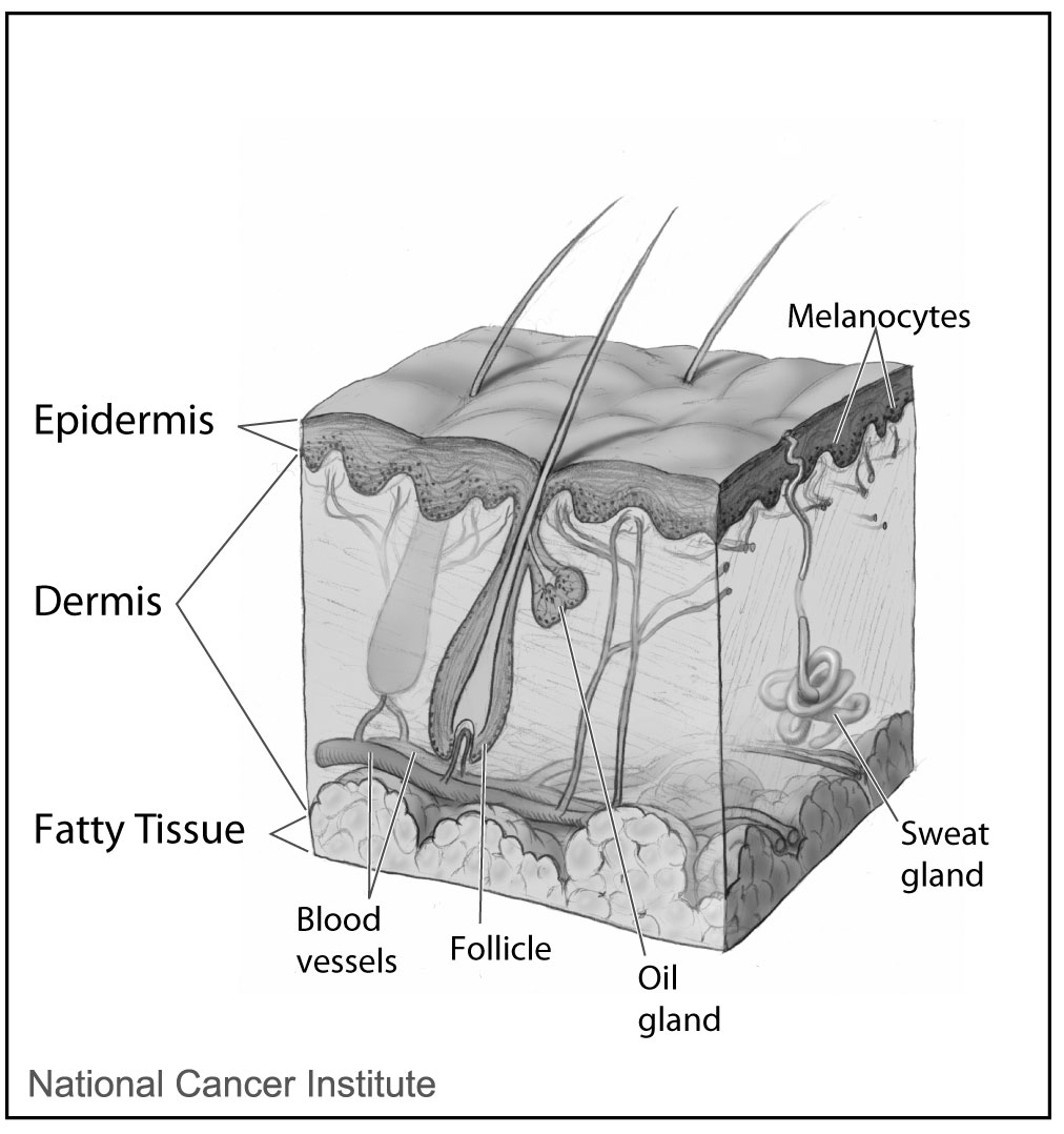
The layers an 18 to 24 MHz probe resolves: the epidermis, the dermis below it and the fatty tissue beneath. This is an anatomical diagram. It is not an ultrasound image. The labels are part of the original (National Cancer Institute).
The probe for skin runs at 18 to 24 MHz, the high end of clinical ultrasound. It reaches only about a centimeter down. Reported figures put its resolution near 80 microns, fine enough to split the epidermis, the dermis and the fat below into separate layers. The layers it separates are thin. The epidermis is a fraction of a millimeter. The dermis runs from one to a few millimeters, depending on the part of the face or body. Both change in measurable ways with age and sun exposure, which is what lets the probe follow the skin across a course of treatment. The fine detail is the whole reason the probe exists. Aesthetic medicine is where it has found its busiest use, since nearly all of that work happens inside the first centimeter the probe can see.
The use that matters more than any other is seeing the facial vessels before a needle goes near them. Filler injected by feel can enter an artery. A blocked facial artery can starve the skin it feeds. Through the connections that run behind the eye, it can even threaten sight. Color Doppler on the high-frequency probe maps where the arteries run in this patient, which is different in every face. The nose carries the highest risk, with the dorsal nasal and angular arteries packed into a small area. The lips and the area between the brows are dangerous for the same reason. A map drawn before the injection lets the injector plan a path that keeps clear of those vessels.
The probe also guides the injection while it happens. One described technique uses an 18 MHz probe in three steps: map the arteries first, inject under live ultrasound, then check that blood is still flowing afterward. A retrospective report described this approach in around 480 patients as a way to lower the chance of a vascular event in high-risk areas. Watching the needle and the filler in real time gives more control than working from surface landmarks alone. The same live view shows the filler spreading into the right plane, clear of the vessels below. The guidance works in reverse later, placing the dissolving enzyme exactly where the screen shows the deposit.
When filler is already in the face, the probe finds it. Hyaluronic acid reads as a dark pocket that looks like fluid on the screen. Old filler often sits deeper, or further from the injection point, than the surface lets on. If a vessel is blocked, ultrasound can steer the dissolving enzyme straight into the deposit and then confirm that flow comes back. The same view tells a lump of filler apart from a swollen vessel or a pocket of infection. A clinic that can see the filler can act on a problem that would otherwise be guesswork.
Past filler, the same probe localizes muscle for botulinum injections, checks the plane for a thread lift, and measures a suspicious skin lesion before referral. The full span of what an 18 to 24 MHz probe does across an aesthetic clinic runs from the first consultation to the rescue of a complication.
The pelvis, scanned from inside
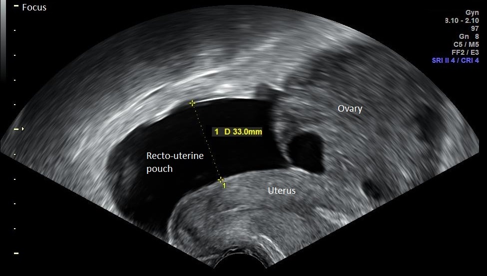
A transvaginal scan of the pelvis: the uterus, an ovary and fluid pooled in the recto-uterine pouch behind the uterus. The probe sits a few centimeters from these organs, which is why they read this sharply. The yellow calipers measure the fluid pocket at 33 mm, a sign of mild ovarian hyperstimulation. It is not a follicle. The labels and the settings at upper right come from the scanner.
The female pelvic organs sit deep. From the lower belly a probe views them across five to fifteen centimeters of bladder and bowel. A full bladder helps by pushing the bowel aside and giving the sound a clear path. The picture still softens over that distance. A probe placed in the vagina sits one to five centimeters from the uterus and the ovaries. It runs at a higher frequency than a belly probe, around 6 to 12 MHz. The short path and the high frequency together give a much sharper view of the same organs, with less artifact in the way. Proximity does for the pelvis what raw frequency does for skin.
This is the probe for the earliest part of pregnancy. A gestational sac of two to three millimeters shows around five weeks from the last period. The yolk sac follows near five and a half weeks. A fetal pole with a heartbeat is reliably seen by about six to seven weeks, often before a transabdominal scan finds anything at all. Measuring the embryo from crown to rump dates the pregnancy closely in these early weeks. The same probe reads the ovaries well enough to count and measure follicles through an IVF cycle. It guides the needle during egg retrieval. Through a stimulated cycle it counts the growing follicles and measures each one, which helps time the trigger and the collection. Outside pregnancy it reads the lining of the uterus, fibroids and ovarian cysts. It is also the test that helps place an ectopic pregnancy early, while there is still time to act. It measures the length of the cervix, a number that matters when a pregnancy is at risk of coming too early.
A probe used inside the body brings a cleaning duty with it. Endocavity probes are sealed to an IPX7 rating, so the whole probe can go through high-level disinfection between patients without water reaching the electronics. A single-use cover goes on during the scan. A wireless dual-head design carries a curved array for the belly and an endocavity array on the same handle, so one device covers both the outside and the inside scan in a single clinic. That combination saves a small practice from buying two separate machines.
When closeness is the deciding factor, the vaginal route wins. The disinfection it demands is part of routine use for any clinic doing transvaginal endocavity scanning with a handheld dual-head probe. The same advantage of proximity returns, on the opposite side of the body, with the prostate.
The prostate and the biopsy needle
The prostate sits just in front of the rectal wall. A probe in the rectum looks at the gland through a few millimeters of tissue, close enough to read its zones and to measure its size. This is how a prostate biopsy is guided. When a blood test shows a raised PSA, or the gland feels abnormal on examination, the needle is steered under ultrasound to sample the prostate in a planned pattern across both sides. Grey-scale ultrasound on its own catches only some cancers, so its main role is to guide the needle and to measure the gland.
The standard sampling takes around twelve cores from mapped positions, so no region goes unsampled. Size matters here too. The gland is measured on three axes. Its volume is estimated with the ellipsoid formula, length times width times height times about 0.52, the method the American and European urology bodies both endorse. A normal prostate runs about twenty to thirty cubic centimeters. Size also steers the treatment of a benign enlargement, since the choice of therapy changes once a gland passes a certain size. The volume then feeds the PSA density, the PSA divided by the size. A density above roughly 0.15 raises the suspicion that an enlarged gland alone does not explain the PSA, which can push the decision toward biopsy. Newer work lines up an MRI taken earlier with the live ultrasound, so the needle can target a spot the MRI flagged, on top of the systematic cores.
The full sampling routine, and the guideline thinking behind it, are the working substance of transrectal prostate biopsy under ultrasound guidance. Both endocavity probes accept a little discomfort in exchange for proximity. One tool in this group leaves ultrasound behind altogether.
Finding veins with infrared light
This tool is not an ultrasound probe. It is an infrared vein finder. It works on light. Near-infrared light, in the 700 to 1000 nanometer range, shines onto the skin and passes a short way into the tissue, up to about a centimeter. Hemoglobin in the blood absorbs that light more strongly than the tissue around it. A vein swallows the light and sends less of it back. The device reads that difference and projects a live map of the veins onto the skin in visible light. A unit may throw that map straight onto the arm, or show it on a small screen for the same purpose. The person drawing blood sees the vein under the pen before the stick, with no screen to look away to.
The point is speed and a better chance of a first-stick success on hard veins, in children, in patients worn down by chemotherapy, in heavier arms or in an emergency. It changes nothing about the vein itself. It only makes a hidden one visible, so a deep or rolling vein is still a difficult stick. It shows surface veins alone and does not reach the deeper vessels an ultrasound-guided line would use. The two tools answer different questions, one for a quick peripheral stick and one for deep access under live imaging. Where the finder fits in difficult access is the subject of the infrared vein finder and how it sees veins through the skin.
Run from a phone, used at the bedside
These exams once meant a cart and a room. A high-frequency skin scan, an endocavity study or a vein search each belonged to a radiology suite or a dedicated machine. A handheld probe moves the same exam to where the patient already is. The aesthetic scan happens in the injector’s own room. An endocavity study fits into the clinic visit. At the bedside, a small stand holds the vein finder. Each connects to a phone or a tablet the clinic already owns, with the screen and the controls living in an app. The probe holds the electronics and sends the picture to the phone.
The cart stayed in radiology. The probe came to the patient.
Wireless heads make the swap simple. A clinic that does several of these jobs can keep more than one probe and run them all from the same app, choosing the head the case calls for. There is no second console to buy or maintain. The vein finder stands apart, since it is a separate device on its own light, not part of the ultrasound system. The shared idea across the group is portability. A specialized exam that used to need a referral can be done in the room, in minutes, on equipment that fits in a pocket or on a small stand. A clinic that already runs handheld scans for the abdomen or the lungs adds one of these heads to a base it owns, with no new machine and no new screen to learn.
None of this lowers the skill the exam needs. A clear picture in the room still has to be read by someone who knows the anatomy. A vein map or a filler plane means nothing without the hands to act on it. Portability changes only where the work is done. The work itself stays the same.
Which one a clinic needs first
Which of these a clinic reaches for first follows from the work it does day to day. An aesthetic practice that injects filler has the strongest case for the high-frequency probe, where the vessel map guards against a serious complication. A fertility service or a women’s clinic relies on the endocavity probe, in the pelvis and through the early weeks of pregnancy. Urology turns to the transrectal probe around the biopsy and the gland measurement. On a busy ward or an oncology unit, the vein finder answers a daily problem of hard sticks. A neonatal service reaches for a micro-convex head for the smallest patients, scanning the brain, the lungs and the hips.
Few clinics buy the whole set at once. A practice usually starts with the one probe its commonest exam calls for. It adds others as the caseload grows. The handheld form keeps that within reach, since each head is bought on its own, not bundled into a fixed console. The base a clinic already carries, a phone or a tablet, does the rest. Trading up later means buying the next head and keeping the same system.
What this group does not replace
None of these is a general-purpose probe. Each does one job well and little else. The standard linear and convex probes still handle the bulk of clinical scanning, from the neck to the abdomen. These are the tools a clinic adds at the edges, for the places the common pair cannot reach.
Common questions about these probes
What makes these different from a standard ultrasound probe?
Each one answers a target the standard linear and convex probes handle poorly. The change is a much higher frequency for skin, a probe that works inside a body cavity, a tighter curve for infants, or in one case a switch from sound to infrared light. Each one fits a single job closely and gives up broad use. The fit is chosen before anything else about the scan.
Is the infrared vein finder an ultrasound device?
No. It works on near-infrared light. Sound plays no part in it. Hemoglobin in the veins absorbs the light, so the veins show up as dark lines in a map projected onto the skin. It finds surface veins for needle access and produces no ultrasound image. A clinic uses it alongside its probes. It is not one of them.
Can one handheld system run all of these probes?
Often, yes, through interchangeable heads or a system that accepts more than one probe on the same app. A clinic picks the heads its work calls for, an aesthetic probe in one room and an endocavity probe in another. The vein finder is a separate device, since it is not ultrasound at all. Buying probes one at a time is part of what makes the handheld approach affordable.
Why is a transvaginal scan sometimes used for the pelvis?
The probe sits one to five centimeters from the uterus and ovaries, far closer than a probe on the belly. It also runs at a higher frequency. The short path and the high frequency give a sharper image. That edge matters above all in early pregnancy, where a heartbeat shows around six to seven weeks, and for a close look at the ovaries during fertility treatment.
How are endocavity probes kept clean between patients?
They are sealed to an IPX7 rating, so the whole probe can be disinfected without water reaching the electronics. A single-use cover is used during the scan. A high-level disinfection follows it. The sealing is what makes repeated internal use safe. This step is as much a part of the exam as the scan itself.
Do these replace the standard linear and convex probes?
No. They sit alongside them. The standard pair still handles the bulk of scanning, from the abdomen to the vessels of the neck. These specialized tools cover the edges the common probes cannot reach. A full clinic keeps both. It reaches for the specialized head only when the target calls for it.
How deep can these probes see?
Not far, by design. The skin probe reaches about a centimeter. The endocavity probes work within a few centimeters of the cavity wall. The infrared vein finder reads only the surface, to about a centimeter. Each one trades depth for the resolution or the access its target needs, so a shallow reach is the expected price here. It is not a fault.
About the Author
Julien Mercier
Senior R&D Engineer · Medical Ultrasound Transducer Development
Senior R&D Engineer with an M.S. in Applied Physics and over 15 years of experience in medical ultrasound transducer development, specializing in the design verification and performance testing of high-frequency imaging transducers. Currently leading the development and verification of the company’s next-generation high-frequency linear-array transducer, responsible for imaging performance evaluation and reliability analysis in preclinical testing. Brings extensive hands-on experience in piezoelectric element tuning, beamforming parameter optimization, and system-level performance testing.