A convex probe reaches deep into the belly where the other heads give out, down to a liver, a kidney, or a pregnancy that sits beyond their range. It runs at a low frequency, trading fine detail for the depth to reach the deepest organs in the largest patients, and draws the wide fan-shaped picture that abdominal scanning is read from. On a wireless handheld, that whole reach rides in one pocket-sized head.
What the convex probe is
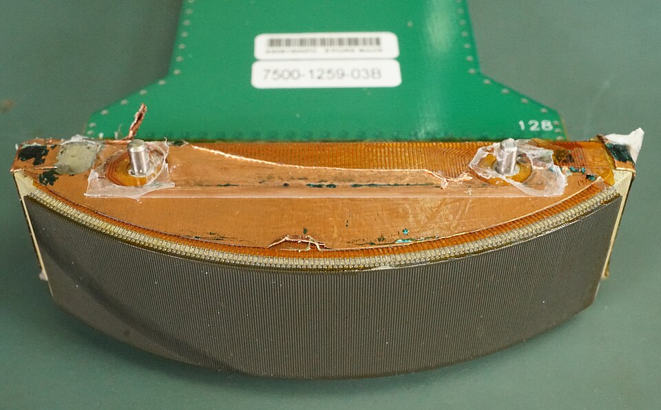
The curved face of a convex probe, opened up. The arc of crystals along the front is what fans the beam into a broad wedge reaching deep; the number stamped on this one, 128, is the count of elements packed along the curve. Image: Binarysequence, CC BY-SA 4.0.
The convex probe has a curved scanning face, a gentle arc of crystals that fans the sound out into a broad wedge reaching deep. The low frequencies it runs at, around two to five megahertz, travel far into the body, which is what lets it reach an organ a hand’s breadth below the skin. The crystals along that curve each send and listen in turn, building the picture line by line across the whole arc. That curved array, packed along the face, is what gives the probe both its reach and its wide field.
That depth is the whole point. The liver, the kidneys, a fetus late in pregnancy, the aorta against the spine all lie too deep for a shallow probe to read. These deep organs are exactly what the convex probe reaches, its fan-shaped picture spreading wide enough to hold a large one in a single view. Its reach runs to fifteen or twenty centimetres, the depth a large patient or a late pregnancy demands of a machine.
One curved transducer, then, opens the entire deep body to ultrasound. The pages below divide that body into four areas, each its own field of work. That a single head serves so wide a range is what makes the convex probe the workhorse of abdominal imaging. From the routine to the urgent, from the first weeks of a pregnancy to a failing kidney, the convex probe is the head a clinician reaches for when the question lies deep. No other single probe covers so much of medicine from one curved face.
Reading the fan-shaped picture
The image a convex probe makes has a shape of its own. It spreads from a narrow top, where the probe meets the skin, into a wide fan, broad in the deep field. A reader learns to think in that wedge, with the target organ sitting somewhere along its spread. The wedge is broad enough at depth to lay a whole kidney or a fetal head across the screen at once, the gift of the curved face. The fan grows wider the deeper it goes, so the deepest part of the picture is also its broadest. A whole organ, laid across that broad deep field, can be taken in at a glance and measured end to end.
Depth is set to the organ in question, shallow for a near one and deeper for a far one, the target set in the middle of the picture, drawn large and plain. A scan that runs too shallow loses the deep organ off the bottom of the screen, so the depth is opened until the whole of the target is in view. The depth is marked in centimetres down the side of the screen, a ruler that doubles as a measuring scale for an organ or a pocket of fluid. A scan that opens the depth too far shrinks the target to a speck in a sea of black, so the depth is trimmed back until the organ fills its share of the screen.
The gain, the brightness of the picture, is balanced next, lifted in the deep field so a far organ does not fade into the gloom. A well-set convex picture is even down its whole length, the surface tissue and the deep organ both legible in the same frame. Time-gain compensation, a row of sliders, lets the operator brighten one band of depth at a time, a fine tuning that pulls a faint deep organ out of the dark.
Orientation is the last habit. A marker on the probe matches a dot on the screen, so the reader always knows which side of the picture is the patient’s right, which is the head, which the feet. On a deep field with few features, that marker is the compass that keeps a scan from getting lost. Convention sets the marker to the patient’s right in a transverse view and to the head in a long one, so any reader picks up the image the same way. A scan read upside down or back to front is a scan that misleads, so the marker is checked before the reading begins. On a deep field with few landmarks to anchor it, that small dot tells top from bottom and one side from the other.
Getting a clean deep picture
Deep imaging asks more of a machine than shallow imaging does. The sound has to travel far and come back, weakening on the way, so a deep organ returns a fainter echo than a near one. Bowel gas scatters the beam, body fat swallows it. A poor patient window leaves a deep organ in shadow. A capable convex probe, with the power to push sound deep and the processing to clean up what returns, holds a clear picture all the way down. The deepest organs in the largest patients are where a weaker machine gives out, the picture dissolving into grain below ten or twelve centimetres, so depth is the truest test of an abdominal scanner. The focus, a marker set down the side of the screen, drops to the depth of the target, so the beam runs narrowest exactly where the organ sits.
The operator helps the machine. Steady pressure pushes gas aside. A held breath stills a moving organ. A change of window, around a rib or through a full bladder, opens a path the first view missed. Harmonic imaging, a mode that sharpens the deep picture, is turned on for the larger patient. Between the probe and the hand that holds it, a clean deep picture is won on a body that gives it up grudgingly. None of these tricks is exotic. They are the daily craft of abdominal scanning, learned at the bedside and carried from one organ to the next. A larger patient asks more of them, a longer search for the window, a firmer hand, a turn to harmonic imaging for the deepest views.
The general abdomen
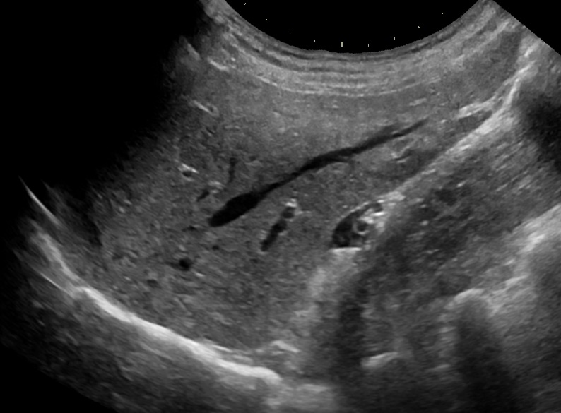
A normal liver on a convex-probe scan. The fan-shaped sector, broad at depth, holds the whole organ in one view: the even grain of healthy parenchyma, the dark branching vessels running through it, the bright line of the diaphragm below. This is the deep, everyday work at the heart of the convex probe’s range. Image: Mikael Haggstrom, CC0.
The broadest area is the general abdominal scan, the systematic look at the organs of the belly. A single sweep takes in the liver and the gallbladder, the pancreas and the spleen, the kidneys behind, and the great vessels down the midline. This is the bread-and-butter of abdominal ultrasound, the work laid out in detail under general abdominal scanning with the convex probe. The order of the sweep is its own discipline, each organ found in turn so none is skipped, the whole belly covered in one unbroken study.
The questions here are the common ones of medicine. Is the liver scarred or fatty. Is there a stone in the gallbladder. Is the aorta widening toward an aneurysm. A convex probe, run in order across the upper belly, settles a great many of them in a single bedside study. A swollen gallbladder, a coarse liver, a kidney shrunk small: each shows itself plainly to a probe that knows where to look. The reading rests on the operator as much as the machine, an organ found, held still, and measured in the right plane giving an answer a clinician can trust.
This area also holds the standards that say what a complete study must cover, the grading of a cirrhotic liver, the screening of the aorta for an aneurysm. Each is the convex probe’s daily work, on organs that sit too deep for any other kind of head. A liver measured and graded, an aorta sized against its threshold, a spleen weighed for enlargement: the convex probe carries the numbers a report is built on. A study without those numbers is half a study, which is why the convex probe’s measurements sit at the heart of an abdominal report. The figures travel with the patient, read the same by the next clinician who opens the record.
For a clinic, this general abdominal scan is the first reason to own a convex probe. It answers the everyday questions of the liver, the gallbladder, and the kidneys, the questions that fill a working day. The answer comes at the bedside in minutes. For a primary-care clinic or a rural post, that single capability, the deep abdominal look, repays the cost of the probe many times over in a year. For the common abdominal questions, the answer now comes at the clinic itself, with no journey to a distant department. That alone widens what a clinic can do, with the deep abdominal look kept in the room.
Pregnancy and the female pelvis
The next area is obstetric and gynecologic work. The convex probe reads a pregnancy through the mother’s abdomen, dating it, measuring a growing fetus, finding the placenta and the fluid around the baby. Across the pelvis it surveys the uterus and the ovaries, in pregnant and non-pregnant women alike. The whole of it, from a first-trimester dating scan to a check on an ovarian cyst, is laid out under obstetric and gynecologic handheld ultrasound. The biometry that dates and sizes a fetus, the placenta read for its position, the fluid gauged around the baby all ride on the convex probe’s reach through the abdominal wall. A growing fetus, too large for any other approach, is read whole through the mother’s belly, where the convex probe’s reach goes deep.
This is a field of its own, paired with a transvaginal probe for the early weeks and the close gynecologic detail. A handheld carrying both heads brings the work to an antenatal clinic, a gynecology room, a midwife on a home visit. The convex probe does the deep, through-the-abdomen part of it, the part that follows a pregnancy from the middle weeks to term. For a midwife or a clinic far from a hospital, that reach brings a pregnancy under the eye of the scan, week by week through to the birth. The transvaginal head, paired with the convex one, carries the early weeks, so a single handheld follows a pregnancy from its first confirmation to term.
The urinary tract
The third area is the urinary tract, the kidneys and the bladder and the path between them. A convex probe finds a swollen kidney, the sign of urine backed up behind a blockage downstream. It measures the urine left in a bladder that will not empty. It reads a stone, a mass, the marks disease leaves on a kidney. The bladder fills the lower belly as a dark, easy target, and the kidneys sit in the flanks, found through the liver on the right and the spleen on the left. A swollen collecting system shows as a dark, branching space in the bright centre of the kidney, one of the plainest findings ultrasound offers. A full bladder, a swollen kidney, a stone with its bright echo: the urinary tract gives up its common troubles readily to a convex probe.
Much of this is urgent, bedside work: a patient who has stopped passing urine, a flank that aches, a kidney that may be obstructed and infected. The full set of it, from a bladder-volume check to the grading of a swollen kidney, sits under urinary tract handheld ultrasound. A scan that finds a swollen, obstructed kidney in a patient who has stopped passing urine sends the team straight to relieving the blockage, an answer that can save the kidney. The same scan reads the bladder below, full or empty. That tells whether the blockage sits at the kidney or at the outlet.
A pocket convex probe suits this work well. The kidney and the bladder are large, steady targets, and the answers they give, a blocked kidney to relieve or a full bladder to drain, change what happens next at once. A catheter passed, a tube placed, a stone confirmed: the convex probe turns a guess about the urinary tract into a plan for it. For a ward or an emergency room, that single answer, obstructed or clear, full or empty, steers the next step in a patient whose kidneys are failing.
The abdomen in an emergency
The fourth area is the emergency belly. When a patient arrives hurt or unstable, a convex probe at the bedside looks for the few things that kill fast: blood loose in the abdomen after an injury, a ballooned aorta close to bursting, fluid gathered where it should be dry. The protocols and the findings, from the trauma scan for free fluid to the read of a leaking aneurysm, are gathered under emergency abdominal handheld ultrasound. Each of these findings is a few seconds’ look with the power to redirect a whole resuscitation toward the operating room. A bedside scan in a crashing patient points the team to the cause of the collapse in seconds.
This is point-of-care work, run in seconds by the clinician at the patient’s side. Here the value of a wireless handheld is plainest. A probe carried into the resuscitation room answers a life-or-death question in the minute it takes to ask, on a patient too unstable to be moved to a department. The FAST scan for free fluid, run in a handful of windows across the belly, is among the first skills taught to a clinician learning point-of-care ultrasound. The aorta is checked in the same sitting, its width measured against the line that calls for a surgeon, in case a ruptured aneurysm lies behind the collapse. These few quick looks, taken together, are the core of the trauma and resuscitation scan.
Why wireless and handheld
What ties the four areas together is the device. A convex probe today is a wireless head the size of a fist, run from a phone or a tablet, carried in a coat pocket to wherever the patient is. The processing now sits in the probe itself and the phone it talks to, the whole machine shrunk to what a hand can hold. A wireless link carries the picture from the probe to the screen, with no cable to tether the operator to a console. The same phone that drives the scan stores the images, attaches them to the patient, and sends the study onward. A bedside look becomes a record the next clinician can open.
The gain is reach. The deep-body work of the convex probe travels now to the ward, the clinic, the rural outpost, the roadside. A single pocket device covers the liver, the pregnancy, the kidney, and the trauma belly, on a patient who never has to be moved to meet it. A scan run where the patient lies spares the sick and the injured the trip to a department, and brings the answer to the room where the care is given. A scan at the bedside of an unstable patient, in the minutes that decide an outcome, is the clearest case for a probe that travels light.
The smaller the device, the lower its price. A wireless convex probe lands within reach of a small clinic or a single practitioner. Deep abdominal imaging, for them, has become affordable for the first time. A probe that costs a fraction of a cart puts deep imaging in the hands of a single doctor, a small clinic, a place a hospital’s reach never touched. The deep abdominal scan becomes a routine bedside tool wherever a clinician carries one. The saving on the device is matched by the saving in time, the scan and its answer arriving in the same visit.
The skill is the same across the device. The convex probe reads a fan-shaped picture the same way in every area, so an operator who learns it for the liver carries that reading into the pregnancy, the kidney, the trauma belly. One probe learned is four areas opened. A clinician who can find the aorta can find the kidney beside it and the bladder below, the same fan-shaped picture read with the same habits in each. The learning curve flattens quickly into a single set of skills that serve the whole abdomen, part of why the convex probe is the first one many clinicians master.
The cleaning and the care follow the form. A sealed wireless head wipes down between patients and charges from a pocket battery, ready for a full clinic round. The convex probe asks little of the room it works in, which is the point of carrying it out of the department in the first place. A device that needs no fixed console and no dedicated room goes where the medicine is needed, which is the whole promise of the wireless form. The same seal that allows a wipe-down lets the probe be rinsed under a tap, the ruggedness a head needs when it travels all day.
One probe, the whole deep body
Step back and the convex probe is a single answer to a wide question: how to see the organs that lie too deep to feel. One curved head, run at a low frequency, reaches them all, the liver and the kidney, the fetus and the bladder, the aorta and the free blood of an injury. The four areas below are the same probe turned to four kinds of question, the routine and the obstetric, the urinary and the urgent, each with its own pages, its own findings, its own skills, all resting on the one transducer and the depth it reaches. That this probe now runs from a wireless device in a pocket is what has carried the deep-body scan out of the imaging room and to the patient. The map starts here, and the four areas follow on the pages below. The four areas share one tool throughout: the curved head, the deep reach, the fan-shaped picture, now carried in a pocket to the patient’s side.
Common questions about the convex abdominal probe
What is a convex ultrasound probe used for?
For deep imaging of the abdomen and the pelvis. Its curved face and low frequency send sound far into the body, reaching the liver, the kidneys, a fetus, the bladder, and the aorta. It is the probe behind general abdominal scans, obstetric and gynecologic studies, urinary scans, and the emergency belly.
What does a convex probe scan in the abdomen?
The solid organs and the great vessels. A single systematic sweep covers the liver and gallbladder, the pancreas, the spleen, the kidneys, the bladder, and the aorta down the midline. The same probe reads a pregnancy through the abdomen and looks for free fluid after an injury.
Can a handheld wireless probe do a full abdominal ultrasound?
For everyday work, yes. A wireless convex probe reads the liver, the kidneys, the aorta, a pregnancy, and a bladder at the bedside, to the depth a cart-based convex probe reaches. The rare, intricate study stays with the high-end console of a referral department.
What frequency does a convex abdominal probe use?
A low band, around two to five megahertz. The lower the frequency, the deeper the sound travels, which is what lets a convex probe reach an organ many centimetres below the skin. That depth comes with a softer picture, lower in fine detail.
Is a convex probe the same as a curvilinear probe?
Yes. Convex and curvilinear are two names for the same curved-array transducer. Its arc of crystals fans the beam into a broad wedge that reaches deep, the shape that holds a large organ in one view.
About the Author
Julien Mercier
Senior R&D Engineer · Medical Ultrasound Transducer Development
Senior R&D Engineer with an M.S. in Applied Physics and over 15 years of experience in medical ultrasound transducer development, specializing in the design verification and performance testing of high-frequency imaging transducers. Currently leading the development and verification of the company’s next-generation high-frequency linear-array transducer, responsible for imaging performance evaluation and reliability analysis in preclinical testing. Brings extensive hands-on experience in piezoelectric element tuning, beamforming parameter optimization, and system-level performance testing.