Handheld urology ultrasound brings one convex probe to the urinary tract at the bedside. The probe sits low on the belly and slides to the flank, with the bladder in view below and the kidneys higher up. A clinician points it at the plumbing to settle a few plain questions. Is the bladder emptying. Is a kidney backing up. Is something blocking the flow, and at what level. The tract itself is a run of pipes and reservoirs from the kidneys down to the outlet. On the screen, the urine shows dark against the grey of the tissue around it. A handheld carries that picture to the person on the couch and answers the everyday urinary questions inside a few minutes. The convex probe is the one tool the job needs, the same beam that reaches deep across the rest of the abdomen. A clinician already handy with belly scanning brings that skill straight over. The tract sits close enough to the surface that a clear answer comes on the first day with the probe in hand.
The bladder gives the quickest answers. Filled with urine, it turns into a dark round pocket that the probe finds at a glance. A clinician sizes it, judges how full it is, and checks what stays behind after a person tries to empty. That last figure, the residual, drives a long run of urinary decisions. The same pocket shows its own wall, its outlet, and any stone or growth lodged inside. No other stop on the tract gives back as much from a single look. The probe needs only the lower belly and a moment, the bladder large, shallow, and all but lit up by the urine inside it. A clinician new to the tract starts here, the one view that forgives an unsteady hand.
The kidneys lie deeper, one tucked high under each lower rib at the back. Bringing one into view takes a moment’s extra work, repaid by what it shows. A kidney swollen with urine that cannot drain flags a block somewhere below it, often the first solid clue that the tract has shut. One sitting covers the bladder and both kidneys, the two together sketching the tract from top to bottom. The handheld follows the urine from the kidneys down to the outlet. The level where it stops moving freely marks the block.
Key numbers in urinary bedside ultrasound
Reading
Number
What it means
Post-void residual
under 50 mL
a bladder emptying well
Post-void residual
over 200 mL
significant retention
Post-void residual
over 500 mL
drainage, as a rule
Bladder volume
W × D × H × 0.52
the figure in millilitres
Hydronephrosis on POCUS
about 76% vs CT
flags the blocked kidney
The bladder: does it empty
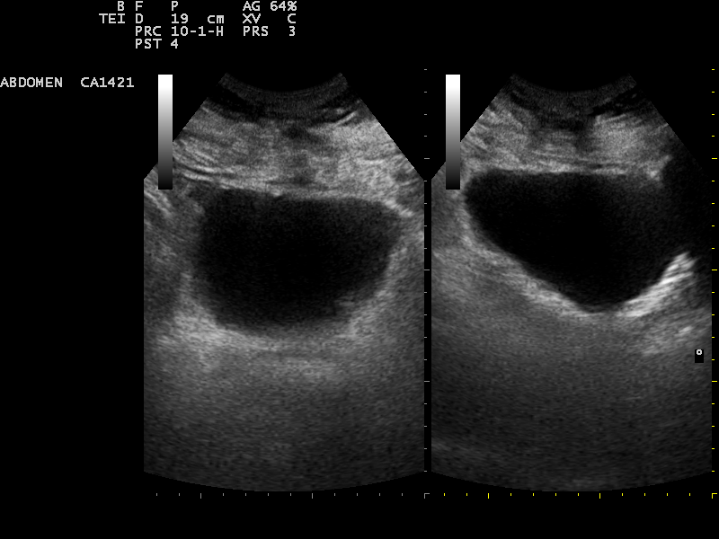
A full bladder on ultrasound, seen in two views. The dark area is urine. A scanner measures across it to size the bladder and read how much is left after a void.
The bladder exam opens on a single question. Does it empty. A bladder that fails to empty keeps urine back after every trip to the toilet. That leftover pool carries a name, the post-void residual. A handheld puts a number on it within seconds. With the person emptied and the probe on the lower belly, the dark of the urine left behind fills the screen. A high residual hides behind a long list of urinary complaints, a blocked outlet, a tired bladder muscle, frequent trips, a leak, or a sense of never finishing. The number puts that vague feeling against the urine still in the tank. It stands at the front of the urinary workup, the first hard fact that sorts a struggling bladder from a sound one.
That residual settles a surprising amount on its own. A low figure puts the worry to rest and closes the workup. A high one launches a hunt for the cause and marks the bladder as something to watch or to drain. The full method for taking and acting on the number sits in how a bladder scanner measures the post-void residual. The setting and the patient’s age set the threshold. The bar sits higher past a certain age. A lone high reading earns a repeat before any catheter, since a rushed void or a long wait can inflate it. The figure that holds high across two honest tries is the one that matters. A second low figure after a proper void clears the worry for good. A figure that stays up sends the search on toward the cause below.
Underneath the residual lies a volume. The machine builds that volume out of the picture. It takes the bladder for a rounded shape, spans it in three directions, and multiplies the three by a fixed factor to arrive at the millilitres. The old way put every caliper in a clinician’s hand. A trained tool now handles that itself, tracing the bladder edge and running the sum in a blink, the step laid out in full under automated bladder volume calculation. What drops out is an estimate in the dress of a measurement. A scarred, slack, or distorted bladder pulls it off the truth, since the rounded model no longer fits. A dedicated bladder scanner runs the same sum behind one button and shows a bare figure. On a handheld the bladder stays on the screen, so the outline behind the number stays open to a check. The volume then feeds the calls that follow, from draining a tense bladder to charting one that empties a shade worse each visit. A clinician weighs the picture behind the figure first, since a tidy number off a bad outline still misleads. The factor itself stays fixed, close to half of the box the bladder would fill if it were square. Two of the three spans come off one cross-view of the bladder; the third comes off a length-view a quarter-turn away. The factor suits a bladder rounded out at a full stretch, the shape the geometry assumes for it. The same bladder measured twice lands within a small margin, steady enough to track a slow change across weeks of visits, and to flag the day a residual starts to climb. The whole sum runs in the instant after the last caliper lands, no slower on a phone-linked handheld than on a cart in a scanning room. A clinician takes the figure off the screen the moment the probe steadies, with the bladder still in view to vouch for it. The number carries its meaning into the note, a millilitre figure a colleague can set against the next scan and the one after.
The volume answers the urgent form of the question too. A bladder that cannot empty at all balloons into a tense dome. The screen shows a large dark sac, the patient straining to pass water. That picture calls for a drain on the spot. The surrounding clues sort a slow problem from a sudden one, the wall, the kidneys above, the story the patient tells. A tense bladder in someone who abruptly cannot void reads as acute and wants quick relief. A roomy bladder in someone leaking for months, with thick walls and quiet kidneys, points to a chronic course and a steadier plan. The split matters from the first minute. An acute block wants a catheter now to spare the kidneys. A chronic one wants a plan that holds the residual down over the long run, since the harm there builds slowly and quietly.
A good bladder study wants a full bladder. A clinician asks the patient to arrive with a comfortable load, since a near-empty bladder folds in on itself and offers the machine little to measure. Bowel gas can curtain the bladder from above. A press of the probe pushes it aside. A heavy belly sinks the bladder deeper. Dropping the frequency reaches it. None of this costs much time. The bladder stays the kindest stop on the tract, and a few minutes there hand back the residual, the volume, the wall, and a first look at the outlet. A clinician who builds the habit takes all four in one smooth sweep. A little patience here buys the fullest picture on the whole tract.
The bladder that will not empty
Acute retention is the urinary emergency the bladder shows plainest. The patient cannot pass water, the bladder swells past its easy limit, the lower belly aches. On screen sits a large dark bladder, often stretched well beyond half a litre, taut against the belly wall. The number turns a hunch into a measure and says how badly the bladder needs a catheter. A bladder pushed toward a litre in a person doubled over makes the case for relief in one frame. The same scan often catches why nothing comes out, a swollen prostate at the outlet, a clot, a stone wedged in the exit. Drainage and the diagnosis come in the one visit. A handheld at the bedside spares a person in real pain the wait for a formal scan. The image makes the case for relief plain to everyone in the room. The catheter goes in on the strength of a picture, with the guesswork gone.
Chronic retention keeps a lower profile. A bladder half-empties for months, banks a steady residual, and stretches by slow degrees with little pain. A routine scan turns it up, a residual that stays high after a genuine effort to void. The real danger lies upstream, since the standing urine pushes pressure back toward the kidneys over months. A high chronic residual sends a clinician straight to the kidneys. Years of carrying that load often print themselves on the bladder, a thickened wall bulked into ridges against the block. The kidneys above can sit quietly dilated, swollen by a pressure that crept up unseen. A look at the comfortable bladder alone misses the harm gathering higher. A bladder that feels fine to the patient can still sit over kidneys quietly going under, a harm only the scan up the flank will catch.
The scan reaches past the size of a stalled bladder. The wall shows the thickening of a bladder that has strained against a block for years. The outlet shows a large prostate bulging up into the base. A stone or a clot can come into view blocking the way out. Tying the residual, the wall, and the cause into one picture runs through the full assessment of urinary retention. The same dark bladder turns up whether a drug holds the urine in, an opioid or an anaesthetic freed once it wears off, or a nerve injury has left the bladder no urge at all and a high residual for life. The reason for it waits in the history alongside the scan.
One column of water
The urinary tract is one linked column of water. A block at any point sets the urine pooling above it, the bladder first, then the ureters, then the kidneys. Stopping at the bladder tells half the story. A handheld trails the column up to the kidneys in the same sitting. The level where the backing-up stops pins where the block lies. A block at the outlet swells the bladder, and only later does the pressure climb to the kidneys. A block in a ureter swells the kidney on that side. The height the swelling reaches points back to where the pipe has closed. The pattern alone narrows the search before any other test runs.
The kidney: a backed-up system
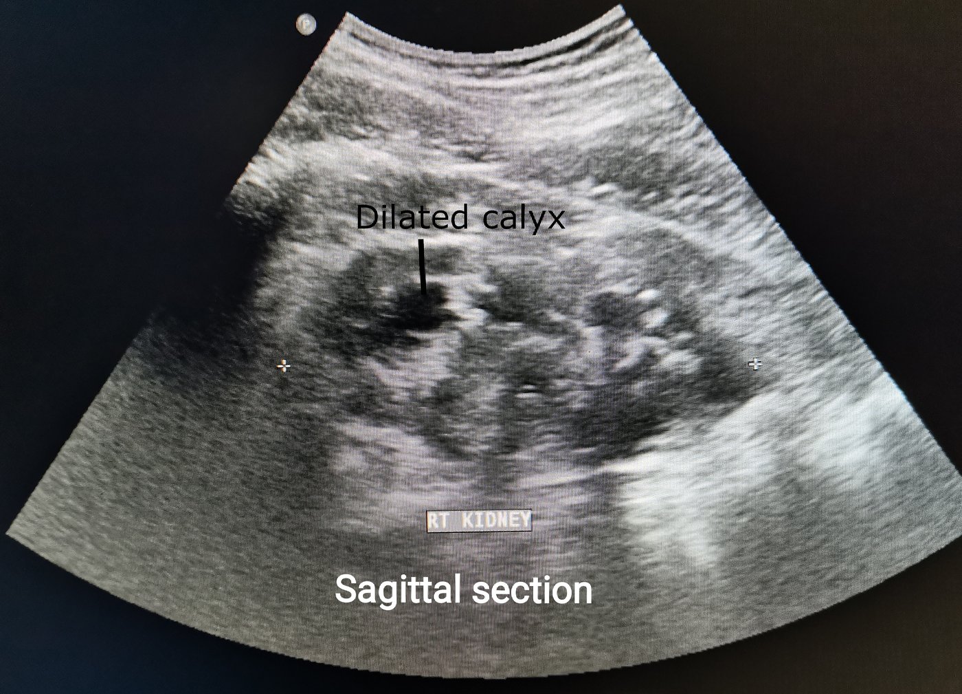
A right kidney in sagittal section. The dark branching spaces are dilated calyces, the look of hydronephrosis, where urine has backed up behind a block. The on-image label marks one dilated calyx.
A kidney comes up as a bean of grey tissue wrapped around a bright core. That bright core is the collecting system, packed tight as long as the urine runs free. A block below pries the core open into dark spaces, the calyces and the pelvis swelling with urine that has nowhere to go. The swelling has a name, hydronephrosis. On screen it spreads as dark fingers branching through the bright centre. The width of that dark spread tracks how far the swelling has gone. A healthy collecting system holds a single thin line of brightness. Urine trapped behind a block opens it into black branches no one can miss. A renal cyst can fool the eye here, a round dark pocket that sits apart from the collecting system. Hydronephrosis follows the branching shape of the calyces, dark spaces that join toward the centre. Telling one from the other means following where the dark leads.
Hydronephrosis grades by how far the swelling has run. A faint split in the central echo counts as mild. Ballooned calyces count as moderate. A kidney blown into a sac of dark fluid, its tissue pared to a rim, counts as severe. The grade signals how urgent and how complete the block is. The rim of kidney tissue carries the wear of a block that has stood a long time. Grading hydronephrosis in full runs to a longer story. Grade and rim travel together, since a thin rim on a badly swollen kidney warns of damage that may not return. A fresh block on a plump kidney calls for fast action, the kidney still able to recover. The grade sets the pace. A mild one earns a watchful wait. A severe one earns a quick referral.
Bedside ultrasound picks up hydronephrosis well enough to steer the next move, surer on the swelling than on the cause behind it. One study of emergency physicians scanning people with renal colic reported a sensitivity near 76 percent for hydronephrosis against CT. A clear hydronephrosis counts as a real finding. A normal-looking kidney does not clear a block, since a fresh or partial block may swell the kidney barely at all. The handheld counts here by catching the backed-up kidney early, often before the bloods stir. A clear hydronephrosis in someone with flank pain and blood in the urine puts the diagnosis in hand at the bedside. The scan marks the kidney that has backed up and steers the workup toward the right side and the right urgency, leaving the stone’s size and name to a CT.
The kidneys ask more of the hand than the bladder does. They lie deep and duck behind ribs and bowel gas. A roll of the patient or a held breath clears a window onto them. A clean scan does not fully clear a block, since a kidney can take hours to swell after a stone drops into the ureter. A clinician still chasing a block after clean kidneys scans again later or sends for the test that settles it. Both kidneys go under the probe every time, since a block can sit on one side alone, the other still draining free. A little patience on the second side guards against passing off a one-sided block as a clear scan. A history of one passed stone, a single working kidney, or a transplant raises the stakes on getting both sides right. The scan is quick enough to repeat through the day. An ache that comes on by evening earns another look.
The bladder wall
The bladder wall draws a thin line around the dark urine. Ultrasound watches that line for any break in it. A growth off the wall stands up into the urine as a lump rooted to one spot. A clot makes a lump too, settling in the low pool of the bladder. Rolling the patient sorts them, since a clot drifts with the roll and a growth holds its place. A stone throws a bright echo and trails a shadow. The full work-up of a bladder mass, and the trick of telling a fixed growth from a clot, takes a closer look than an overview allows. The thing that stays put is the thing to fear. Colour flow can light up a vessel feeding a growth, a sign a clot never gives. Size feeds in as well, since a lump grown well past a centimetre carries more weight than a fleck on the wall. The picture gathers shape, base, movement, and flow into one judgement before anyone calls it.
A handheld turns these up on the same scan that took the residual. A high residual already invites a look at the wall in the same pass, since a thick wall, a pouch, or a growth so often hides behind a bladder that will not empty. The find can swing the path, from a stone passed within a day to a growth booked for a camera and a biopsy. Wall, contents, and outlet all surface in the one window that already showed how full the bladder was. A wall thickened all over speaks of a bladder that has pushed against a block for years. A single lump speaks of a growth to chase. The whole wall deserves a pass before the bladder is called clear, since a small growth near the outlet can tuck in behind the prostate.
One probe, the whole tract
One probe handles urinary ultrasound, the low-frequency convex probe that sees deep into the belly. The same probe that covers the liver and the aorta covers the kidneys and the bladder. A handheld folds that probe into a wireless wand the size of a marker, tied to a phone or a tablet. A clinician carries it to the bedside, the clinic room, or the home visit, and works the urinary tract where the patient already sits. The convex face sends the beam deep, the right tool for an organ a hand’s breadth under the skin. A linear probe stays in shallow tissue and cannot reach the kidneys. This one convex probe takes in the whole urinary tract, the same way it takes in the rest of the belly. No probe change, no second machine, no move to another room enters into it. One wand and one preset cover the kidneys, the bladder, and everything the beam meets between them.
A dedicated bladder scanner runs one errand, a bladder volume and the number, with no picture behind it. A handheld delivers that volume inside the whole scene around it, the wall, the outlet, the kidneys, any free fluid. Cost against reach is the trade. For a clinic that wants the whole tract from one device, the handheld makes the case on its own, answering the volume and the next ten questions.
A picture answers what a number cannot.
Handheld urology ultrasound will not stand in for the scans that settle the hard cases. A CT still grades a stone and a block more finely. A formal study still maps a tumour for the surgeon. What the handheld adds is the answer at the bedside, in the minutes a decision gets made, with no radiation and no wait. The handheld settles the urgent questions on the spot, sends the cases that need more along, and saves the rest a trip they never needed. The tract comes clear top to bottom in one short scan. The deeper studies behind each finding fill in what an overview only gestures at. This page maps what the urinary scan can answer. The focused pieces carry the how of each one. Bladder, kidneys, and wall each repay a closer look. Together they make the handheld a first answer for the whole tract, given at the bedside in the minutes that count. The point is a fast answer to the urgent cases and a clear next step for the rest.
Common questions
What can a handheld ultrasound check in urology?
It surveys the bladder and the kidneys at the bedside. At the bladder it gauges fullness, the residual left after a void, the wall, and the outlet. At the kidneys it hunts for hydronephrosis, the swelling of a kidney backed up behind a block. One convex probe spans the tract from top to bottom.
Can a handheld measure post-void residual?
Yes. The patient empties as best they can. The probe gauges the urine left behind within seconds. The machine sizes the bladder and works the volume from three measurements. A low residual settles the worry. A high one flags a bladder that fails to empty.
Is bedside ultrasound accurate for hydronephrosis?
It catches the swelling of a backed-up kidney well. On the swelling it is surer than on the cause behind it. One study of emergency physicians put the sensitivity near 76 percent against CT. A clear hydronephrosis counts as real. A normal-looking kidney does not clear a block, so a second scan later or a referral follows.
Why use a handheld instead of a dedicated bladder scanner?
A bladder scanner gives a volume and the number, no picture attached. A handheld delivers the same volume with the scene around it, the bladder, the wall, the outlet, the kidneys above. The picture catches what a bare number buries, from a pouch faking a low reading to a tumour on the wall. One device spans the whole urinary tract.
About the Author
Julien Mercier
Senior R&D Engineer · Medical Ultrasound Transducer Development
Senior R&D Engineer with an M.S. in Applied Physics and over 15 years of experience in medical ultrasound transducer development, specializing in the design verification and performance testing of high-frequency imaging transducers. Currently leading the development and verification of the company’s next-generation high-frequency linear-array transducer, responsible for imaging performance evaluation and reliability analysis in preclinical testing. Brings extensive hands-on experience in piezoelectric element tuning, beamforming parameter optimization, and system-level performance testing.