Handheld ultrasound brings imaging to the cot-side for the smallest patients, the newborns and infants whose bodies suit it especially well. A small probe, held against a baby, gives a real-time, radiation-free look into the brain, the lungs, the belly, and the hips. The newborn is, in many ways, the patient ultrasound fits best. A handheld scanner is how that fit reaches the incubator and the cot.
What it is for a child
Ultrasound shows the inside of the body with sound, in real time, on a screen. For a child, that simple idea carries extra weight. A small body holds small organs close to the surface, within easy reach of a probe. Sound passes through a baby’s thin tissues cleanly. The pictures a clinician gets from a newborn are often sharper than anything the same machine shows in an adult. Ultrasound reads a premature baby, the patient scanned most often, with particular clarity. Held still by a parent or asleep in a cot, most babies take a scan without complaint.
The first question is which probe to use. A baby is not a small adult. A probe made for a grown abdomen is the wrong tool for a newborn: too large to sit on a small body, too low in frequency for organs so near the surface. The choice of ultrasound probes that work for neonates and infants comes down to a small footprint and the right frequency. A probe made for small bodies is where pediatric scanning starts.
From the right probe, a handful of scans open up. The brain through the soft spot. The lungs through the chest wall. The bowel and the appendix through a soft belly. The hips before the bones harden. Each uses the same handheld machine, moved from one part of the small body to the next. A single probe covers the lot, swapped between regions with no second machine to wheel in. A clinician moves through whichever parts the baby’s problem calls for.
These scans share a setting too. Many happen on a sick newborn in intensive care, where moving the baby to a scanner is risky. A handheld machine comes to the incubator. The clinician scans at the bedside, reads the picture on the spot, and acts on it. The baby never leaves the warm cot. That alone can decide whether a fragile newborn is imaged at all.
The probe for small bodies
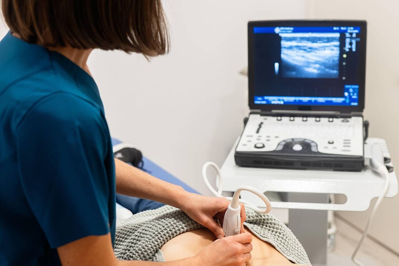
A clinician scanning with a convex probe on a portable machine. The micro-convex probe used on babies is a smaller version of this shape, sized for a newborn’s body.
The probe for a baby is a micro-convex one. It has a small curved face, far smaller than the convex probe used on an adult abdomen. That small face is the point. It sits in the space a newborn offers: on a soft spot the size of a coin, between two little ribs, on a hip no wider than a thumb. A footprint that small lies flat on a newborn and keeps full contact with the curved skin. Smaller than a fingertip, the curved face still opens a fan of view wide enough to hold a whole organ.
A tight curve is what lets it do this. The probe’s face curves over a small radius, around twenty millimetres, so it fans the sound out into a wide view from a tiny contact patch. The advantages of an R20mm radius micro-convex probe are exactly this: a small touch on the skin that opens a broad picture underneath. The shape suits bodies that give a probe very little room. Curving the face over so tight a radius is what packs a wide view into so small a touch.
Frequency is the other half. A micro-convex probe for children runs at a higher frequency than an adult belly probe, often in the range of five to ten megahertz. The higher frequency draws fine detail from the shallow organs of a small body. The probe pairs a small reach with a sharp picture, the combination a child needs. Depth is rarely a problem in a baby, since the organs sit so close that even a higher-frequency probe reaches them with ease. Frequencies in this range trade a little depth for the fine detail a small body rewards.
Handheld ultrasound in the newborn and child, at a glance
Scan
What it shows (key figure)
Brain (newborn)
through the anterior fontanelle; bleeding in and around the ventricles
Lung
line patterns separate wet lung, consolidation, and pneumothorax; no X-ray
Bowel (NEC)
bowel-wall thickening, gas in the wall, gas in the portal veins
Hip (DDH)
alpha angle about 60° or more is normal; best before about 6 months
Appendix (child)
swollen past 6 mm, non-compressible
Probe
micro-convex, about 20 mm radius, around 5–10 MHz
The brain through the fontanelle
A newborn skull is not yet sealed. The bones leave a soft gap at the top, the anterior fontanelle, where only skin and membrane cover the brain. That gap is a window. A probe placed on the soft spot sends sound straight into the brain. No bone stands in the way. Sound crosses that soft gap as cleanly as it would through water.
Through that window, a clinician sees the whole brain. Neonatal cranial ultrasound through the fontanelle shows the fluid spaces, the tissue around them, and the vessels running between. The picture is clear enough to find the problems a premature baby is most at risk of. It is the routine first look at a newborn brain. A clear scan can also reassure a team that a fragile brain looks normal so far. A normal scan, noted in the chart, settles a worried team as much as any finding does.
The main thing it looks for is bleeding. A premature brain is prone to bleeding in the fluid spaces, into and around the ventricles. Ultrasound shows that blood plainly, fresh or settling. Doctors grade the bleeding by how far it spreads: a small amount in the lining, or a larger collection that swells the ventricle. A scan in the first days of life catches it early, while there is still time to act on it. Grades run from a small bleed in one spot up to a large one that fills and swells the ventricle.
It watches for other things too. Built-up fluid can swell the ventricles, a sign the scan tracks over days. The brain tissue itself can show injury in a premature baby. A clinician repeats the scan through the early weeks, following any change at the bedside.
All of it happens at the cot. The baby stays warm in the incubator, undisturbed. A radiation-free scan of a few minutes can be repeated as often as the baby’s care needs. For a fragile premature newborn, that bedside, repeatable look at the brain is hard to match.
The newborn lung
Ultrasound reads a newborn’s lungs from the surface. A baby’s lungs give a pattern of lines on the screen. The pattern shifts in known ways when something is wrong. Neonatal lung ultrasound tells wet lungs from a collapse, or a pneumonia from a pneumothorax, by the look of those lines. A screen crowded with bright vertical lines points to fluid in the lungs of a newborn. A breathless newborn can be sorted at the bedside in minutes. Read in series, the line pattern shows a treatment taking hold within the hour.
Breathing trouble is common in the nursery. Lung ultrasound answers it at the cot in minutes, a radiation-free look the clinician can repeat. A clinician scans a struggling baby, sees the cause, and checks an hour later that treatment is working.
The infant belly
Sound passes easily through a baby’s thin belly wall. The most feared problem in a premature gut is necrotizing enterocolitis, where the bowel wall is dying. Necrotizing enterocolitis on neonatal ultrasound shows what an X-ray cannot: the thickness of the bowel wall, gas within the wall itself, gas tracking to the liver, and whether the bowel still has blood flow.
Those signs guide real decisions. Gas in the bowel wall, or in the veins to the liver, marks the disease. Falling blood flow in the wall warns that the bowel is failing. The scan can be brought back to the same belly through the day, tracking a gut that may be turning. A scan at the cot, repeated when the baby’s belly changes, helps a team judge how far the disease has gone.
In an older child, the belly question is often appendicitis. A child with a sore abdomen needs a safe answer quickly. Pediatric appendicitis on ultrasound is the first test: it looks for an appendix that is swollen past six millimetres, firm, and tender under the probe. In a child, ultrasound is the recommended first imaging for a suspected appendix. It spares many children a CT scan.
The same soft belly that helps in a newborn helps in a child. The organs sit shallow, the wall is thin, the probe reaches them easily. A radiation-free handheld scan answers the urgent belly questions of childhood at the bedside: the dying gut of a newborn, the hot appendix of a school-age child. Shallow organs and a thin wall make the young belly an easy read.
The infant hip
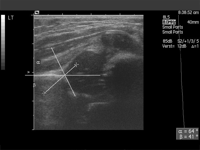
An ultrasound of a newborn hip. The drawn lines measure the socket angles, the alpha and beta angles; an alpha of about 60 degrees or more, as in this normal hip, means a well-formed socket.
A baby’s hip is mostly soft cartilage, the kind of tissue ultrasound shows perfectly. In the first months of life, the ball and socket of the hip are still forming. Developmental dysplasia of the hip on infant ultrasound checks that the socket is deep enough and the ball sits properly in it. A clinician measures the angle of the socket, the alpha angle, where a normal hip reads about sixty degrees or more. The scan works best before the bones harden, in the first few months, which is why a baby with a risky hip is scanned early. Caught in time, a shallow hip can be guided to grow well, often with a simple harness. Scanning in the first months, before the joint hardens, is what keeps that simple fix within reach. Measured against the bony rim, the alpha angle puts a number on how deep the socket sits.
Scanning to a standard
A pediatric scan is worth most when it is done the same way every time. Professional bodies set out how each scan should be performed, what to record, and how to keep a small patient safe. The AIUM pediatric ultrasound practice parameter is one such guide, a written standard for scanning children. Following it keeps the quality even from one clinician to the next.
A standard covers the practical care a child needs. It sets out how to clean a probe between babies, how to keep a newborn warm during a scan, and how long a study should take. On a fragile newborn, these details keep the scan safe. A warmed gel and a gentle touch keep a newborn settled through the study. Small as they seem, these steps are what hold a scan steady on a fragile newborn.
It also says what a good scan includes. For each kind of study, the standard lists the views to capture and the structures to check, so nothing is missed. A clinician who follows it produces a complete record, one another doctor can read and trust. A labelled set of images, saved with the baby’s record, lets the next clinician pick up exactly where this one left off. The standard turns a quick bedside look into a documented study. Filed with the notes, the images let any later reader follow what was seen.
Handheld scanning needs a standard all the more. A small device at the cot makes it easy to take a quick look in passing. A written parameter holds that bedside look to the same level as a study done in the imaging department. A baby gets the same careful scan wherever it happens.
Ultrasound at the cot
The handheld part is what makes all of this fit a child. A small machine goes where the baby is, to the incubator, the cot, the emergency room. The sick newborn who cannot be moved is scanned where it lies. The frightened toddler is scanned on a parent’s lap. A device the size of a tablet or a small laptop tucks beside an incubator without crowding it. The machine comes to the child. Wheeled or carried to the bedside in seconds, it fits the tempo of a busy nursery.
Bringing the machine to the child saves more than a trip. The baby stays in its warm cot, on its monitors and drips, through the whole scan. The clinician gets the picture they need right there at the bedside.
Speed is part of it as well. A handheld scan gives an answer in minutes, at the moment a decision has to be made. A clinician can scan, see, and act in one visit to the cot. For a baby whose state can turn quickly, an answer in minutes is worth a great deal. A scan, a look, and a decision can follow one another in a single pause at the cot. On a crashing newborn, those minutes change the outcome.
Why it suits the smallest
Ultrasound fits a baby for reasons that all line up. The first is radiation, or the lack of it. Ultrasound uses sound, so it carries no ionizing radiation at all. A baby has a whole life of cell division ahead, which makes it the patient most worth sparing from radiation. A child may need a series of scans over weeks. Ultrasound carries that load safely, every time. Counted across a hospital stay, a baby may have a dozen scans, each adding nothing to track.
Bedside use is the second reason. A handheld scan happens where the baby is. It needs no transport and no delay. A review of point-of-care ultrasound in pediatric and neonatal intensive care describes it as imaging brought to the bedside, used and read by the clinician on the spot. For a fragile patient, staying in the cot is a benefit in its own right. The clinician who orders the scan is often the one who performs and reads it on the spot. No request travels to another department. No report comes back hours later.
Repeatability is the third. A scan that is quick, safe, and bedside can be done again and again. A clinician follows a brain bleed, a sick gut, or a struggling lung across hours and days, watching it change. A brain bleed can be checked again the next morning, a lung an hour after a treatment, the same probe each time. Few other tests can be repeated so freely on a newborn.
The small body itself is the fourth reason. A newborn’s organs lie shallow, under thin tissue, close to the probe. Sound reaches them easily and comes back as a clear picture. A baby’s organs sit so shallow that the machine sees them with room to spare. Shallow organs give back an especially clean picture.
Put together, these make ultrasound the first imaging most children get. No radiation, at the bedside, repeatable, a clear view of a small body: four reasons that together put the case beyond doubt. A handheld scanner in a nursery or a children’s ward is a tool that fits its patients almost perfectly.
What this collection covers
This sub-pillar walks through handheld ultrasound for newborns and children, one topic at a time. It starts with the probe, the micro-convex shape that fits a small body, and the frequency that draws detail from shallow organs. From there it follows the scans: the brain through the fontanelle, the lungs through the chest, the gut and the appendix through a soft belly, the hip before it hardens.
Each topic has its own page, with the detail a clinician or a parent might want. The pages cover what each scan shows, how it is done, and what its findings mean. One page covers the standard for doing pediatric scans well. Another lays out why the small micro-convex probe suits a child. Read together, the pages build a full picture of handheld scanning from the newborn nursery to the children’s clinic. Taken in order, they move from the probe through each scan to the standard behind them.
The thread through all of it is a simple fit. A small probe on a small body, real-time pictures, no radiation: ultrasound suits a child the way few tools in medicine do. Handheld scanning puts that fit in a clinician’s hand, at the cot, in the ward, wherever a small patient needs a look inside. The pages that follow take each use in turn.
Common questions
What probe is used for ultrasound on a baby?
A micro-convex probe. It has a small curved face, much smaller than the convex probe used on an adult abdomen, so it fits the little spaces a baby offers: a soft spot, a gap between ribs, a small hip. It runs at a higher frequency, often five to ten megahertz, which draws fine detail from organs that sit close to the surface. The small shape and the higher frequency are what make it right for a small body.
Why is ultrasound used so much in newborns?
Because a baby is the patient ultrasound fits best. A newborn’s organs are small and shallow, easy for sound to reach and show clearly. Ultrasound uses no radiation, which is a real benefit for a patient with a whole life ahead. The scan is quick and works at the cot-side, so a sick newborn need not be moved. Those reasons together make it the first imaging in most nurseries.
What can ultrasound see in a newborn?
A great deal. Through the soft spot on top of the head, ultrasound sees the newborn brain and any bleeding in it. On the chest, it reads the lungs and sorts out causes of breathing trouble. On the belly, it checks the bowel for necrotizing enterocolitis in a premature baby, or the appendix in an older child. On the hip, it checks that the joint is forming properly. All from a handheld probe at the bedside.
Is ultrasound safe for a baby?
Yes, as safe as imaging gets. Ultrasound is made of sound waves, so it carries no ionizing radiation at all. That is the very kind of radiation a baby is most vulnerable to. The sound levels used for imaging are low and well within safe limits. A scan can be repeated as often as a baby’s care needs. Nothing has to be tracked. No exposure has to be limited. This safety is a large part of why ultrasound is the first choice for children.
Can a handheld scanner really do pediatric scans?
Yes. A handheld scanner pairs a small probe with a screen. For the shallow organs of a child, that is enough for a clear, useful picture. It does the scans a nursery and a children’s ward rely on: the brain, the lungs, the belly, the hip. Done to a recognised standard, a handheld scan at the cot reaches the same quality as a study in the imaging department. The small size is the point, since it lets the machine come to the child.
About the Author
Julien Mercier
Senior R&D Engineer · Medical Ultrasound Transducer Development
Senior R&D Engineer with an M.S. in Applied Physics and over 15 years of experience in medical ultrasound transducer development, specializing in the design verification and performance testing of high-frequency imaging transducers. Currently leading the development and verification of the company’s next-generation high-frequency linear-array transducer, responsible for imaging performance evaluation and reliability analysis in preclinical testing. Brings extensive hands-on experience in piezoelectric element tuning, beamforming parameter optimization, and system-level performance testing.