Neonatal Cranial Ultrasound Through Fontanelle Handheld Micro Convex
Cranial ultrasound looks at a newborn’s brain through the soft spot on top of the head. A newborn’s skull has a gap there, the anterior fontanelle, where the bone has not yet closed. That gap is a clear window for sound. A small probe placed on it sends sound straight into the brain and brings back a picture of the fluid spaces, the tissue, and the vessels inside. The scan is the routine first look at a newborn brain, done at the cot, most often to find bleeding in a premature baby. It is quick, safe, and repeatable, which is why it runs in every neonatal unit.
The fontanelle window
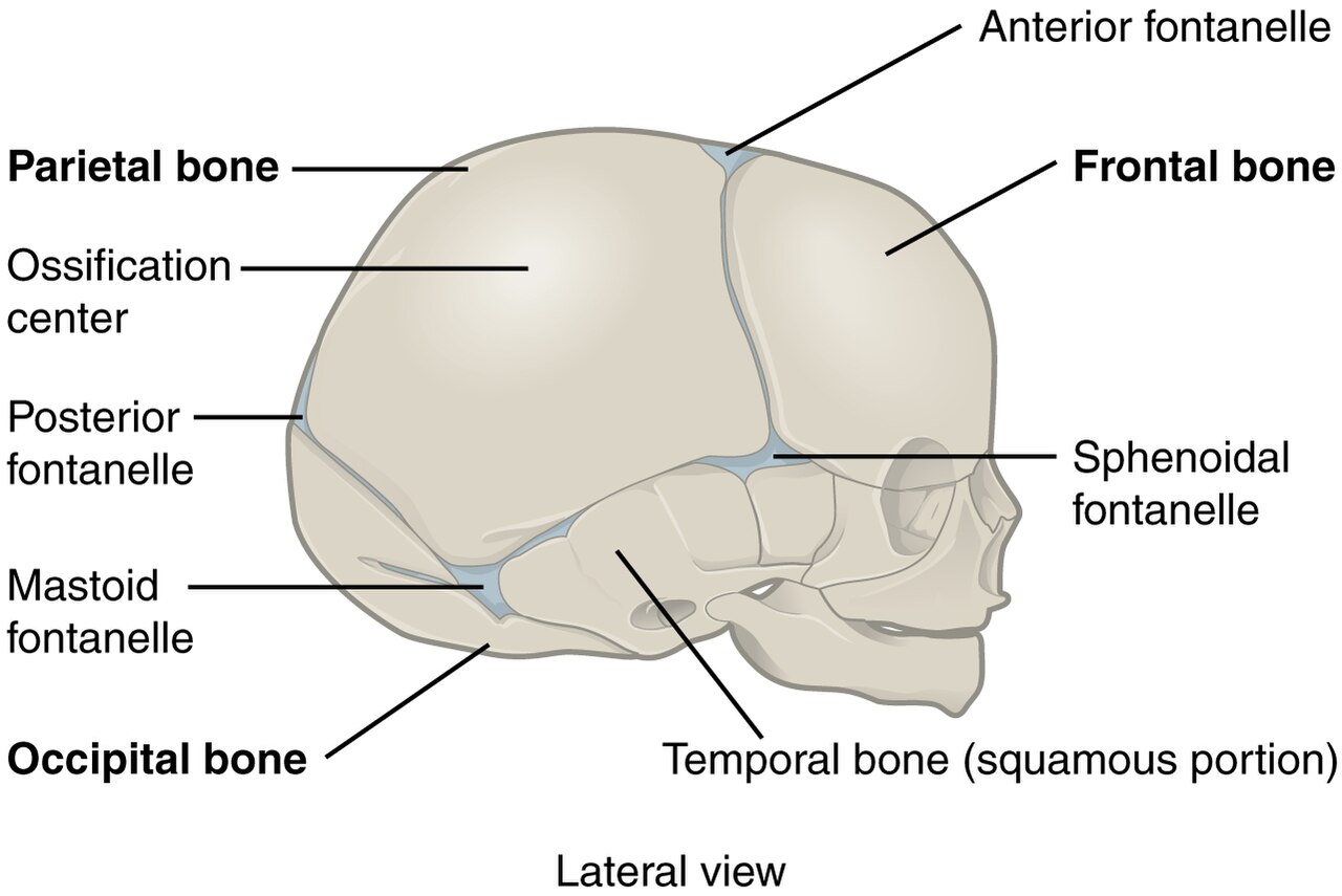
A newborn skull from the side. The plates of bone have not yet fused, leaving soft gaps called fontanelles; the largest, the anterior fontanelle at the crown, is the window ultrasound uses to reach the brain.
The newborn skull is built in plates that have not yet fused. Between the frontal and parietal bones, at the crown of the head, the plates leave a diamond-shaped gap covered only by skin and a tough membrane. This is the anterior fontanelle, the soft spot a parent is told not to press. It stays open for the first year or more of life. For those months it is an open door for ultrasound, a patch of skull with no bone to turn the sound away. Through it, the brain lies open to sound for as long as the bone stays apart.
Bone is the enemy of ultrasound. Sound bounces off the hard skull and never reaches the brain behind it. This is why ultrasound plays no part in scanning a grown head. A baby’s open fontanelle removes that barrier. A probe set on the soft spot sends its sound through skin and membrane straight into the brain. The window lasts only as long as the fontanelle stays open.
Timing follows the fontanelle. It is widest in a newborn, widest of all in a premature one. Over the first year it narrows, until the skull bones meet and close it for good. The early months, when a premature baby is most at risk, are exactly when the window is widest. By the first birthday the scan has done its work. The job then passes to other tools.
Who is scanned, and when
Not every newborn needs a brain scan. The babies who do are the premature and the sick. Every baby born before about thirty-two weeks, or under about fifteen hundred grams, is screened, because the most common bleed strikes the immature brain. A baby with seizures, a difficult birth, or signs of something wrong in the head is scanned whatever its age. The scan goes to the babies whose brains are most at risk. Such a baby may look well from the outside, so the scan is run as a routine screen on every baby in the risk group.
Timing of the first scan is set by the bleed it looks for. The bleeding that threatens a premature brain happens in the first days of life, most of it within the first week. A first scan in the first few days catches it. A baby who stays well still gets the scheduled scans, since a bleed often gives no outward sign. Units scan on a schedule: a first look in the first days, then repeats at set points over the following weeks. A bleed is caught early, then watched over the days that follow.
How the scan is done
The scan is done at the cot, with the baby left where it lies. A clinician warms the gel, sets the small probe on the fontanelle, and watches the screen. The baby can stay in the incubator, on its monitors, asleep. The whole study takes a few minutes. Nothing about it disturbs a fragile newborn beyond a little warm gel on the head.
The probe sweeps the brain in two directions. Held across the head, it takes a fan of coronal slices, front to back, like cutting a loaf. Turned ninety degrees, it takes sagittal slices, from the midline out to each side. The two sets of slices together build a full picture of the brain in three dimensions. Every part of the brain above the window is covered by one sweep or the other. Tilting the probe forward and back, then side to side, lets a clinician walk through the whole brain in a couple of minutes.
A high frequency gives the detail. A probe of seven to ten megahertz resolves the fine structures of a small brain: the thin walls of the fluid spaces, the tissue around them, the vessels that run in the grooves. The shallow brain of a premature baby sits well within the probe’s reach. The picture is sharp enough to show a bleed of a few millimetres.
Reading the scan follows a set order. The clinician works through the same views every time, naming the structures and checking each one. The fluid spaces, the ventricles, are measured and compared side to side. The tissue is checked for bright or dark patches that should not be there. A standard run-through means nothing is missed. One clinician’s scan can then be read by the next. A picture of each standard view is saved, so the next scan can be set beside it.
The normal newborn brain
A normal newborn brain has a familiar look on ultrasound. The ventricles, the fluid spaces deep in the brain, show as thin dark slits, matched on the two sides. The brain tissue around them is an even grey. Down the middle runs a bright line where the two halves meet. The choroid plexus, a bright fold inside each ventricle, is a landmark a reader uses to stay oriented. A clinician learns this normal picture first, so anything out of place stands out at once.
The look depends on the baby’s age, in a known way. In a very premature baby the brain is smooth, its surface still forming. Closer to term, the folds and grooves deepen into the familiar pattern. A clinician reads the brain against the baby’s age, since each gestational age has its own normal.
Knowing normal is most of the skill. The bleeds and injuries the scan looks for all show as a change from this baseline: a bright patch where the tissue should be grey, a dark space grown too wide, a midline pushed off centre. A clinician who carries the normal picture in mind spots the abnormal at a glance. The scan rewards a reader who has seen many healthy brains.
Bleeding, the main target
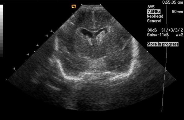
A coronal cranial ultrasound, taken through the anterior fontanelle. The dark spaces near the centre are the fluid-filled ventricles; a bleed shows up as a bright patch where none should be. The text at the top is the machine’s own settings.
Bleeding is the main thing the scan looks for. A premature brain holds a fragile patch of tissue near the ventricles, the germinal matrix, rich in delicate vessels. In the stress of early life those vessels can burst. The bleeding starts there and can spread into the ventricles, sometimes into the brain tissue itself. Ultrasound shows the blood plainly, as a bright patch where none should be. The germinal matrix shrinks away in a maturing brain, which is why the bleed belongs to the early weeks of prematurity.
The bleed is graded by how far it has spread. A review of cranial ultrasound for germinal matrix and intraventricular hemorrhage sets out the Papile grades still used today. Grade one is bleeding held in the germinal matrix alone. Grade two is blood reaching the ventricles, their size still normal. Grade three is blood in ventricles that have swollen. Grade four is blood in the brain tissue beside them.
The grade carries weight because it tracks with outcome. The higher the grade, the heavier the risk it carries for the baby’s later development. A low-grade bleed often settles with little lasting harm. The number a clinician writes down shapes how closely the baby is watched and what the family is told. A grade set on the first scan gives the family and the team an early read on what lies ahead.
The Papile grades of germinal matrix and intraventricular hemorrhage
Grade
What the scan shows
Grade I
bleeding held in the germinal matrix alone
Grade II
blood in the ventricles, their size still normal
Grade III
blood in ventricles that have swollen
Grade IV
blood in the brain tissue beside the ventricle
Who is screened
every baby born before about 32 weeks or under about 1500 g
Catching the bleed early changes the care. A scan in the first days finds a bleed at its freshest. From there the team watches it, ready to act if the ventricles start to swell. Early knowledge lets a unit plan the next steps in good time. The scan turns a hidden event into one the team can see and follow.
Most bleeds are handled by ultrasound alone. The scan finds them, grades them, and follows them, all at the cot. Only the unusual case, a bleed that behaves oddly or a question the scan cannot settle, goes on to MRI. For the common bleed of a premature baby, cranial ultrasound is the whole story, from the first sign to the last follow-up.
Scanning again and again
A cranial scan is rarely a single event. A bleed in a premature brain is not a fixed thing. It appears, grows or holds over days, then settles or goes on to swell the ventricles. One scan catches a single moment. A series of scans, spread across the early weeks, follows the whole course. A unit scans on a set schedule: a first look in the first days, then repeats at set points after. A bleed is found early and watched through the days that follow. Each scan in the series is read beside the ones before it, so a slow change shows up that a single picture would miss. The repeat scans cost nothing in radiation and little in time, so a fragile baby can be followed as closely as its care needs. That ability to scan again and again, safely, at the cot, is much of what makes ultrasound the right tool for a newborn brain.
Ventricles and swelling
The ventricles are the brain’s fluid spaces. The scan watches them closely. In a healthy brain they are thin slits. After a bleed, blood can block the drainage of that fluid. The ventricles then swell, because the blocked fluid has nowhere to go. This swelling, post-hemorrhagic ventricular dilatation, is the chief danger that follows a serious bleed.
The scan measures the swelling, so it can be tracked. A clinician puts callipers on the ventricle and reads its width against a known normal for the baby’s age. The same measure, repeated on each scan, shows whether the ventricles are holding steady or growing. Two simple measurements, the ventricular index and the width of the front horn, put numbers on the swelling. A number that climbs scan after scan is the warning the team watches for.
A swelling that keeps growing needs treatment. A growing pool of fluid presses on the brain and raises the pressure in the head. The team steps in when the measure crosses a set line, draining the fluid to relieve the pressure. The scan is what tells them when that line is crossed, and how fast it is rising.
Most swelling settles on its own. After a smaller bleed, the ventricles often widen a little, then return toward normal once the blood clears. The scan follows that settling, scan by scan, and spares the baby any treatment it does not need. Only the swelling that keeps climbing calls for the team to act.
The white matter
Beyond bleeding, the scan watches the white matter. The tissue around the ventricles, the periventricular white matter, can be injured in a premature baby, even in one with no bleed at all. On the scan it shows first as a bright patch, brighter than the normal tissue around it. It tends to sit in the white matter behind and above the ventricles, the part most vulnerable in a preterm brain. This injury, periventricular leukomalacia, can mark a risk to the baby’s later movement.
White matter injury asks for the same serial scanning. The early bright patch can fade, or it can break down into small fluid cysts over the weeks that follow. The cysts, when they form, are what the scan watches for, since they carry the clearer warning. Following the white matter over time tells more than any single scan.
What else it finds
The scan does more than hunt for bleeds. It checks the basic build of the brain, that the structures are present and in their place. A baby born with a brain that formed unusually can be picked up on the first scan. The midline, the fluid spaces, and the major structures are all checked against the normal plan.
It looks for signs of infection and injury too. An infection that reached the brain before or after birth can leave bright streaks or scattered flecks the scan can see. A brain starved of blood or oxygen around birth can show swelling or patches of damage. A calcified fleck left by an old infection stays bright on every later scan, a lasting mark of what happened. The scan gives the team a first read on a brain that may have been harmed.
Term babies get the scan as well, when there is reason. A full-term baby with seizures, a hard birth, or signs of raised pressure in the head can be scanned through a fontanelle that is still open. In a term baby the fontanelle is already narrowing, leaving a smaller window. It still serves for a first look. Where the question runs deeper, the scan points the way to MRI.
Where MRI takes over
Cranial ultrasound is strongest at the centre of the brain. The structures around the ventricles, where bleeds and white-matter injury happen, sit right in the beam’s best reach. For the premature brain’s commonest troubles, the scan is the first and often the only tool needed. It stands as the routine screen for a newborn brain. Day after day, the same probe at the same window builds a record no other tool could gather so easily.
Some parts of the brain are harder to reach. The surface just under the skull, and the back of the brain low in the head, sit at the edge of the window. A small bleed or a subtle injury in those corners can be hard to see from the fontanelle alone. A clinician reads those areas with extra care. When a doubt remains, another view settles it.
This is where MRI comes in. For the fine detail of the white matter, for the deep and posterior parts, and for a question the ultrasound leaves open, MRI gives the fuller picture. Many units scan a high-risk baby with MRI once, near term, to round off what the serial ultrasound has shown. Between them, the two tools cover ground that neither covers alone.
Knowing the limits is part of using the scan well. A clinician leans on the ultrasound for what it does best, the central brain and the common bleed. Where the ultrasound runs short, MRI takes the question on. Used this way, the scan carries the bulk of the work. The deeper tool is saved for the cases that truly need it. The fontanelle scan does not have to do everything to be the right first tool.
What the scan gives
For a newborn brain, cranial ultrasound is the tool that fits. The open fontanelle gives it a clear window. The shallow brain sits in easy reach of a high-frequency probe. The scan runs at the cot, harms nothing, and can be repeated as often as a fragile baby needs. No transport, no sedation, no radiation: the scan asks almost nothing of a baby already under strain. Everything about a newborn points to ultrasound as the first look at the brain.
Its main work is to find and follow bleeding in a premature brain. A scan in the first days catches the bleed, the grade marks how far it has spread, and serial scans follow it through to its settling or its swelling. From that one tool a team gets the warning it needs, early enough to act. The early warning it gives is what lets a team prepare, talk to the family, and plan the weeks ahead. The scan brings a once-hidden event into plain view.
The window lasts only as long as the fontanelle stays open. For those months, ultrasound owns the newborn brain. It is safe, quick, and ready at the cot. It answers the question that comes first in a premature baby: is the brain bleeding, and how much. For a newborn, the soft spot is a doorway. Cranial ultrasound is what walks through it.
Common questions
Why can ultrasound see a baby’s brain but not an adult’s?
Because a baby’s skull is not yet closed. A newborn has a soft gap at the top of the head, the anterior fontanelle, where the bone has not grown over. Sound passes straight through that gap into the brain. A grown skull is solid bone, so sound bounces off it and never reaches the brain inside. The open fontanelle is the one window ultrasound has, and it closes around the first birthday.
When is a newborn’s head scanned?
When a baby is premature or unwell. Every baby born before about thirty-two weeks, or under about fifteen hundred grams, is screened, because the bleeding the scan looks for strikes the immature brain. A baby of any age is scanned after a difficult birth, with seizures, or with signs of trouble in the head. The first scan comes in the first days of life. Repeats follow at set points over the next weeks.
What is the scan mainly looking for?
Bleeding, above all. A premature brain holds a fragile patch of vessels near the ventricles that can bleed in the stress of early life. The scan finds that bleeding, grades how far it has spread, and follows it over the weeks. It also checks the fluid spaces for swelling, the white matter for injury, and the whole brain for anything built or growing wrong. Bleeding is the main target. The rest is checked on every scan.
What do the bleed grades mean?
They mark how far the blood has spread. Grade one is bleeding held in the germinal matrix, the fragile patch where it starts. Grade two is blood reaching the ventricles, their size still normal. Grade three is blood in ventricles that have swollen. Grade four is blood in the brain tissue beside them. The higher the grade, the heavier the risk it carries, so the number guides how closely the baby is watched.
Does the scan replace an MRI?
No, it works alongside it. For the common bleed of a premature baby, cranial ultrasound does the whole job, finding it, grading it, and following it at the cot. MRI comes in for the fine detail of the white matter, for the deep and back parts of the brain, and for a question the ultrasound leaves open. Many units add one MRI near term to round off the picture. The two tools cover between them what neither covers alone.
About the Author
Julien Mercier
Senior R&D Engineer · Medical Ultrasound Transducer Development
Senior R&D Engineer with an M.S. in Applied Physics and over 15 years of experience in medical ultrasound transducer development, specializing in the design verification and performance testing of high-frequency imaging transducers. Currently leading the development and verification of the company’s next-generation high-frequency linear-array transducer, responsible for imaging performance evaluation and reliability analysis in preclinical testing. Brings extensive hands-on experience in piezoelectric element tuning, beamforming parameter optimization, and system-level performance testing.