AIUM Pediatric Ultrasound Practice Parameter Handheld Probe
An ultrasound practice parameter is a written standard for how a scan should be performed, recorded, and read. For children, the American Institute of Ultrasound in Medicine sets these standards together with the American College of Radiology and the pediatric and radiology societies. The parameters cover who may scan, what the machine must do, how to keep a child safe from too much sound energy, and how the images and report are stored. A handheld micro-convex scanner is held to the same standards as a full cart system.
A pocket-sized probe brings the scan to the cribside, the clinic, or the emergency department. The standards stay the same in every location. A child scanned with a handheld probe deserves the same trained operator, the same safe settings, and the same recorded images as a child scanned in the radiology suite. That principle runs through every parameter below.
What the pediatric practice parameter sets out to do
A practice parameter is guidance for clinical practice, written to be applied with judgment. It describes what a high-quality pediatric ultrasound examination should contain, so that a child gets a consistent standard of care from one clinic to the next. The document still leaves room for clinical judgment in an individual case. Its purpose is to set a floor for quality, to train new practitioners against a shared standard, and to give a clear reference when a scan is later reviewed.
The pediatric parameters are written by several bodies at once. The American Institute of Ultrasound in Medicine develops them together with the American College of Radiology, the Society for Pediatric Radiology, and the Society of Radiologists in Ultrasound. Drawing on radiologists, sonographers, and pediatric specialists gives the standards weight across the field. For a handheld user, the value is the same: a single, agreed reference for what a good pediatric scan looks like, whatever machine produces it.
Who may perform and read the scan
The first thing the parameters address is people. An ultrasound image is only as good as the person who makes it and the person who reads it. For a pediatric scan the parameters call for a physician qualified to interpret the study, working with a sonographer trained to perform it. The two roles can sit in one person at the bedside, such as an emergency physician who both scans and reads. The qualification behind each role stays the same.
A sonographer who performs pediatric scans is expected to hold a recognised credential. In many countries that means registration through a body such as the American Registry for Diagnostic Medical Sonography, earned by training and a written examination. The credential shows that the operator knows the physics of the machine, the anatomy of a child, and the safe handling of acoustic output. A handheld device does not lower this bar. A pocket-sized probe in untrained hands produces a pocket-sized picture of little value.
The physician who reads the study carries the matching duty. Interpretation calls for training in pediatric ultrasound findings, the range of normal across ages, and the conditions that bring a child to a scan. The parameters expect this reader to have documented education and ongoing experience in the field. Reading a child’s scan is not the same skill as reading an adult’s, since the anatomy, the common diseases, and the normal measurements all change from one age group to the next.
Reaching competence takes a measured number of supervised scans. Professional bodies put rough figures on it: emergency physicians are often asked for twenty-five to fifty good scans in each type of study before working unsupervised. Some critical-care groups ask for fifty. A pediatric review of point-of-care ultrasound stresses that a raw count is not enough on its own. Competence is judged by watching the operator scan, reviewing stored images, and giving feedback over time.
A handheld scanner makes this discipline matter more. The ease of pulling a probe from a coat pocket can tempt an untrained hand to scan and act on what it sees. The parameters answer that plainly: the person scanning a child needs the training, whatever the size of the device. A clinic bringing handheld ultrasound to the bedside builds the same credentialing and supervision around it as it builds around a cart system.
The written request and the indication
Every scan should answer a question that someone has asked. The parameters expect a written request with a clear clinical indication before a child is scanned. A request that names the problem — a swollen scrotum, a limp, a bulging fontanelle — points the scan at the right area and the right protocol. A scan ordered without a reason wastes the child’s time and the clinician’s.
The indication decides how the scan is done. A question about the hips in a newborn sets up a different study from a question about the appendix in an eight-year-old, with its own positions, measurements, and views. Matching the protocol to the question is part of what the parameters ask for. A handheld user at the bedside follows the same logic, choosing the preset and the sweep that fit the clinical question in front of them.
Writing the indication down also protects the child. It records why a scan carrying acoustic energy was performed, which matters most in the youngest patients. The reader can then judge the images against the actual question. A clear thread runs from the clinical concern to the scan to the report, one the next clinician can follow. The request is the first line of a well-kept record.
Equipment the parameter calls for
The parameters describe the machine in general terms. A pediatric scan needs a real-time scanner with a transducer suited to the size of the patient and the part being examined. Frequency follows a simple trade-off: the higher the frequency, the finer the detail, so an operator picks the highest one that still reaches the depth the scan needs. In a small child that depth is shallow, so high frequencies do the work well.
Different probes suit different jobs in a child. The infant hip and a superficial lump call for a high-frequency linear probe. The abdomen and the soft window of a fontanelle call for a curved or micro-convex probe that reaches deeper. The small head of a micro-convex probe slips into the tight spaces on a small body. The parameters ask for the probe that matches the task at hand.
A handheld micro-convex scanner meets this equipment point squarely. Its probe runs at frequencies suited to small bodies. Its presets hold the focus and the dynamic range for a pediatric abdomen or a neonatal head. The image quality of a good handheld now covers many pediatric questions at the bedside. The parameters set the target the device has to reach: a clear, real-time picture at a frequency suited to the child.
The parameters also expect the equipment to be kept in good order. A scanner should meet the relevant safety standards, stay in proper working condition, and pass documented quality-assurance checks at least once a year. A handheld device carries the same duty. A pocket scanner needs its probe checked, its software kept current, and its image quality confirmed against a known standard, the same as any machine that touches a patient.
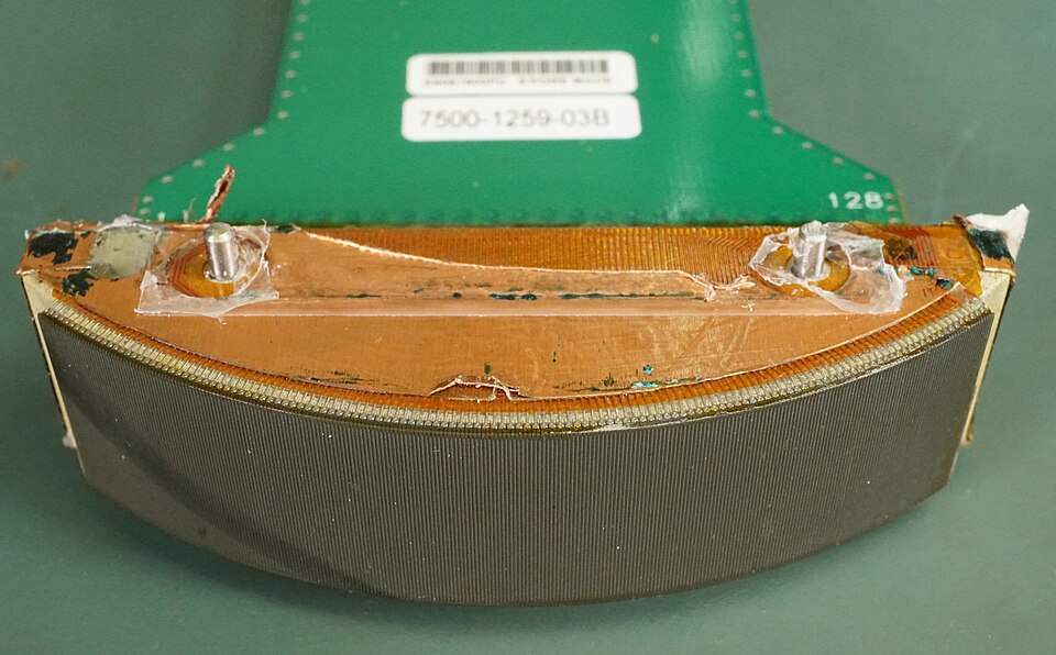
A curved-array transducer with its cover removed, showing the gently curved strip of elements that fans the beam into a wide view. A curved or micro-convex probe of this kind reaches deeper than a flat linear probe and fits the small abdomen of a child. The green board behind the strip holds the probe’s electronics.
ALARA and the child
Ultrasound is among the safest imaging a child can have. It works with sound, so the radiation dose that limits how often a child can be x-rayed or scanned by CT simply does not apply. Sound energy still has real effects. At high enough output it can warm tissue or stress it mechanically, which is why every parameter folds in a safety principle called ALARA.
ALARA stands for As Low As Reasonably Achievable. The idea is to use the lowest output and the shortest scan time that still answer the question. Two numbers on the screen guide it: the thermal index, an estimate of heating, and the mechanical index, an estimate of mechanical stress. A careful operator keeps both low, trims the dwell time over any one spot, and never runs more power than the image needs. The parameters expect every operator to know these numbers and to act on them.
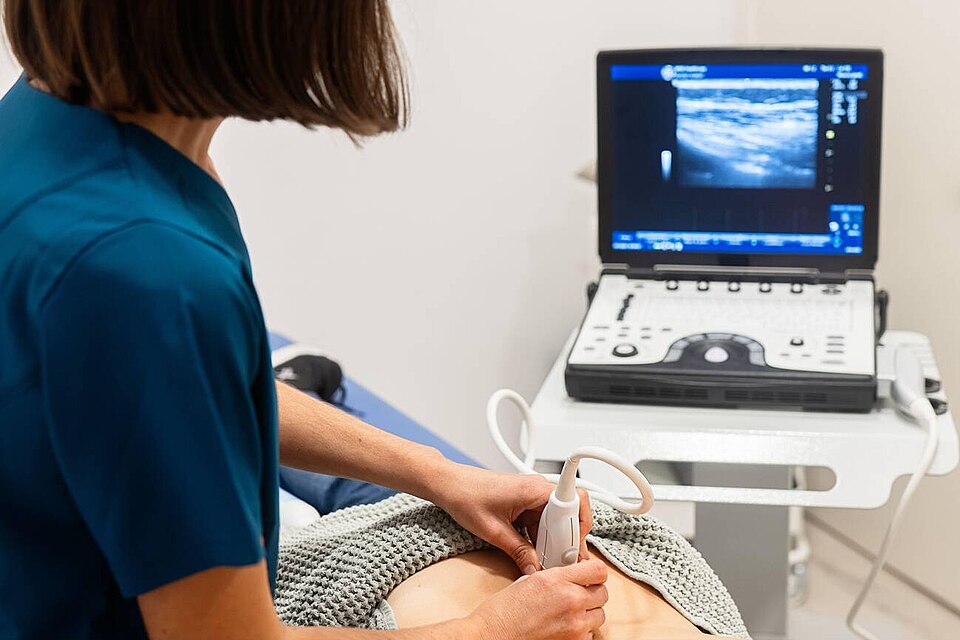
A clinician performs a bedside ultrasound with a convex probe, the portable machine showing the live image. The small readouts in the upper corners of the screen include the thermal and mechanical index, the numbers an operator watches to keep the output as low as the question allows.
The eye and the newborn brain — the strictest limits
Some pediatric scans call for extra caution. The eye and the newborn brain are the most sensitive targets, so the safe output limits for them are set lower than for the rest of the body. Knowing this is part of scanning a child safely. An operator who scans these areas keeps the output well down and the exposure brief.
The eye has the tightest limits of any routine scan. Regulators cap the acoustic output for an eye examination far below the general ceiling: a mechanical index no higher than about 0.23 and a time-averaged intensity around 50 milliwatts per square centimetre. The general limit sits near 720. The lens and retina have little blood flow to carry heat away, so they warm more easily. An operator scanning a child’s eye turns the output down to these low settings before the probe goes near it.
The newborn brain gets similar care. A neonatal head scan runs through the soft fontanelle, straight onto developing brain tissue, so the safe thermal limit for it is set low, around 0.7 on the thermal index. The scan is quick. The output stays gentle. Even so, ultrasound remains the first look at a newborn’s brain in the unit, exactly because it is gentle enough to repeat at the cot side.
Keeping to these limits is mostly a matter of habit. The operator checks the thermal and mechanical index on the screen, starts low, and lifts the output only as far as a clear image demands. A scan of the eye or the newborn brain stays short. The probe comes off the moment the question is answered. None of this slows a skilled operator down, and it keeps a child’s most delicate tissues clear of any avoidable stress.
Pediatric ultrasound — the figures behind the safety and equipment standards
Item
Figure
Note
ALARA display indices
TI (thermal) and MI (mechanical), on screen
Keep both low
Eye exam — mechanical index
≤ ~0.23
Strictest limit
Eye exam — intensity (ISPTA.3)
≤ ~50 mW/cm²
vs general ~720
General diagnostic — intensity
≤ ~720 mW/cm²
Regulatory ceiling
General diagnostic — mechanical index
≤ 1.9
Regulatory ceiling
Neonatal head / spine — thermal index
≤ ~0.7
Developing tissue
Competence — supervised scans
~25–50 per study type
Plus image review
Equipment QA testing
At least annually
Documented
Minor’s record retention
To adulthood + years
Set by local law
ALARA at the bedside with a handheld
A handheld scanner sits naturally inside the ALARA principle. Its acoustic output is modest, its examinations are short and aimed at one question, and the same thermal and mechanical index appear on its screen for the operator to watch. The portability that brings the probe to the cribside also keeps each look brief, aimed at a single question, which is the heart of ALARA. A smaller machine does not loosen the safety rule. A handheld operator reads the indices, keeps the output low, and limits the dwell time over a newborn’s brain or eye exactly as the parameters ask, carrying the full weight of the standard in a device that fits a pocket.
Documenting the examination
A scan that is not recorded barely happened. The parameters expect a permanent set of images from every examination, saved to the child’s record. Each image needs labelling: the patient’s identity, the date, the side of the body, and the orientation of the probe. Measurements taken during the scan are stored with the pictures. The record lets another clinician see what was found, and it lets the same scan be compared with a later one.
A written report turns the images into an answer. The parameters expect a report that states what was examined, what was found, and what it means for the clinical question. The report becomes part of the medical record, signed by the clinician responsible for it. For a child, that report often guides the next step, whether that is reassurance, a repeat scan, or a referral. Clear wording in it carries the whole value of the scan forward.
Good labelling matters more in a child than people expect. A tiny structure measured a millimetre out can shift a diagnosis, so the record has to show exactly what was measured and where. A left hip and a right hip look alike on a screen, so the side has to be marked. The parameters press on these details because a clear, well-labelled record is what makes a scan trustworthy days or weeks later.
Getting handheld images into the record
This is the hardest part of the standard for a handheld device. On a cart system in a radiology room, images flow into the hospital archive and the report by default, with no extra effort. A handheld scanner starts outside that plumbing. The path from its probe to the permanent record has to be built on purpose.
Modern handheld systems close that gap by design. Most pair with an app on a phone or tablet that captures the images, labels them with the patient’s details, and sends them into the hospital record over a secure link. Some connect straight to the picture archive that radiology uses. Setting this up is part of bringing a handheld into a service, done before the probes go into daily use.
Labelling deserves the same care on a handheld as anywhere. An image saved to a phone needs the child’s identity attached, the date, the side, and the orientation, the same fields a cart system records. A picture sitting unlabelled in an app, or saved only to the device, does not meet the standard. The discipline of naming and filing each image at the moment it is taken keeps a handheld scan as trustworthy as any other.
The report requirement holds for a handheld scan too. A look at a child’s kidney at the bedside still needs a note in the record of what was seen and what it means. A scan can guide a real decision. With nothing written down, that decision leaves a gap in the child’s care. The convenience of a quick bedside look does not remove the duty to record it. A short, clear note saved with the images satisfies the standard.
Bringing a handheld into a children’s service is as much about the workflow as the device. The probe answers the clinical question. The app, the labels, the archive, and the report turn that answer into a permanent part of the child’s care. A service that plans this path from the start gets the speed of bedside scanning and a complete record. The parameters set the bar. The technology to clear it already sits in the device.
Keeping the equipment and the exam quality up
A standard means little without a way to keep meeting it. The parameters ask each service to run a quality programme: regular review of stored images, feedback to operators, and maintenance of the equipment. Reviewing a sample of scans against the standard catches drift early and keeps the whole team sharp. A pediatric review of bedside ultrasound found quality assurance to be the weakest link in many programmes, behind training and credentialing.
Records of a child’s scans are kept longer than an adult’s. A minor’s images and reports are often held until the child reaches adulthood, plus a set number of years, the exact period set by local law. The reason is practical: a scan taken in infancy can matter for a condition that surfaces years later. A handheld service stores its images under the same retention rule, in the same archive, for the same length of time as the rest of the department.
Infection control between patients
A probe touches one child after another, so cleaning it matters. The parameters expect a clear infection-control routine: the transducer wiped or disinfected between patients, the level of cleaning set by how the probe was used. A probe that touched intact skin needs a low-level wipe. A probe used near broken skin or a body opening needs more.
A handheld probe raises the stakes a little. It travels from room to room in a pocket, moving between children faster than a fixed machine does. That mobility makes a cleaning routine matter more. A handheld probe is wiped down between every child, the same as any shared probe, and a disposable cover is used where the situation calls for one.
Children in hospital are often the most vulnerable to infection. A newborn in intensive care, a child with a weak immune system, an infant with a fresh surgical wound — each can be harmed by a germ carried on an unclean probe. The infection-control step in the parameters protects exactly these patients. A clean probe is part of safe scanning, as much as a low acoustic output is.
In practice the routine is simple to keep. A wipe rated for medical probes sits beside the workstation or in the bag with the handheld. The probe gets cleaned the moment a scan ends, before it goes back in the pocket. A cover goes on for any contact near a wound or a mucous surface. None of this takes long, and it keeps a shared probe from carrying anything from one child to the next.
Where the parameter and the handheld meet
The pediatric parameters are device-neutral by design. They describe what a good scan of a child requires, regardless of the machine that delivers it. A handheld scanner meets every one of those requirements when it is used with the same care as a cart system: a trained operator, a clear indication, a suitable probe, safe output, a clean transducer, and a recorded, reported result.
Reading the parameters as a checklist helps a handheld service stay honest. Who is scanning the child, and are they trained for it? Is there a clear reason for the scan? Does the probe suit the patient’s size? Is the output kept low, the eye and the newborn brain handled with extra care? Is the probe clean? Is every image labelled, stored, and turned into a report? A yes to each is a scan that meets the standard.
Ultrasound now reaches well beyond the radiology department. A written standard matters more than ever for that reason. A scan of a child happens in a clinic, an emergency bay, or a neonatal unit, in the hands of clinicians from many fields. The pediatric parameters give all of them one shared definition of a good scan. A handheld probe carries that definition to the bedside in full. The standard is what keeps a pocket-sized scan of a child as safe, as careful, and as well-recorded as one done on the largest machine in the hospital.
Common questions
Does an AIUM practice parameter apply to handheld ultrasound?
Yes. The parameters describe what a good pediatric scan requires, regardless of the machine. A handheld device is held to the same standards as a cart system: a trained operator, a clear indication, a safe acoustic output, a clean probe, and a labelled, stored, reported set of images. The size of the device does not change the standard.
Who is allowed to perform a pediatric ultrasound?
A pediatric ultrasound is performed by a trained sonographer or a clinician with documented ultrasound education, and it is read by a physician qualified to interpret pediatric findings. The two roles can sit in one person at the bedside, such as an emergency physician. Reaching competence takes a set number of supervised scans, often twenty-five to fifty in each type of study, judged together with image review and feedback.
What is ALARA, and why does it matter more in children?
ALARA means As Low As Reasonably Achievable. An operator uses the lowest acoustic output and the shortest scan time that still answer the question, watching the thermal and mechanical index on the screen. It matters most in children because growing tissue reacts more to acoustic energy. The eye and the newborn brain carry the strictest output limits of any routine scan.
How does a handheld scan get into the medical record?
Most handheld systems pair with an app that captures the images, labels them with the child’s details, and sends them into the hospital record over a secure link. Some connect straight to the picture archive that radiology uses. The clinician then writes a short report of what was found. Setting up this path is part of bringing a handheld into a service, so that a bedside scan is stored and reported the same as any other.
Educational information on ultrasound practice standards. It does not replace formal guidance or assessment by a qualified clinician.
About the Author
Julien Mercier
Senior R&D Engineer · Medical Ultrasound Transducer Development
Senior R&D Engineer with an M.S. in Applied Physics and over 15 years of experience in medical ultrasound transducer development, specializing in the design verification and performance testing of high-frequency imaging transducers. Currently leading the development and verification of the company’s next-generation high-frequency linear-array transducer, responsible for imaging performance evaluation and reliability analysis in preclinical testing. Brings extensive hands-on experience in piezoelectric element tuning, beamforming parameter optimization, and system-level performance testing.