A sudden severe belly pain hides a short list of killers. Ultrasound sorts them at the bedside in minutes, on a wand that fits a coat pocket. The probe goes to the part that hurts and brings the trouble onto the screen. A swollen appendix, an inflamed gallbladder, a blocked gut, a stuck stone, and a widening aorta each leave a mark. The scan rules a danger in or moves the search on. The acute abdomen rewards a fast look over a long wait. Imaging that once waited on a department now rides in a pocket to the bedside. The shift hands a first answer to the clinician who meets the patient, in the first minutes that count. A scanner the size of a phone reaches the belly of a patient too sick to move. The picture comes to the patient, on the trolley or the resus bed, where a fixed machine never could. A handheld asks for no prep, no contrast, and no wait for a free slot.
Speed changes the outcome in the worst of these. A ruptured aorta or a dead loop of gut counts its danger in minutes. An early scan moves the person to surgery before the harm spreads. The picture also calls off a false alarm, and a clear gallbladder over a soft belly sends a person home with advice. At the bedside the first scan often beats the bloods back from the lab. The probe asks for no booking, no transfer, and no dye. A nurse runs it on a ward round. An emergency team runs it in the resus bay while the lines go in. A clinic far from a scanner runs it on the one machine it owns. The handheld answers the first question fast and hands the grade and the stage to the formal scan. The bedside paradigm rests on one idea: the picture goes to the patient, where the patient already lies.
The pain narrows the danger before the probe touches skin. Site and story point to one organ. A few sweeps confirm or clear the lead. One probe covers the whole belly: the liver, the gallbladder, the gut, the kidneys, the bladder, and the aorta. The window moves from one organ to the next with no change of tool. The acute abdomen turns into a short ordered search. The probe joins the exam as another sense, the eye reaching where the hand stops.
Where the pain sends the probe
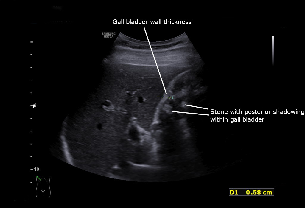
Acute cholecystitis on ultrasound. The image is labelled by the original author: the upper line marks a thickened gallbladder wall, measured at 0.58 cm, and the lower line a gallstone casting a shadow.
Pain low on the right pulls the probe to the appendix. Pain high on the right pulls it to the gallbladder. Flank pain pulls it to the kidney and the ureter. A distended belly pulls it to the loops of gut. Collapse in an older patient pulls it straight to the aorta. The site of the worst pain opens the first window in seconds. The map holds the deadly causes on the list even when the belly feels soft. A quiet belly can sit over a leaking aorta and give little to the hand. The hardest cases hide the danger behind a complaint that looks routine, so the map runs past the obvious to the lethal every time.
The story sharpens the site. Pain that began at the navel and settled low on the right fits the appendix. Pain after a fatty meal fits the gallbladder. Pain in waves that shoots to the groin fits a stone on the move. A tense belly with no wind fits a blocked gut. Back pain and a faint pulse in an older patient fit the aorta. Pulse and pressure steer the order as hard as the story. A racing pulse with a low pressure drives the aorta and the free-fluid windows to the front. The worst danger gets the first sweep whenever the numbers look unsafe. The age of the patient bends the odds too, and a wide aorta climbs the list with every decade past fifty. A history of an old aneurysm, a past stone, or a gallbladder already known to hold stones tilts the first look before the probe moves. The story does half the work. The scan confirms or overturns it in a sweep.
The order runs by danger. The loudest pain waits its turn behind the deadliest cause. An older patient in shock gets the aorta swept from the ribs to the navel first, since a burst aneurysm kills in minutes. Right-sided pain brings the appendix and the gallbladder next. The probe presses the sore spot for a non-compressible tube, then slides up for the stones and thick wall of an angry gallbladder. A distended belly brings the gut, fanned across the loops for dilated bowel that has stopped moving. Flank pain brings the kidneys, each checked for the swelling that marks a stone, with the bladder and the aorta swept after to clear the mimics. A hand stays on the patient and an eye on the monitor through every sweep. A falling pressure cuts the search short and sends the patient to theatre on the aorta alone. Position opens each window. A left roll floats the gallbladder forward. A held breath drops the kidneys below the ribs. A bent knee softens the belly for the appendix. The windows run in a fixed habit from one patient to the next. Only the order shifts with the story. A set routine of windows misses fewer dangers under pressure than a frightened scramble. The deadliest cause stays at the front of the line until the scan clears it. A first pass that comes back clean on a worrying belly earns a second pass an hour on, once the picture has had time to declare itself. The order is the discipline of the acute abdomen. The order keeps the quiet aorta from hiding behind the loudest pain. A run through the dangers in turn catches the killer before it declares itself in blood. The map gives a frightened room a fixed routine to fall back on. The deadliest danger holds the front of the queue until a clean sweep clears it.
A clear sweep settles many a frightening belly. A calm gallbladder, a soft belly, and a normal aorta point a person toward the door, away from the theatre. The bedside look saves a bed, a scan slot, and a needless operation. A normal scan over an easing pain often closes the visit with a review booked and a safety net spelled out. The few who need more go on to the CT and the surgeon. The value of the scan runs as much through the dangers it clears as the ones it finds.
The deadliest goes first
An older patient in shock gets the aorta scanned before anything else. A burst aneurysm kills in minutes.
What each danger shows
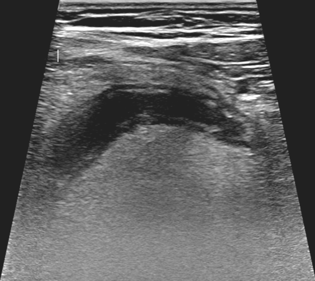
Acute appendicitis on ultrasound. The blind-ending tube of an inflamed appendix runs through the right lower belly. A tube like this will not squash flat under the probe, the mark of inflammation.
Acute appendicitis shows a blind-ending tube in the right lower belly, wider than six millimetres, that will not squash flat under the probe.
Stones cast bright shadows in the gallbladder up under the right ribs. A wall past three millimetres and a sore spot right over the gallbladder fill in the rest of the picture of acute cholecystitis. Pain on a press straight down onto the gallbladder seals the call.
Loops of small bowel wider than two and a half centimetres, swinging their contents back and forth on the live scan, mark a small bowel obstruction. A flat collapsed loop beyond the swollen ones points to the block.
On the painful flank, a kidney swollen behind a stuck stone gives the indirect sign of renal colic. A twinkle of colour behind a bright dot at the bladder floor names the stone itself.
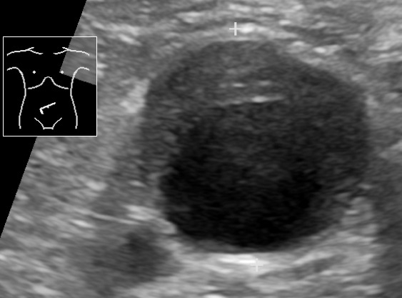
A round vessel in front of the spine wider than three centimetres is an aneurysm. A ruptured aortic aneurysm in an older patient with pain and a low pressure goes to theatre on the spot. The bleed of the rupture itself often hides behind the belly lining, out of the probe reach, so the wide vessel and the shocked patient carry the call.
Five abdominal emergencies: the bedside figure
Emergency
Number on the scan
What it points to
Appendicitis (lower right)
tube over 6 mm, non-compressible
inflamed appendix
Cholecystitis (upper right)
wall over 3 mm, stones, sore spot
inflamed gallbladder
Small bowel obstruction
loops over 2.5 cm, to-and-fro
a blocked gut
Renal colic (flank)
hydronephrosis, twinkle behind a stone
a stone in the ureter
Aortic aneurysm
aorta over 3 cm, repair past 5.5 cm
a surgical emergency
The unstable patient
An abdominal aortic aneurysm in cross section. The calipers mark the width of the vessel. The dark centre is the flowing channel; the grey rim around it is clot packed against the wall.
A collapsing patient turns the scan into a hunt for blood. Four windows catch free fluid where it pools. The space between the liver and the right kidney fills first, so the right upper window leads. The pocket around the spleen comes next on the left. Behind the bladder, the lowest dip of the belly traps blood in the pelvis. Under the breastbone, the sac around the heart shows its own bleed. Dark fluid in any of these, over a wide aorta, names a rupture in full flow. The same probe drops to the big vein beside the heart for a sense of the fluid state. A flat vein in a shocked patient backs heavy blood loss. A vein that does not collapse points the other way, toward a heart that cannot keep up. Lungs join the sweep too, checked for a collapsed edge or fluid above the diaphragm. The whole emergency look runs from the neck veins to the pelvis on one wand, a head-to-belly survey of the bleeding and the pump in a single pass. The heart sits at the centre of the same protocol, scanned for a weak squeeze or a sac of fluid choking it. A rim of dark fluid around the heart in a shocked patient names a tamponade in need of a needle. The bleeding, the tank, and the pump come together in one look that guides the resuscitation as it runs. The four corners, the great vein, and the heart come up in the time a line takes to thread. The protocol turns a blind crash into a mapped one in under two minutes.
Free fluid and a wide aorta together send a patient straight to surgery, ahead of any CT. The minutes the scan saves decide who lives. The vascular team meets the patient with the size of the aorta already in hand. Blood runs through wide lines while the pressure stays low on purpose, to keep a clot from breaking loose before the repair. A hard push of fluid can pop that clot and turn a contained leak into a free bleed, so the resuscitation stays light and the pressure stays on a leash. The bedside scan sets that whole machine in motion with one measured width. Speed beats the bleed only when the answer comes at the bedside. A team that scans early loses no minute to a trip down the corridor to a fixed machine. Blood and clotting factors run in by the unit as the bleed is named. A massive-transfusion call goes out on the same breath as the surgeon. The picture on the screen turns a chaotic resus into an ordered run to theatre, each step timed to the bleed. Two clinicians work the patient at once, one scanning and one driving the resuscitation. The scan steers the fluids as they run.
The handheld carries the look to the sickest patient, who cannot travel to it. A clinic with no scanner of its own works up a crashing belly where the patient lies. A photo saved from the scan travels ahead to the team that will operate and warns the surgeons to stand ready. A remote post becomes the first link in a chain that ends in theatre. The probe runs off a wall socket or a power bank between calls, on a battery that holds where the power comes and goes. A telephone and a wand turn a single room far from a hospital into a place that can name a vascular emergency. The picture moves faster than the patient and buys the receiving team time to prepare. A wand that costs a fraction of a cart machine brings emergency imaging to a clinic that never had a scanner. The reach of the tool widens the gap it can close, from a city resus bay to a rural post a day from a hospital. The picture sent ahead turns a long transfer into a planned arrival, with the surgeons warned and waiting. Imaging in the reach of a handheld lands in clinics that a cart machine will never serve. A village post, a refugee camp, and a ship at sea each gain a first look at a deadly belly. The tool meets the need where the need runs deepest, far from the scanners of a big hospital.
What the bedside scan misses
A clear scan does not clear every danger. Gas in the gut, a heavy build, and a tense belly all blunt the picture. Some dangers hide by their nature. Free air from a burst gut scatters the sound and hides in the gas of the belly. The first hours of a dying loop look near normal before the wall swells. A bleed deep behind the belly lining sits where the probe sees poorly. A perforation, an early ischaemia, and a retroperitoneal leak each ask for a CT when the story fits. A poor scan on a sick patient earns the CT, not a second blind sweep of the same gassy belly. The yield rests on the hand that holds the probe and the body under it, and a tired operator over a gassy belly lowers what the scan can show. The scan rules a danger in with confidence and rules one out with caution. A clean picture lowers the worry only so far. The story drives the next test when the doubt holds. A CT remains the map for the level, the cause, and the plan that the bedside look only points toward. The handheld and the scanner work as a pair. The wand comes first for speed. The CT comes after for the full map. A belly that worsens against a soft scan still goes to the surgeon on the strength of the exam and the blood.
The scan opens the workup and stays in it to the end. The picture joins the exam, the bloods, and the story in one judgement. A rising white count and a worsening belly push toward surgery even over a soft scan. A single frame catches one moment of a moving illness. A repeat scan an hour on catches the loop that has turned or the fluid that has gathered. The bedside look earns its weight by starting fast and watching close. A clinician names the blind spots out loud and plans around them, so a clean scan never closes a worrying case on its own. A second scan an hour later is the cheapest test in medicine, free of cost and free of dose. The trend across two looks tells more than either look alone. A belly watched over hours gives up its secret to the same wand that started the search.
Children, pregnancy, and the dose
A child or a pregnant patient has the strongest case for a scan with no radiation. Ultrasound reaches the appendix, the kidney, and the gut in a small body without a dose of CT. A first look at a child belly often settles a worry on the spot or sends the child on for a careful watch. The growing appendix of a slim child sits close under the probe and shows well to a patient hand. A scan spares a young body the radiation of a CT that might otherwise come on a thin story. The same care guards a baby in the womb from a dose the mother might otherwise take. A swollen appendix in a child can tip into a burst within a day, so the early look carries real weight. The slim build of a young child brings the bowel close to the probe and rewards a patient hand. A scan that finds the tube spares a child a CT and its dose.
A pregnant patient with belly pain brings a longer list of causes and a softer margin for radiation. Ultrasound clears the gallbladder, the kidneys, and the free fluid first, before any thought of a CT. The probe finds the swollen kidney of a stone or the stones of a sore gallbladder with no harm to the baby. The swelling of a kidney in late pregnancy can sit there from the womb alone, so the read leans on the pain and the side as much as the picture. A clinic reaches for the wand first in the young and the expecting, and turns to the scanner only when the picture stays unclear. The dose saved across a lifetime of scans weighs heaviest in a body with years ahead of it. A woman of childbearing age with belly pain brings a pregnancy into the picture. A scan checks for the dangers of early pregnancy before a CT comes near. The same probe that clears the gallbladder looks for the free fluid and the empty womb that change the whole plan.
One probe, one fast decision
One probe covers every danger in the acute abdomen and turns next to the chest, the heart, and a pregnancy on the following patient. A clinic buys a single tool and covers a department range of emergencies. The cost sits far below a cart machine for the same work, and a small clinic reaches imaging it could never house as a fixed system. Point-of-care ultrasound finds an abdominal aneurysm in about ninety-eight of a hundred people who carry one, a measure of how far one wand reaches in the right hands. The acute abdomen stops waiting on a distant scanner. A saved clip drops into the record, a picture a colleague can open and a second eye can check long after the patient moves on. The saved image carries weight in a handover and in a record, a finding the team can see for itself. In a clinic far from a hospital, that one wand stands in for a whole imaging department. The reach of a pocket scanner has put a vascular emergency on the screen in places that never held one before.
The skill grows on the same probe that does the work. A nurse, a junior doctor, or a rural clinician builds the five looks one at a time and gains speed across the weeks. A learner scans alongside the work of the shift and carries the eye into every belly that comes through the door. One device holds the work and the teaching together. The five windows of the acute abdomen make a clear syllabus, each look short, each finding plain, each one built into a habit by the tenth scan. A training programme can build a whole clinic of scanners around these five looks. The eye that learns the acute abdomen carries into the chest, the lungs, and the pregnant belly on the same wand. A short course and a busy shift turn a beginner into a confident hand within a season.
The pain sets the window. The window shows the danger. A swollen appendix, a stone in the flank, a blocked gut, an inflamed gallbladder, and a wide aorta each turn from a guess into a finding in the few minutes a handheld takes. The fuller account of each one waits on its own. A single anxious guess gives way to a clear finding. The bedside scan gets the right patient to the right care first.
Common questions
What can a handheld ultrasound find in the acute abdomen?
A handheld finds the main surgical dangers of a sudden severe belly. It catches an inflamed appendix, an inflamed gallbladder with stones, a blocked small bowel, a kidney swollen behind a stone, and a widening aorta. It also hunts free fluid and bleeding in a collapsing patient. The probe goes to the site of the pain and brings the cause onto the screen in minutes.
Which danger does a clinician scan for first?
The deadliest cause goes first. An older patient in shock, or with belly and back pain, gets the aorta scanned before anything else, since a ruptured aneurysm kills in minutes. A falling pressure with free fluid sends the patient to surgery at once. A steadier patient gives time for an ordered sweep through the appendix, the gallbladder, the gut, and the kidneys.
Can a normal scan rule out a surgical emergency?
No. A handheld misses an appendix behind the bowel, a small stone, free air from a burst gut, and a rupture leaking behind the belly lining. A clear scan over a convincing story still goes on to a CT or a spell of watching. The scan rules a danger in fast. It does not clear every danger on its own.
How accurate is point-of-care ultrasound for an abdominal aneurysm?
Point-of-care ultrasound finds an abdominal aortic aneurysm in about ninety-eight of a hundred people who carry one. The probe shows a vessel wider than three centimetres in front of the spine. The rupture itself, a bleed behind the belly lining, hides from ultrasound. A clinician acts on the width of the vessel and the state of the patient.
About the Author
Julien Mercier
Senior R&D Engineer · Medical Ultrasound Transducer Development
Senior R&D Engineer with an M.S. in Applied Physics and over 15 years of experience in medical ultrasound transducer development, specializing in the design verification and performance testing of high-frequency imaging transducers. Currently leading the development and verification of the company’s next-generation high-frequency linear-array transducer, responsible for imaging performance evaluation and reliability analysis in preclinical testing. Brings extensive hands-on experience in piezoelectric element tuning, beamforming parameter optimization, and system-level performance testing.