A ruptured abdominal aortic aneurysm is a burst in the body’s largest vessel. Handheld ultrasound finds the swollen aorta behind that catastrophe in seconds, on a probe pressed to the midline above the navel. A wide round vessel in front of the spine marks an aneurysm ready to give way. The scan catches that width in one look. The bedside study answers the one question that counts in the first minute: how wide is the aorta. A wide aorta in a shocked older patient names the cause of the collapse and calls the surgeon. The handheld runs this look in the resus bay, at the trolley, or in a clinic far from a scanner. The probe finds the aneurysm, measures it, and starts the race against the clock. A few seconds of scanning give up the cause a collapse hides. A person bleeding inside the belly cannot afford a wait for a free scanner. The answer comes to the patient where the collapse happens.
Speed decides the outcome of a ruptured aorta. Half of the people with a burst aorta die before they reach a hospital. Each minute the diagnosis waits, the death rate climbs higher. An aneurysm found early moves the patient toward surgery before the bleeding wins. The scan cuts the time to the diagnosis against a workup with no early ultrasound. One report put the time to the answer near fifty minutes with a bedside scan, less than half the wait of a workup without one. A wide aorta in a shocked older patient earns trust and action at once. The bedside look turns a guess into a call in the time the probe takes to reach the belly. A bleeding aorta leaves no minutes to spare for a queue. The handheld brings the answer to the patient at the place of the collapse. A scan in the first minute buys the head start that decides who lives.
The bedside scan finds the danger the body hides. A burst aorta sends a pain that wanders to the back, the flank, or the groin. A pulsing mass in the belly turns up under the hand only half the time. The physical exam alone misses a burst aorta in more than a third of cases. The probe reaches the aorta directly and sees the width the hand cannot feel. An older patient with belly pain and a low pressure earns a scan of the aorta every time. The wide aorta on the screen settles a picture the exam leaves open. The probe sees through the build of a heavy patient to the vessel in front of the spine. A light hand over a tender, pulsing belly avoids a press on a fragile aorta.
A handheld brings this scan to the patient. A clinic with no scanner of its own checks an aorta at the bedside. A rural clinician finds a burst aneurysm and arranges a desperate transfer with the picture in hand. An emergency team scans in the resus bay as the lines go in. The probe needs only the midline of the belly and a steady hand. The width of the aorta and the side of the pain go into the note. A saved clip of the wide vessel travels to the vascular team. One device covers the aorta, the kidneys, and the free fluid in a single sweep. The surgeon gets a measured aorta before the patient leaves the bay. A collapse of unknown cause turns into a named emergency in seconds.
Finding the wide aorta
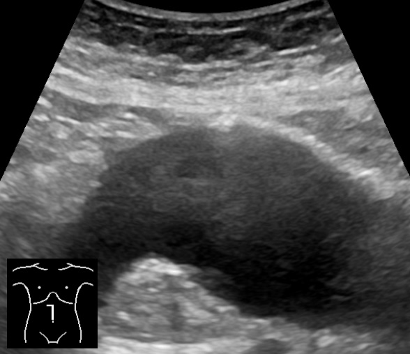
An abdominal aortic aneurysm in long axis. The aorta runs along the spine and balloons into a wide dark sac. A vessel this wide in an older patient with pain points to an aneurysm near bursting.
The probe lays across the midline above the navel. The aorta shows as a round vessel in front of the bright line of the spine. A press through the bowel gas brings the vessel into a clear view. The aorta runs down the belly from the ribs to the navel, where it splits into the two leg vessels. The whole length comes under the probe to catch a bulge at any point. A wide stretch anywhere along the aorta marks an aneurysm. The vessel throbs under the probe, driven by the beating heart. The thick wall and the strong pulse set the aorta apart from the thin vein beside it. Two directions, across and along, measure the vessel true. The round dark aorta sits just left of the midline against the spine. A slow pass from below the ribs down to the split at the navel covers the run. A bulge at any point along that sweep marks an aneurysm to measure. The upper aorta below the ribs can hide behind stomach gas. A steady press pushes the gas aside and brings the vessel out. The lower aorta near the split comes up more readily through a calmer lower belly. A roll of the patient or a slow lean on the probe clears the gas off the vessel. The aorta sits deep in a large belly and asks for a lower frequency to reach it.
The aorta gets measured from outer wall to outer wall. The bedside measurement runs across the full width of the vessel, since a clot lining the wall hides the true size. The calipers sit on the outer edges of the wall on each side. A clot packed against the wall fills part of the vessel and narrows the channel of flowing blood. The dark channel alone can fool a quick eye into taking the vessel for a smaller one. The whole vessel, clot and all, gives the real width. The outer-wall reading gives the size the surgeon plans against. The widest point across the short axis catches the true peak. A borderline width earns a second view before the call of an aneurysm. A long-axis sweep confirms the length of the swollen segment for the surgeon. The short axis, cut square across the vessel at its widest, carries the true width. The two iliac vessels past the split take the same measure, since an aneurysm can sit there too. A wide iliac adds to the map the surgeon works from.
What the scan shows and what it hides
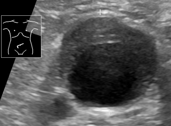
An abdominal aortic aneurysm in cross section with clot lining its wall. The dark round sac can look like one wide channel, since the clot runs nearly as dark as the flowing blood inside it. The calipers cross the whole vessel for the true size. The small diagram at the top left marks where the probe sits on the belly.
Ultrasound finds the aneurysm with near-perfect certainty. The bedside scan shows a wide aorta in about ninety-nine of a hundred people who carry one. The vessel comes onto the screen in seconds, its width noted. No other bedside test names the aneurysm faster. A wide aorta on the screen stands as the cause of a collapse in an older patient. The width settles the call when the story and the pressure point to a burst. The handheld answers the first question with a speed no scanner in another room can match. The aorta comes up through the belly wall in a sweep or two. The vessel sits where the probe expects it, against the spine and just left of centre. The aorta holds its round shape and a strong pulse under the probe, the marks that set it apart from any vein nearby. A glance at the vena cava shows the volume the patient runs on. A flat, collapsing cava in a shocked patient marks a tank run low by a bleed.
The rupture itself hides from the probe. A burst aorta leaks blood backward into the space behind the belly lining. That retroperitoneal space sits deep and shows poorly on ultrasound. The leaking blood of a rupture rarely shows on the bedside scan. The scan shows the aneurysm and leaves the bleed unseen. A wide aorta and a shocked patient drive the call without a wait to see the rupture. A wide aorta in a person with pain and a low pressure goes to surgery as a rupture. The unseen bleed changes nothing about the urgency. The absence of free blood gives no comfort over a wide aorta in shock. The bleed behind the lining drains a life out of sight.
Free blood shows where the scan can find it. A rupture that breaks forward into the belly spills blood the probe can see. The corners of the belly come under the probe for free fluid around the liver, the spleen, and the pelvis. Dark fluid in those spaces over a wide aorta confirms a leak in full flow. The free-fluid sweep adds a finding when the rupture spills into the belly. Free fluid and a wide aorta together mark a burst aorta bleeding fast. The two signs together leave no room for delay. The free-fluid sweep runs on the same probe in the same minute as the aorta.
The aneurysm carries the signs of a vessel under strain. A clot lining the wall can crack and let blood track into the wall itself. A wide aorta with pain and tenderness over it warns of a wall about to give. A tender aneurysm with pain over it stands as a rupture in the making. The size of the vessel and the pain of the patient drive the call. A wide aorta with pain moves toward the surgeon on the strength of those two alone. The scan opens the door, the clinical picture sets the pace. A contained leak can hold the pressure for a while. It can give way without warning at any moment. A wide aorta with pain is a bleed waiting to happen. A holding pressure gives no safety against a contained leak.
The bedside look takes its place in the workup of a collapse. The handheld names the aneurysm and starts the clock. The exact plan stays with the surgeon and the formal scan. The width on the screen and the pressure on the monitor sort the path ahead. A pressure that holds leaves time for a CT angiogram that maps the burst for the repair. The CT names the neck and the leak for the operation. A pressure that falls sends the patient straight to theatre on the bedside scan alone. A scan, a decision, and a move follow in the time the bleed allows. The team gives blood to a falling pressure and holds the patient on the edge until the surgeon takes over. A call for blood goes out early, run through wide lines into the patient. A massive transfusion of blood and clotting factors holds the patient together on the way to theatre. The fluids stay light and the pressure stays low enough to feed the brain, since a hard push of fluid can pop the clot and turn a contained leak into a free bleed. Permissive hypotension is the name for that careful low pressure, a brake on the bleed until the clamp goes on. The team moves as one: the scan at the belly, a nurse drawing blood, a runner calling the surgeon and the theatre. Every step times to the bleed, with no minute wasted on a test that changes nothing. The bedside scan sets that whole response going with one measured width. The patient reaches the vascular team with the aorta sized and the blood already running. The result goes to the surgeon in plain words: a wide aorta of a measured size in a shocked older patient. The surgeon meets the patient already braced for the repair. A chaotic resus turns into an ordered run to theatre.
No time to wait
A burst aorta loses ground every minute. A wide aorta found at the bedside calls the surgeon that second.
Abdominal aortic aneurysm on ultrasound: the numbers
What is measured
Value
What it points to
Normal aorta
under 3 cm
no aneurysm
Aneurysm
3 cm or wider
a AAA
Repair threshold
over 5.5 cm in men, 5.0 in women
high rupture risk
Ultrasound for an aneurysm
~99% sensitive
found in seconds
Retroperitoneal rupture
often unseen on ultrasound
act on size and state
Ruptured AAA, mortality
~80 to 90%
minutes matter
Measuring the danger
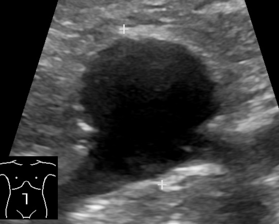
A saccular abdominal aortic aneurysm in cross section. The calipers measure the wide dark vessel from outer wall to outer wall. A bulge to one side like this carries a high risk of bursting.
The width of the aorta sets the risk. A normal aorta runs under three centimetres across. A vessel of three centimetres or wider is an aneurysm. A vessel past five and a half centimetres in a man carries a high risk of bursting. A vessel past five centimetres in a woman crosses the same line. The widest point of the aorta gives the size that drives the plan. The bigger the aorta, the closer it sits to a burst. A vessel past the danger width counts as a threat on its own, with no rupture needed. The width on the screen carries weight on its own in an older patient with pain. The true figure comes from the widest bulge. A tapered end gives a figure below the peak. The single widest figure carries the weight in the call to the surgeon. A wider aorta bursts more often each year. A vessel near five centimetres bursts in a small share of people each year, the share climbing steeply past six centimetres. A growth of more than half a centimetre in a year pushes a borderline aorta toward repair. The width and the speed of growth together set the timing of a planned fix.
The size guides the path even before a burst. A known aneurysm under watch grows over the years toward the line for repair. A quiet aneurysm gets measured at each visit, its slow climb tracked. A vessel that crosses the repair line goes to the surgeon for a planned fix ahead of any burst. A fast-growing aorta moves forward before it reaches the danger width. The bedside scan follows a known aneurysm between the formal scans with no dose at all. A person who carries an aneurysm and turns up with pain earns a scan on the spot for a change in the vessel. A known aneurysm climbing past the repair line goes on before it bursts. A screening scan offered once to older men catches a quiet aneurysm long before it bursts, the one-time look proven to save lives. A handheld brings that same screening look to a clinic with no other scanner.
The shape of the aneurysm adds to the picture. A vessel that balloons evenly along its length forms the common fusiform aneurysm. A vessel that bulges to one side forms a saccular aneurysm, with a higher chance of bursting. The shape, the width, and the length of the swollen segment go to the surgeon. A clot, a bulge, and a tender belly together raise the worry on any wide aorta. The whole vessel from the ribs to the split comes under the probe for the full picture. The aneurysm can reach down into the leg vessels and add to the work of the repair.
The aorta behind the colic
A burst aorta wears the disguise of lesser troubles. The pain of a rupture can pass for a kidney stone, a back strain, or a bout of gut trouble. The aorta of every older patient with flank or back pain comes under the probe before a stone gets the blame. A wide aorta found behind a suspected stone changes the whole emergency in one look. The scan strips the disguise from a rupture an exam would miss. The killer stays in mind behind a complaint that looks routine. The aorta and the kidney come under the probe in the same breath on any older flank pain. The two looks together rule the stone in or the aorta out. The pain of a rupture can throw to the testicle, the hip, or the thigh and pull the eye to the wrong organ. A faint that came and then lifted can mark a contained leak that sealed for the moment. A wide aorta on the screen carries the danger on its own, whatever the belly feels like.
The aorta stays on the list for an older belly. A person past fifty with sudden pain and a faint pulse earns a look at the aorta first. The vessel comes onto the screen before any talk of a stone or a strain. A wide aorta turns a routine pain into a dash to the operating room. The scan catches the killer hiding behind a common complaint. A minute at the bedside on the aorta saves a life now and then.
Scan and go
A collapsing patient gets the aorta and the free fluid in one sweep. The wide aorta comes onto the screen, the belly checked for blood. A wide aorta with a low pressure and pain calls the surgeon at once, ahead of any CT. The scan buys the minutes that decide who lives. An unstable patient goes straight to theatre on the bedside scan alone. The vascular team meets the patient with the size already in hand. A measured aorta and a clear story hand over in the time the transfer takes. A crashing patient turns into a surgical plan in seconds. The blood bank and the theatre get the call alongside the surgeon. The bedside scan stays under a minute so the race to theatre loses no time.
A stable patient earns a fuller map before the repair. A wide aorta with a holding pressure goes for a CT angiogram. The CT maps the neck of the aneurysm, the leak, and the leg vessels for the surgeon. That map sets the choice between a keyhole repair through the groin and an open operation. The CT times to the pressure and the pain of the patient. A holding pressure buys the minutes for the scan that plans the safest repair. The bedside scan starts that path with the width that raises the alarm. A stable patient still earns a close watch, since a contained leak can give way at any moment.
A handheld fits the places a burst aorta strikes. A clinic far from a hospital finds an aneurysm and calls for the transfer that gives a chance. A pocket scanner has named a burst aorta where little else stood to hand. A clinician in a remote post reaches the aorta with a wand and a phone. The scan brings a vascular emergency into view where no scanner stood before. A saved clip travels ahead to the team that will operate. The picture moves faster than the patient and warns the surgeons to stand ready. A remote clinic becomes the opening move in a transfer that ends in theatre.
One probe holds the whole emergency look at the bedside. The aneurysm found, measured, and checked for free blood, the team gets the call on the same wand. A clinic with no scanner works up a collapse and refers the burst aorta on. The aorta comes up in seconds, with no bill and no radiation. An older belly pain gets scanned on the spot, a rupture ruled in or out. A nurse, a junior doctor, or a rural clinician each learn the wide-aorta look. The same probe that found the aneurysm sends the patient to the room that can save them. The skill takes a handful of scans to build and serves for a career.
The bedside scan earns its place in the worst few minutes of a life. The aorta comes up faster than any other test, the danger named on the spot. The width on the screen and the pressure on the monitor set the speed of the response. The patient, the team, and the theatre fall into line behind one fast look. The handheld carries that look to the bedside of a patient who cannot wait. A burst aorta meets a probe and a plan where once there was only delay. The surgeon gets the patient with minutes to spare. One wide aorta found in time turns a near-certain death into a fighting chance.
Common questions
Can ultrasound detect a ruptured abdominal aortic aneurysm?
Ultrasound finds the aneurysm itself in about ninety-nine of a hundred people who carry one. The probe shows a wide aorta in front of the spine in seconds. The rupture itself, a bleed into the space behind the belly lining, hides from ultrasound. A wide aorta with pain and a low pressure gets treated as a rupture, with no wait to see the bleed.
How wide is an aortic aneurysm on ultrasound?
A normal aorta runs under three centimetres across. A vessel of three centimetres or wider is an aneurysm. A vessel past five and a half centimetres in a man, or five centimetres in a woman, carries a high risk of bursting. The aorta gets measured from outer wall to outer wall, since a clot lining the wall hides the true width.
Why does a ruptured aneurysm get missed?
The pain of a burst aorta can pass for a kidney stone, a back strain, or gut trouble. The physical exam alone misses a burst aorta in more than a third of cases. The bedside scan reaches the aorta directly and shows the width the hand cannot feel. The aorta of every older patient with belly or back pain and a low pressure comes under the probe.
What happens with a wide aorta in a collapsing patient?
A wide aorta with pain and a low pressure goes straight to surgery as a rupture. The vascular team gets the call at once, ahead of any CT. The bedside scan hands the surgeon a measured aorta in the time the transfer takes. A stable patient with a holding pressure earns a CT angiogram to map the repair.
About the Author
Julien Mercier
Senior R&D Engineer · Medical Ultrasound Transducer Development
Senior R&D Engineer with an M.S. in Applied Physics and over 15 years of experience in medical ultrasound transducer development, specializing in the design verification and performance testing of high-frequency imaging transducers. Currently leading the development and verification of the company’s next-generation high-frequency linear-array transducer, responsible for imaging performance evaluation and reliability analysis in preclinical testing. Brings extensive hands-on experience in piezoelectric element tuning, beamforming parameter optimization, and system-level performance testing.