Renal colic is the sharp pain of a stone moving down the ureter. Ultrasound shows the kidney behind that pain at the bedside in minutes, on a probe pressed into the flank. A stone lodged in the ureter dams the urine and swells the kidney upstream. The swelling catches the eye first, the dark of backed-up urine spreading through the bright centre of the kidney. The scan turns a writhing patient into a clear picture of the kidney and its drainage. A bedside look runs at the trolley, in the clinic, or on a ward round. The probe finds the swollen kidney, hunts the stone at the bladder, and weighs the danger of the block. A few minutes of scanning give up the cause the flank pain hides. The workup starts here, long before a slot on the big scanner comes free. A patient doubled over with colic gets an answer where they lie. The colicky pain comes in waves, a writhing patient unable to find a still position. The scan asks for no prep beyond a flank to lean on. The picture lands in the first minutes of the pain.
The bedside scan works by an indirect sign. A small stone buried in the ureter behind the bowel gas rarely shows on ultrasound. The scan finds the kidney swollen above the block. A swollen kidney on the painful side points to a stone holding up the urine below. The indirect sign carries the diagnosis when the stone itself stays hidden. A stone the size of a grain of rice can hide behind a loop of gas, beyond the reach of the probe. The kidney above it speaks for the stone the scan cannot see. The pressure of the dammed urine writes the diagnosis in the dark spaces of the kidney.
The probe lays into the flank, angled under the ribs, and brings the kidney into a clean window. A held breath or a roll onto the side drops the kidney clear of the ribs. The dark of backed-up urine stands out against the grey of the kidney. The flank window opens in a sweep or two. A heavier build sinks the kidney deep and asks for a lower frequency to reach it. A flank pain in an older patient also brings the aorta under the probe before any talk of a stone, since a wide aorta gives the same pain. The kidney, the bladder, and the aorta come into one short sweep. The deadly cause clears first. A stone rests easy only once the rest come clean.
A handheld brings this scan to the patient. A clinic with no scanner of its own checks a flank pain at the bedside. A rural clinician arranges the next step with the picture in hand. The swollen kidney, the side, and any stone seen at the bladder go into the note. One device covers the kidney, the bladder, and the aorta in a single visit. A saved clip of the dark kidney travels with the referral to the urologist. A child with a flank pain, a rarer event, comes to the same probe, the swollen kidney seen the same way at any age.
The swollen kidney behind the pain
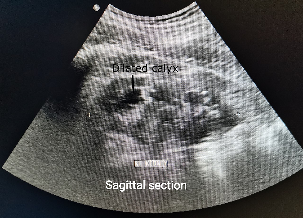
Moderate hydronephrosis of the right kidney, a dilated calyx labelled on the image. Urine backed up behind a blocked ureter opens the dark calyces in the kidney bright centre. The swelling is the indirect sign the bedside scan looks for in renal colic.
The swollen kidney sits at the heart of the bedside look. A blocked ureter backs urine up and opens dark spaces in the kidney’s bright centre. The branching dark of trapped urine shows there. A higher pressure behind the stone opens it wider. The depth of that dark, weighed against the calm kidney opposite, settles the obstruction. The degree of swelling gives a rough measure of the stone below, the full grading of a kidney a study of its own. A heavy swelling on the sore side warns of a larger or stuck stone. The dark pool followed out toward the rim shows how far the pressure has spread. The dark of obstruction branches through the centre of the kidney where the urine should drain in a tight bright line. A swollen kidney on the sore side, checked against the other, leaves little doubt. The comparison of the two sides is the surest call the bedside offers in colic.
Bedside ultrasound finds the swelling in about three of four people with a stone in the ureter. A larger stone gives a surer sign. A small stone can pass with little swelling, the scan near normal. A convincing story keeps the search alive past a calm kidney. A moderate or worse swelling carries a high chance of a real stone behind it. A larger stone and a surer hand both raise the yield. Two planes of the kidney catch a swelling a single view would miss. A faint swelling shows up plainly on a slim patient. A heavier build buries the kidney and the swelling with it. A urine test for blood, checked alongside the scan, backs the picture of a stone on the move. Many stones put blood in the urine, the dipstick a quick partner to the scan. A stone that blocks the ureter fully can shut the blood off, so a clean dipstick never clears a stone on a convincing story.
The swelling depends on how many hours the pain has run. A kidney scanned in the first hour of a fresh stone can look near normal before the pressure builds. A scan a few hours on shows the swelling at its fullest. A first look that comes back clean on a convincing story earns a second scan. The swelling fades once a stone passes and the urine drains free, so a falling swelling on a repeat scan marks a stone on its way out. The kidney tells the story of the block through the swelling it carries. A scan caught too early, before the pressure builds, is the one that fools, the kidney calm above a stone just lodged. A repeat a few hours on, with the pain still raging, shows the swelling the first scan missed.
Finding the stone itself
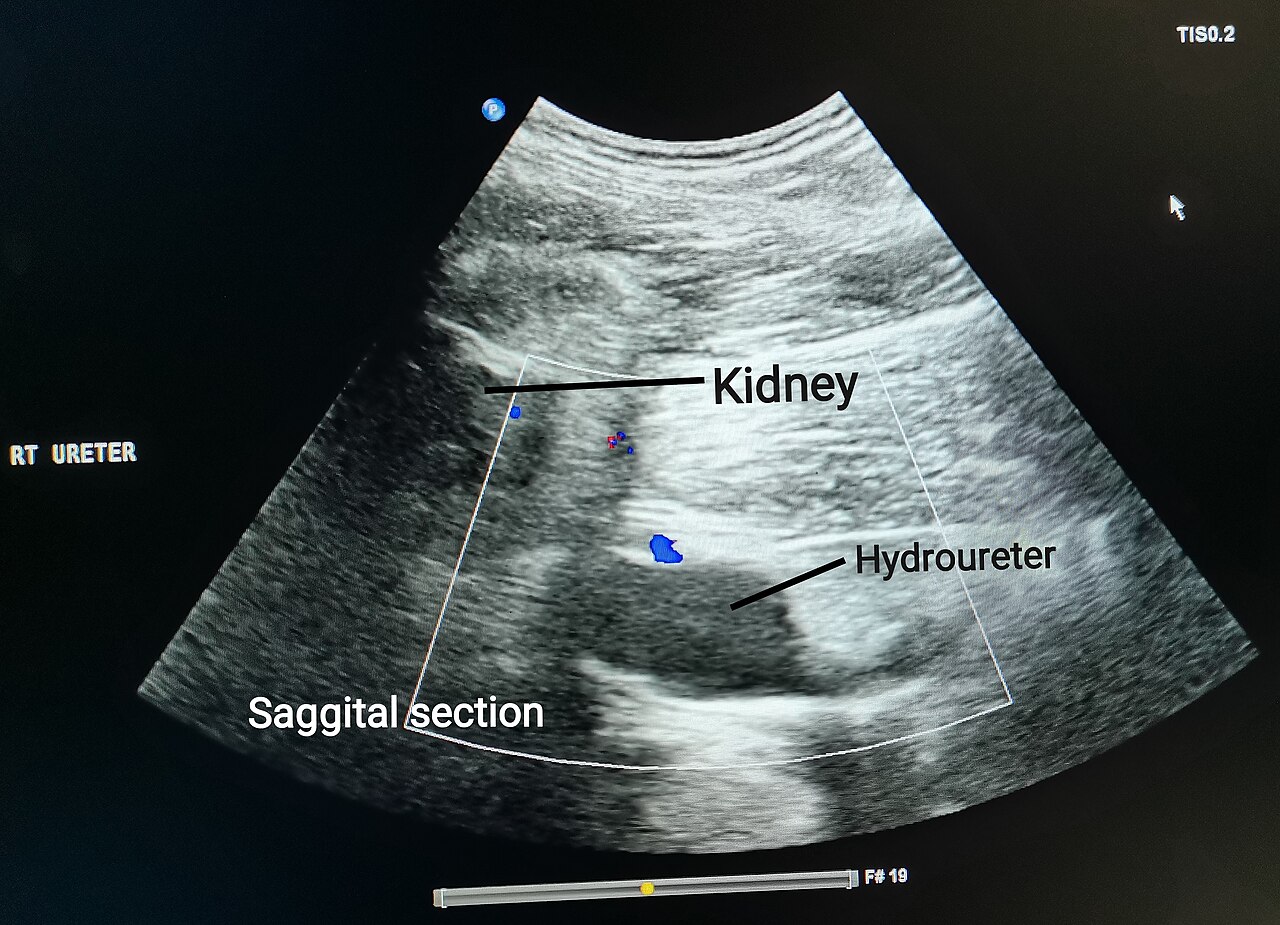
Hydronephrosis with a dilated ureter on a sagittal scan, the kidney and the hydroureter labelled by the original author. The dark tube tracking down from the kidney is the swollen ureter above a stone. Colour Doppler shows no flow inside it, the sign of a urine-filled ureter.
The stone hides where it tends to lodge. Three narrow points along the ureter catch it: the join with the kidney, the crossing of the pelvic rim, and the entry to the bladder. The entry to the bladder catches more stones than the other two. A full bladder turns the lower ureter into a target the probe can reach. A bright stone at the ureter opening shows against the dark urine of the full bladder, a dot with a dark shadow behind it. The shadow is the giveaway of a hard stone. The probe angles down toward each ureter opening and studies the wall for a lodged stone. A bladder held full gives the clearest window onto the stone. The three narrow points sit where the ureter pinches: at the kidney’s outlet, where it crosses the vessels at the pelvic brim, and where it tunnels into the bladder wall. A stone rides down the ureter until a narrow point stops it. The lowest narrowing, at the bladder, traps the greatest share of stones and sits closest to the probe through a full bladder. A stone caught there glows bright at the floor of the dark bladder. A stone sat right at the mouth of the ureter, where it pokes into the bladder, can lift the wall into a small bump the probe picks out. The bump, the shadow, and the twinkle together pin the stone at the junction.
Colour Doppler pulls the stone out of the background. Over a suspected stone at the bladder floor, the colour box throws a burst of mixed colour behind a rough stone, a flicker called the twinkling sign. The twinkle marks a stone the grey picture alone can miss. A high colour gain brings it out behind a stone the eye would skip. A faint bright dot, too small to throw a shadow, turns into a sure stone under that flicker. The flow of urine carries its own sign. A jet of urine spurts from each ureter opening into the bladder on a working side, caught on colour Doppler. A jet that spurts freely on the sore side argues against a complete block. A jet gone missing on the sore side backs a tight obstruction. The colour box held at the bladder floor for several seconds catches a slow jet. A weak, late jet tells of a stone choking the flow. The presence of a jet brings comfort that the ureter still drains past the stone. The swollen ureter adds a third thread. A blocked ureter swells into a dark tube tracking from the kidney toward the bladder, traced to the point where it narrows over a stone. Colour Doppler tells the swollen ureter from a nearby vessel, a tube with no flow inside it marking a dilated ureter past a stone. The dilated ureter leads the eye toward the lodged stone like an arrow. The twinkling sign comes of the rough surface of a stone scattering the colour signal, a quick mosaic of red and blue behind a dot too small to shadow. A turn of the colour gain and the wall filter brings it out where the grey image shows nothing. The three threads, the twinkle, the jet, and the swollen ureter, weave one picture of the stone the kidney could only hint at.
Four signs build the picture of the block together: the swollen kidney, the dilated ureter, the bright stone, and the twinkle. A stone seen at the bladder, measured across its width, sets the odds of it passing on its own. A clip of the stone and the twinkle travels with the referral. Each sign on its own leaves room for doubt. Stacked together, the four name a stone with little left to question. The whole picture costs nothing and carries no dose. A clip saved of the twinkle and the stone hands the urologist a head start. The whole study runs in the few minutes a flank window takes to open.
Ultrasound shows a stone of any make, the soft uric acid kind a plain X-ray lets through. A radiolucent stone hidden from a film shows plainly to the probe. The scan works by echo and catches what the X-ray cannot. The bedside picture names the stone, its size, and the grip it holds on the kidney. A phlebolith, a small calcified vein low in the pelvis, can sit near the ureter and mimic a stone, its place off the ureteric line and the calm kidney above it telling the two apart.
Both kidneys come under the probe on every colic. A person who makes one stone often carries another in the quiet kidney. The far kidney, scanned too, gives up a silent stone hiding without pain. Bright flecks scarring a kidney mark a person who builds stones again and again, a history folded into the plan for the stone at hand. The bedside sweep finds the silent stone alongside the noisy one. A clot or a tumour in the ureter blocks the urine the same way a stone does, a rarer cause the scan keeps in view when no stone shows. The bright shadowing dot of a stone, absent on a swollen kidney, turns the search to the wall of the ureter itself.
The mimic that can kill
A wide aorta mimics a stone in an older patient. The aorta comes under the probe before any flank pain is called a stone.
Renal colic on ultrasound: the numbers
What is measured
Value
What it points to
Stone under 5 mm
passes in ~2 of 3 to 9 of 10
watch and wait
Stone 5 to 10 mm
passes less often
a closer watch
Stone over 10 mm
rarely passes alone
plan to remove it
Hydronephrosis, sore side
~70% sensitive
the indirect sign
Moderate or worse swelling
~94% specific
a larger or stuck stone
Ultrasound first vs CT
~10 vs 17 mSv
less radiation, same outcomes
Stone size and the wait
The width of the stone sets the plan. A stone under five millimetres passes on its own in two of three to nine of ten cases. A small stone sends a person home on fluids and pain relief to pass it over days. A stone of five to ten millimetres passes less often and asks for a closer watch. A stone over ten millimetres rarely passes on its own and goes to a urologist for removal. The width of any stone at the bladder, measured at the bedside, weighs the wait against a procedure. A small stone with a settling pain earns a watch and a repeat look in a week or two. A larger stone or a tight block books an earlier visit to the urologist. The smaller the stone, the freer its pass; the wider it grows, the surer the wedge. A stone of a few millimetres rides the ureter out on a flush of fluid over days to weeks. The width measured at the bedside, against the body’s narrowest point, weighs a quiet pass against a trip to the urologist.
The site of the stone shifts the odds too. A stone low in the ureter near the bladder passes more freely than one high near the kidney. The side, the size, and the place of the stone go into the note for the urologist. A drug that relaxes the ureter, given for a stone in the lower third, eases the passage and shortens the wait. A sieve catches the stone as it drops. The lab names its make, calcium or uric acid or the rest, and steers the plan that holds the next stone off. A flush of fluid through the days washes the small stone along its way. A stone that has not passed in four to six weeks earns a closer plan, the wait on a small stone not open-ended.
The pain guides the watch alongside the scan. A pain that eases with simple relief, over a small stone, points to a stone on its way out. A repeat scan and a check on the swelling bring the person back to the bedside. A falling swelling and an easing pain mark a stone that has passed or sits close. The handheld follows the stone over days with no trip to the radiology room. A pain that keeps climbing, or a swelling that grows, sends the person back for a CT and a urologist. A stone declares itself over a day or two. A pain that sharpens and holds points to a stone wedged fast. A handheld at each visit tracks the swelling down to nothing and times the calm of a passed stone.
When a blocked kidney turns dangerous
A fever over a blocked kidney is an emergency. A stone that dams the urine and an infection above it build a closed system of infected urine under pressure. A swollen kidney in a feverish patient brings the urologist at once. The trapped infected urine needs draining within hours to save the kidney and the patient. Bright specks of debris swirling in the dark urine of a blocked kidney, in a fever, mark pus under pressure. The scan brings that danger to the front of the queue. Pus trapped above a stone, an obstructed infected kidney, is the one true emergency of colic. A tube up the ureter or through the back drains the kidney within the hour. The fever, the swollen kidney, and the debris in the urine together name a system that must be opened fast. A stone left to dam an infected kidney costs the kidney, the patient soon after. A forniceal rupture lets urine leak from an over-pressured kidney into the fat around it, a rim of dark fluid that vents the pressure and eases the pain. The rim of fluid flags how tight the block had grown.
The kidney weighs against the whole patient. A swollen kidney with a rising marker of failing kidneys raises the stakes. A block in a person with one working kidney leaves no spare. A tight block, a fever, or a failing kidney moves along the urgent path. A pregnant patient with the same pain gets the same care with no radiation, the natural swelling of a kidney in pregnancy weighed against the pain and the side. A kidney already weak, or a person down to a single kidney, has no margin for a block. The womb itself presses the ureter, so a swelling can sit there in pregnancy with no stone at all. The call leans on the pain and the side as much as the dark. The bedside scan carries a pregnant patient through a colic with no dose at all. A blocked kidney in a transplant, sat low in the pelvis, comes under the same probe, its swelling judged against no twin on the far side.
One probe in the flank-pain workup
The scan sits inside the workup of a flank pain. The swollen kidney confirmed, the stone hunted, the aorta cleared, the bedside look points the way in minutes. A CT then maps the exact stone and its size for the urologist. A scan-first path matches the outcomes of a CT-first path with far less radiation over a year. The wand comes first in a young person, a pregnant patient, or a repeat stone former who has scanned many times. A CT keeps its place for the older patient, the unclear scan, or a stone that needs its exact size. A scan that answers in minutes moves a colic out of the imaging queue and back to the ward, the CT saved for the cases that earn it. The scan-first path, tested against a CT-first path, reached the same outcomes with far less radiation over the months that followed. A young stone former, scanned again and again across the years, gains the greatest benefit from a wand that adds no dose.
A stone former returns through the years. A scan-first habit holds the dose down each visit. A lifetime of stones counts the saved radiation up to a real number. The handheld lowers the cumulative dose for a person who builds stones again and again. A removal by shock waves from outside, or a scope passed up the ureter, clears a stone too wide to pass, the scan tracking the kidney before and after. A settled kidney on a follow-up scan, its swelling gone, marks a clearance that held. The small radiation of a CT weighs against the clear answer it gives in a hard case.
One probe holds the whole flank-pain look, the block found, the stone hunted, the aorta cleared, the pass tracked. A clinic with no scanner checks a flank pain and refers the dangerous ones on. The scan repeats over days at no cost and no dose. A kidney watched through a stone’s slow journey to the bladder gives up its trend. A nurse, a junior doctor, or a rural clinician each learn the swollen-kidney look. The same probe that found the block follows it to the day the stone drops free. A handheld stored on a charger waits for the next flank pain. The same wand that named the stone tracks it down the ureter over the days. It steps in again the moment a fever or a rising marker turns the case. One probe carries the colic from the first wave of pain to the stone in the sieve.
Common questions
Can ultrasound find a kidney stone in renal colic?
Ultrasound finds the swollen kidney above a stone more readily than the stone itself. A blocked ureter backs urine up and opens dark spaces in the kidney, the indirect sign of a stone. The scan catches a stone at the bladder opening as a bright dot with a shadow. Colour Doppler throws a twinkle behind it. Bedside ultrasound shows the swelling in about three of four people with a ureteric stone.
What is the twinkling sign?
A rough stone under colour Doppler throws a burst of mixed colour behind it, a flicker called the twinkling sign. The twinkle marks a stone the grey picture alone can miss. The colour box sweeps the bladder floor to light up a small stone at the ureter opening. The sign turns a faint bright dot into a sure stone.
Will a kidney stone pass on its own?
A stone under five millimetres passes on its own in roughly two of three to nine of ten cases. A stone of five to ten millimetres passes less often and asks for a closer watch. A stone over ten millimetres rarely passes alone and goes to a urologist. A stone low in the ureter near the bladder passes more freely than one high near the kidney.
When is renal colic an emergency?
A fever over a blocked kidney is an emergency. A stone that dams the urine and an infection above it build a closed system of infected urine that needs draining within hours. A failing kidney, or a block in a person with one working kidney, also raises the stakes. The aorta comes under the probe in an older patient, since a wide aorta can mimic a stone.
About the Author
Julien Mercier
Senior R&D Engineer · Medical Ultrasound Transducer Development
Senior R&D Engineer with an M.S. in Applied Physics and over 15 years of experience in medical ultrasound transducer development, specializing in the design verification and performance testing of high-frequency imaging transducers. Currently leading the development and verification of the company’s next-generation high-frequency linear-array transducer, responsible for imaging performance evaluation and reliability analysis in preclinical testing. Brings extensive hands-on experience in piezoelectric element tuning, beamforming parameter optimization, and system-level performance testing.