BLUE Protocol Bedside Lung Ultrasound Handheld Step By Step
A patient arrives unable to breathe and the cause must be named in minutes. Fluid in the lungs, a pneumonia, a tightened airway, a clot in the lung, air collapsing a lung from outside: five common causes of acute breathlessness, each needing a different, often opposite, treatment. The BLUE protocol turns the probe into a decision tree, a fixed sequence of looks that sorts the five at the bedside in about three minutes. Bedside Lung Ultrasound in Emergency is the name behind the letters; a fast, ordered answer is the gift.
Five causes, one ordered sweep of the probe.
The genius of the protocol is order. It does not ask a clinician to recognise a disease at a glance. It asks them to check a few plain signs in a set sequence and follow the branches to a name, a path even a beginner can walk under real pressure.
The question and the clock
Acute respiratory failure gives no time for the slow workup. A chest film waits for a porter and a reading. The blood gas and the markers come back in their own time. The patient, meanwhile, is drowning, or starving of air, or bleeding into a chest, and the treatments for these pull hard in opposite directions. Fluid poured into a flooded lung sinks it further; the same fluid withheld from a clot changes nothing at all. The cost of a wrong guess is paid in minutes, sometimes in a life. Each minute of the wrong treatment is a minute lost, the lung filling or the airway closing as a disease the patient does not have goes untreated.
The probe answers at the bedside, before the slower tests return. It reads the lung directly, the air and the water and the sliding, turning a frightened guess into an ordered look. The protocol exists to make that look fast, repeatable, hard to get wrong even in untrained hands. It is the rare test that grows more useful the sicker the patient, since the crashing chest is exactly the one a slow workup cannot serve.
Finding the points
The protocol scans a fixed map, the same spots on every patient. It locates them with the hands, never a tape measure. Two hands laid on the chest, side by side, fingers together, the upper hand’s little finger tucked under the collarbone, the lower hand’s edge along the bottom of the lung: this simple gesture marks the standard points without measuring a thing, quick and repeatable in any pair of hands that has learned it.
The upper point sits on the front of the chest, high, over the second or third rib space. The lower point sits where the lower hand rests, near the lung’s bottom edge. These two anterior points catch the front of the lung, the place free air rises to in a patient lying flat and the place the flooding of a failing heart shows itself first. A scan that begins high on the front meets the two findings that matter soonest in a crashing patient: the air of a pneumothorax and the water of a failing heart, both gathered at the top of a supine chest.
The third point lies further back, where the chest meets the bed, the posterolateral spot the protocol calls PLAPS. It catches what gravity pulls down and back: a consolidation of pneumonia, a pool of pleural fluid, the findings the anterior points sail past in a patient lying on their back. Reaching it means rolling the probe down the side until the lung, the diaphragm and the dark of any pooled fluid come into one view, the deepest corner the protocol asks the clinician to find.
Two points in front, one behind, both legs after.
The signs it reads
At each point the protocol reads a short list of plain signs. Is the lung sliding, the shimmer of a lung touching the wall and moving with the breath. Are the lines below the pleura horizontal A-lines, the mark of air, or vertical B-lines, the mark of water in the tissue. Is there a consolidation, a patch of lung gone airless and tissue-like. Is there free fluid pooling behind. Each sign is bold, learned in an afternoon, read in seconds at the bedside. None of the four asks for fine judgement on its own. A line slides or it does not; the artifacts run flat or stand upright; the lung is airy or solid; fluid is there or absent. The protocol leans on exactly that bluntness, building its tree from signs too plain to argue over.
Reading the profiles
The protocol’s power lies in combining those few signs into named profiles. Reading the profile is the verdict. The signs alone say little; their combination, at the right point, says a great deal. Begin at the anterior points. A-lines with sliding on both sides is the A-profile, a dry, aerated front of lung. B-lines with sliding on both sides is the B-profile, both lungs wet to the front, the signature of fluid backed up from a failing heart. The B-profile, in a breathless patient, names cardiogenic pulmonary edema almost on its own. When the sides disagree, A-lines over one and B-lines over the other, the picture is the A/B profile, an uneven flooding that points at pneumonia, not the even tide of a failing heart. A patch of airless, tissue-like lung at the front is the C-profile, a consolidation pressed to the chest wall, pneumonia again. Now the slide itself. A-lines with the sliding gone, at the top of a supine chest, is the A-prime profile, the warning of a pneumothorax, confirmed when the probe finds the lung point. B-lines with the sliding gone is the B-prime profile, a wet lung stuck to the wall, pneumonia once more. The anterior look has already split the field wide. A wet front means the heart or a pneumonia; a still front means a pneumothorax; a dry, sliding front sends the search onward. That dry A-profile is the hinge of the whole tree. It rules the flood out and turns the clinician to the legs and the back, to the two diagnoses a dry front cannot exclude: a clot in the lung, or an airway disease that leaves the surface looking well. Each profile is a sentence in a language of six or seven words, and the protocol is the grammar that turns those sentences into a diagnosis at speed.
The BLUE protocol as a decision tree: anterior points, then the legs, then the back, each branch ending in a diagnosis. Original illustration.
Walking the tree, step by step
The sequence runs the same way every time. First the anterior points, both sides. B-lines there, with sliding, end the search early: the B-profile is edema, and the treatment turns at once to pulling fluid off and easing the heart. A consolidation or an uneven A/B split at the front names a pneumonia. A still pleural line, A-lines without the slide, raises a pneumothorax, sent to the lung point for its proof. Three of the five causes can fall out at this first anterior look, before the probe has moved past the front of the chest, the quickest answers reached first.
A dry, sliding A-profile at the front clears the early branches and moves the search down. The flood is out, the pneumothorax is out, the obvious pneumonia is out. Two suspects remain, and the legs decide between them. The probe leaves the chest for the veins behind the knee and in the thigh, hunting a clot in the same exam. This is the branch that sets the protocol apart from a plain lung scan: a dry, well-sliding front does not end the search, it redirects it, sending the probe to organs the lung windows never showed.
A dry front turns the probe to the legs.
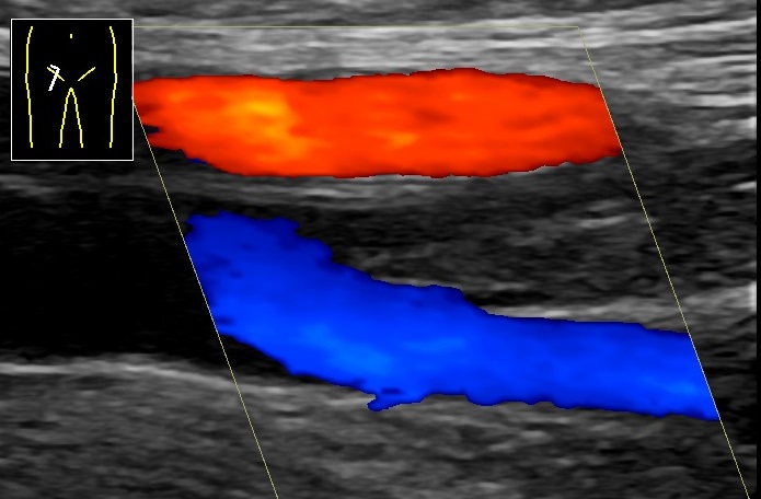
Doppler ultrasound of a clot in the femoral vein. Behind a dry lung, a leg clot points to pulmonary embolism. Wikimedia Commons, CC0.
A clot in a leg vein, behind a dry-fronted breathless chest, names a pulmonary embolism, the lung dry since the trouble sits in its vessels. No clot sends the probe to the PLAPS point at the back. A consolidation or a pool of fluid found there is the last face of pneumonia, hidden from the front, caught from behind by the one posterior look. Pneumonia, in the end, can announce itself at any of the points, in front as a consolidation or a split, behind as a PLAPS, which is why the protocol checks them all before it rests.
With the front dry, the legs clear, and the back clear, one suspect is left standing. The nude profile, a dry sliding lung and nothing else anywhere, names an airway disease, the asthma or the chronic lung that tightens the pipes. The tree has reached its last leaf, and the answer stands. Five causes have been sorted by a fixed walk of the probe, no step of it requiring a judgement a beginner could not make.
Where the tree bends
The protocol is fast and accurate, never infallible. It reads the dominant pattern, and a patient with two diseases at once can hide the quieter one behind the louder. A septic patient in heart failure, a pneumonia on top of a worn lung: the tree shows the strongest branch and can fall silent on the second, leaving a clinician to find it by other means. The diagnosis of embolism is inferred, never seen directly, resting on a dry lung and a leg clot, never a direct look at the blocked vessel in the chest. A clinician who treats the embolism branch as proof, in place of a strong pointer, leans harder on the leg veins than they can always bear.
The tree is read with the patient, never alone. It is built for acute breathlessness, never the slow grumble of a chronic disease. A clinician who knows its branches and its blind spots uses it as a fast first sort, then weighs the answer against everything else the patient and the history bring to the bed. Used inside its limits, as a first sort and never a final word, it is among the strongest three minutes a clinician can spend on a breathless patient.
Learning the sequence
The protocol is learned as a path, never a pile of facts. The signs come first, each bold and quick: sliding, A-lines, B-lines, consolidation, free fluid, a leg clot. Then the sequence that strings them, the fixed order of the points and the branches that run between them. A clinician who drills the tree until it runs without thought can sort a crashing, breathless patient in the minutes that decide the outcome, the path doing the remembering when the mind is busy elsewhere. The order is the safeguard against panic: the same points in the same sequence on every patient, ensuring a step is never skipped in the rush of a resuscitation.
Learn the signs, then drill the order until it runs itself.
What it changed
Before the protocol, acute breathlessness was a guess refined slowly, treatments started in parallel as the films and the bloods caught up. The clinician hedged, gave a little of everything, waited to see what helped and what harmed. The BLUE protocol put a fast, ordered answer in front of them, a near-certain name for the cause inside three minutes, with a probe already in the hand from the trauma survey. A diagnosis that once leaned on a chest film, a blood gas and an anxious wait now arrives in the time it takes to lay a probe across a chest and roll it to the back.
The deeper change was turning a hard recognition into a simple sequence. The protocol asked not for the eye of an expert, only for the discipline to follow a tree. That shift, from judgement to algorithm, is what let lung ultrasound spread from the few to the many, a crashing chest read the same way in any pair of trained hands, anywhere a probe could reach a patient. What had been the preserve of a few skilled hands became a routine any trained clinician could run, the same crashing chest read alike in a teaching hospital and a rural clinic.
About the Author
Julien Mercier
Senior R&D Engineer · Medical Ultrasound Transducer Development
Senior R&D Engineer with an M.S. in Applied Physics and over 15 years of experience in medical ultrasound transducer development, specializing in the design verification and performance testing of high-frequency imaging transducers. Currently leading the development and verification of the company’s next-generation high-frequency linear-array transducer, responsible for imaging performance evaluation and reliability analysis in preclinical testing. Brings extensive hands-on experience in piezoelectric element tuning, beamforming parameter optimization, and system-level performance testing.