Small Parts Ultrasound Handheld Superficial Imaging
Small parts ultrasound is the high-frequency scan of the small, shallow structures of the body: the testes, the salivary glands, the lymph nodes, and the lumps that rise under the skin. A linear probe held over any of them draws fine detail at a finger’s depth. Colour Doppler reads the blood that runs through them. The field answers two plain questions at the bedside. Is this an emergency, and is this lump dangerous? A handheld carries that answer to wherever the patient is.
The superficial organs
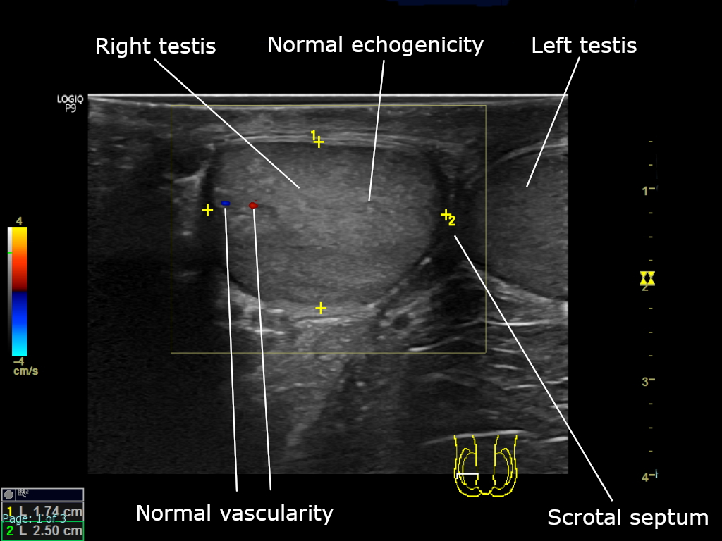
A normal scrotum on ultrasound, both testes seen together in one transverse view. The even grey of normal echogenicity, the colour dots of normal flow, and the midline septum are all labelled. Reading the two sides together is the heart of the small-parts method. The labels, arrows, and calipers are the author’s. Image: Cerevisae, CC BY-SA 4.0.
The field is held together by depth. The structures it reads sit within a centimetre or two of the skin, each small enough that a high-frequency probe draws it in fine grain. A testis, a parotid gland, a neck node, a lump in the fat, all share that shallow seat. The probe that reads one reads them all with no change of setting. The scrotum is the busiest of the small parts, a testis and its cord and the layers around it all read in one scan, the two sides laid side by side for the comparison that drives the field. The breast and the thyroid are small parts too, each grown into a field of its own with its own pages. What remains here is the rest of the superficial body, the scrotum, the glands, the nodes, the lumps, gathered under one method. The skill is one across the whole breadth of it, a reader who learns it on the scrotum carrying that skill to the neck, the parotid, the lump in the arm, with little to relearn. The whole field lives in the first few centimetres. A structure deeper than that passes to a lower-frequency probe and another kind of scan, the small parts the province of the high-frequency head alone.
The organs are an odd assortment. A testis hangs in the scrotum, a salivary gland sits in the cheek and the jaw, lymph nodes string down the neck and the groin, and a soft-tissue lump can rise anywhere on the body. What they share is the surface, a shallow window that suits a small linear probe as well as any tissue does. Each organ brings its own normal. A testis reads as an even mid-grey oval, a parotid as a smooth slightly brighter gland, a node as a flat oval with a bright core. A reader holds each normal in mind and reads the lump that breaks it. The lump a patient brings is the start of every scan, a thing felt under the fingers that the probe turns into a thing seen. The hand finds the lump; the probe reads it. The salivary glands sit just under the skin of the face and the neck, the parotid before the ear, the submandibular under the jaw. A stone in a duct, a swollen gland, a lump within one, all read on the same superficial scan.
The questions they raise rhyme across the organs. A swelling that may be an emergency, a lump that may be a cancer, a gland that may be inflamed, all come to the same probe for the same kind of read. The skill of one small part carries to the next, the patterns of the normal and the abnormal echoing from organ to organ. The patient brings the same handful of complaints to each organ: a lump felt, a pain come on, a swelling noticed. The scan meets the complaint where the organ sits and reads it the same way each time. A patient rarely knows which organ a neck lump comes from. The scan that reads the lump often names the organ behind it, a node from a gland, a cyst from a duct, a mass from the muscle.
The probe for the job
The work calls for a high-frequency linear probe, the same flat-faced, fine-grained head that reads a thyroid or a tendon. A high frequency, scaled to the shallow depth, draws the fine structure of a small organ that a deeper probe would blur. The cost of that detail is reach, of no concern where the targets all lie within a centimetre or two of the skin. The probe runs at the high end of the range, often above ten million cycles a second, the figure climbing for the shallowest work. A small footprint helps in the tight spots, the curve of a jaw or the fold of a groin, where a long probe cannot sit flat. One probe covers the field, a single linear head swapped between a scrotum and a neck and a forearm without a thought, the tool as portable as the method. Some small parts are read in motion, a testis as it is squeezed for its reflex, a gland as a sour taste sets its duct flowing, a tendon-side lump as the joint bends, the live picture catching what a frozen frame would miss.
Colour Doppler is half the field. The flow in a small part tells as much as its shape. A testis with no flow is twisting, a node with flow at its rim is suspect, a mass packed with chaotic flow is a tumour. A probe that reads the grey and the colour both is the tool the field is built on. The grey picture maps the structure, its size, its shape, its texture. The colour lays the blood over that map, the flow the structure cannot show on its own. A reader runs the grey first, then the colour, the two together settling the call. The depth is set shallow and the focus laid on the structure, the whole screen given to the first few centimetres, a structure that fills the picture read in its finest grain. The colour setting is tuned for the slow flow of a small part, set sensitive enough to catch the trickle in a small node, with the noise of an over-eager setting kept from showing as flow. A reader keeps the box small and laid over the spot in question.
The one method
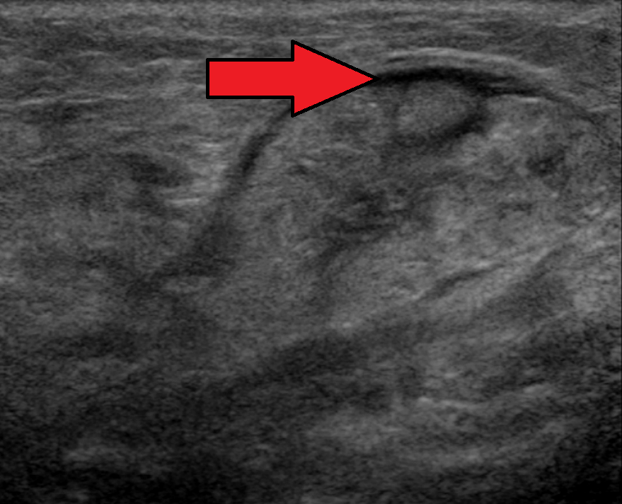
A submandibular salivary gland on ultrasound, one of the small parts the field covers. The red arrow, added by the author, marks the abnormal area in an inflamed gland. Image: James Heilman, MD, CC BY-SA 4.0.
Every small-parts scan runs the same few steps.
The scan begins by reading the structure for what it is. A normal organ has a look the eye learns by heart: the even grain of a testis, the smooth body of a parotid, the bright fatty hilum of a node. A lump is read against that normal, its shape, its edges, its texture, and its echoes all weighed for how far they stray from it. The probe sweeps the whole structure in two planes, the long view and the cross, nothing left to hide in a single slice. The next step is the comparison. The sound side is read against the sore one, the right testis against the left, the swollen node against its calm neighbours, the lump against the tissue around it. The other side is a ruler each patient carries, calibrated to their own normal, a difference between the two standing out where a single view would pass for ordinary. Then comes the colour. Doppler laid over the structure reads the blood, the pattern of that flow often settling the question the grey picture only raised. Flow gone from a painful testis, flow crept to the rim of a node, flow run wild through a mass, each turns a suspicion into a finding. The last step is the decision, always one of a short list: leave it, watch it, sample it, or send it on now. The whole method, learned once, runs the same over every small part the probe is laid on. The order holds across the field: look, compare, colour, decide. A reader who keeps that order misses nothing, the sequence the guard against the eye that jumps to a guess. The few steps cost a minute and miss nothing, the speed of the read no enemy of its care, the same eye that reads one organ reading the next. Measurement runs through it. A lump is sized in three planes, a node’s short axis read against its long, a testis measured against its mate, the numbers going on the record for a later scan to read the structure against its old size. The sweep is total. A reader covers the whole organ end to end, every face of a lump, the length of a node chain, since the one patch left unscanned is the one that hides the finding.
Knowing the normal is the whole foundation. A reader who has scanned a hundred healthy testes, glands, and nodes reads the abnormal one at a glance, the change leaping out against the remembered normal. The field is learned from the normal up, the odd one named by how it breaks the familiar pattern. The testis runs an even speckle, the parotid a fine bright body, the node a dark rind around a bright core. A reader who carries these in the eye reads the break from them in a moment. A reader builds the library of normal by scanning the healthy, the routine scan that finds nothing the one that teaches the eye best. The eye that knows a hundred normal nodes names the odd one without a second thought. The normal varies with the patient, larger in a big adult, smaller in a child. A reader holds that range in mind, a structure at the top of normal for one patient reading plainly enlarged in another, the comparison to the other side the surest guide.
Colour earns its place across the field. The flow it shows is the sign grey-scale alone would miss: the dead testis that still looks grey, the live tumour that hides in a bland lump, the inflamed gland that reads only a little dark. A flick of the colour box turns a maybe into a yes or a no on scan after scan. The colour is read for its pattern as much as its presence. Flow crept to the rim of a node worries the reader. Flow filling a mass in a chaotic tangle marks a tumour. Where the flow runs is read as closely as whether it runs at all. A node lit at its centre is the calm one. The reader reads the place of the flow before its amount, a rim of colour the louder warning.
The method outlasts the organ. A reader fluent in it moves from a scrotum to a neck to a forearm without losing the thread, the same characterise, compare, colour, and decide carried from one to the next. The skill compounds, each small part learned making the next quicker to read. The decision is written plainly for the next reader: leave, watch, sample, or refer. A clear call on the page guides the hand that acts on it, the scan ending in a clear verb.
The emergency and the lump
One question the field answers is whether a swelling is an emergency. A testis that has twisted on its cord loses its blood supply. The hours to save it are few. Colour Doppler that finds no flow in a painful testis, read against the full flow in its mate, is the finding that sends a patient to the operating room without waiting on anything else. The scan there is the difference between a saved testis and a lost one. A swelling that has come on fast and hurts is read for the emergency first, the torsion ruled in or out before anything else. The torsion scan is read fast and acted on faster. A clinician with a probe in a busy department can have the answer in the minute it takes to lay colour over both testes, the saved hours bought at the bedside. A testis robbed of its blood for too many hours cannot be saved. The scan that confirms the twist early is the one that beats the clock. A high suspicion does not wait on the scan when the picture and the pain already agree.
The other question is whether a lump is dangerous. A solid mass in a testis, a hard node with no fatty hilum, a deep lump packed with flow, each carries the marks that send it for a needle or a knife. The field reads those marks the same way wherever the lump sits, the shape and the flow weighed against the look of the benign. A mass that reads solid is the worry, in the testis above all, where a solid mass is a cancer until proven otherwise. The probe tells solid from cystic in a second, the first fork on the road to the answer. The testicular mass is the gravest of the lumps, a solid mass inside the testis treated as a cancer from the first, the scan starting a path that runs to surgery. The probe that finds it early finds a cancer at its earliest. The two grave questions, the emergency and the cancer, are the ones the field exists to answer. A torsion caught saves an organ; a cancer caught early saves a life. The rest of the work is the reassurance that neither is present. The field stands ready for the worst and meets the ordinary, the same probe laid on the twisted testis and the harmless cyst alike.
Far more often, the answer is reassurance. A simple cyst, a soft lipoma, a reactive node, a normal gland, the scan names the harmless thing and sends the patient home easy. The field spares more needles than it calls for, the plain benign read its commonest gift. A cyst, a lipoma, a reactive node, a normal gland, named and released, make up the bulk of a clinic’s small-parts work. The scan that reassures saves a patient a fright and a needless test. For every torsion or cancer, a clinic reads scores of cysts, lipomas, and reactive nodes, each named and let go, the daily work of the field the quiet lifting of a worry far more than the catching of a disaster.
Between the two sits the inflamed organ. A swollen tender gland, a hot node, an abscess in the fat, each reads as an angry structure lit up with the flow of inflammation. The scan names the inflammation and points to the cause, a course of antibiotics or a drain. No cancer to fear sits behind it. The history of pain and heat, read beside the picture, settles it, the angry tissue and the fever telling the same story. An inflamed gland or node settles with treatment, the scan following it back toward normal over the weeks. A swelling that grows under treatment is the one the reader looks at again, the failure to improve a flag of its own.
Ultrasound sorts and points; it rarely names the cells. A solid testicular mass, a hard node, a deep lump, each reads as a structure under suspicion until a needle or a surgery reads what it holds. The scan’s job is to flag the lump and steer the test, the final word left to the pathologist. The scan ranks a lump from clearly benign to clearly suspect. The needle reads the cells the picture only guessed at. The two together, the look and the cells, settle a small part that neither could settle alone. The scan that knows what it cannot tell is the trustworthy one. A reader who writes likely benign, or cannot exclude, names the limit honestly and lets the needle close the gap. The picture and the needle finish the work the one starts, neither the whole answer on its own.
The overlap is the limit. Cancer and inflammation can wear the same grey face, the picture not always able to part the two. A reader who leans on the grey too hard is the one a cancer fools. Humility, and a low bar for sending the doubtful on, keep a reader on the safe side.
The handheld earns its place inside those limits. It catches the lump, names the clear cases, flags the doubtful, and starts the test, the work no imaging department does any better at the first look. The scan that knows its own edge is the safe one.
On a handheld
Small parts is the ideal home for a handheld. The targets all sit shallow, square in the sharp range of a small linear head. The exam leans on the probe and the colour more than on any setting a cart adds. A clinician who feels a lump, or meets a swollen, painful organ, reads it on the spot, the answer given in the same visit. The exam asks nothing of the patient but to bare the part. No fasting, no dye, no wait, the scan done where the complaint is brought. The probe goes to the patient in the bed, the chair, the cot, no trip to a scanning room asked of a sick or a frightened one. A worried patient with a lump leaves the room with an answer, the wait for a distant scan spared, the relief of a benign read given on the spot the field’s quiet daily good. The small parts are where a pocket scanner does its plainest good, a clear answer to a felt lump in the time of a single visit.
The reach carries the scan to the bedside, the clinic, the field. A twisted testis is caught before a trip to a far hospital costs the organ, a worrying lump marked for a referral on the spot, a reactive node sent home with reassurance. The image saves to the phone, ready for a specialist to read from afar. A clip saved of a normal and an abnormal organ teaches the next learner the difference, the even testis beside the twisted one there to study away from the patient. The cost has fallen far enough that a clinic, an emergency room, or a rural post can own the probe that reads its own small parts, the work that once meant a referral done in the room where the lump is found. A pocket probe reads the thyroid, the node, the lump in the same visit a hand first finds them.
Common questions about small parts ultrasound
What does small parts ultrasound cover?
The small, shallow structures of the body: the testes and scrotum, the salivary glands, the lymph nodes of the neck and the groin, and the lumps that rise under the skin. A high-frequency linear probe reads them all in fine detail. Colour Doppler reads the blood that runs through them.
Which probe is used for small parts?
A high-frequency linear probe, the same flat-faced head used for the thyroid and the tendons. Its high frequency draws the fine structure of a small organ at a shallow depth. The detail comes at the cost of reach, which costs nothing where every target lies within a centimetre or two of the skin.
Why does colour Doppler matter in small parts?
The flow tells as much as the shape. A testis with no flow is twisting, a node with flow crept to its rim is suspect, a mass packed with chaotic flow is a tumour. The flow the grey picture cannot show, the colour reads, and a reader who runs both together is doing what the field is built on.
Which small parts scan is the true emergency?
The scan for a twisted testis. A testis that has turned on its cord loses its blood supply. Only a few hours stand between a saved testis and a lost one. Colour Doppler that finds no flow in a painful testis sends the patient straight to surgery, the scan the fastest path to the answer.
Can a handheld ultrasound do small parts work?
Yes. It suits the field as well as any. The targets sit shallow, within easy reach of a small linear probe. The exam leans on the hand and the colour more than on the machine. A clinician reads a lump or a painful organ at the bedside and reaches an answer in the same visit.
About the Author
Julien Mercier
Senior R&D Engineer · Medical Ultrasound Transducer Development
Senior R&D Engineer with an M.S. in Applied Physics and over 15 years of experience in medical ultrasound transducer development, specializing in the design verification and performance testing of high-frequency imaging transducers. Currently leading the development and verification of the company’s next-generation high-frequency linear-array transducer, responsible for imaging performance evaluation and reliability analysis in preclinical testing. Brings extensive hands-on experience in piezoelectric element tuning, beamforming parameter optimization, and system-level performance testing.