A cervical lymph node ultrasound reads the lumps in the neck to sort the harmless from the ones that need a needle. A high-frequency linear probe sizes a node, looks for the fatty core that marks it as reactive, and reads the flow inside it on Doppler. The shape, the core, and the pattern of vessels together place a node on a scale from plainly benign to clearly suspect. Nearly every node a neck scan turns up is reactive and needs nothing more than a second look.
The node that reassures
A reactive node has a look to know by heart, since it is the look that lets the rest go unsampled. It is an oval, longer than it is wide, with a thin rind of darker tissue, even all the way round, wrapped about a bright streak at its core. That bright streak is the fatty hilum, the gateway where the vessels enter. Its presence is the surest reassuring sign a node can show. A node like this, swollen by a passing infection, is the commonest finding in any neck. Nodes run in chains down the neck, scores of them in a healthy person, each a small oval the body keeps as a filter. A node’s job is to filter. A busy filter swells. A neck fighting a cold fills its nodes with cells, every one along the drain enlarging together, the swelling that comes and goes with an illness the mark of a node doing its work. A swollen node that hurts and settles is the everyday work of a healthy neck, nothing the scan need flag. A reader thinks nothing of a string of little ovals with bright hila, the reassurance in the pattern repeated down the chain. The reading starts with the easy ovals and pauses only at the one that breaks the pattern, the eye trained to slide past the ordinary and stop at the odd.
Doppler completes the friendly picture. A reactive node carries its flow from the hilum outward, a neat spray of vessels fanning from that central core. The flow is orderly, branching the way a tree branches from its trunk. An oval node with a bright hilum and this central flow is a node a reader keeps under a calm watch. The dark rind around the hilum is the cortex, the working tissue of the node. In a reactive node the cortex stays a thin, regular band the whole way round, swelling a touch in an infection, smooth from edge to edge.
The features that worry
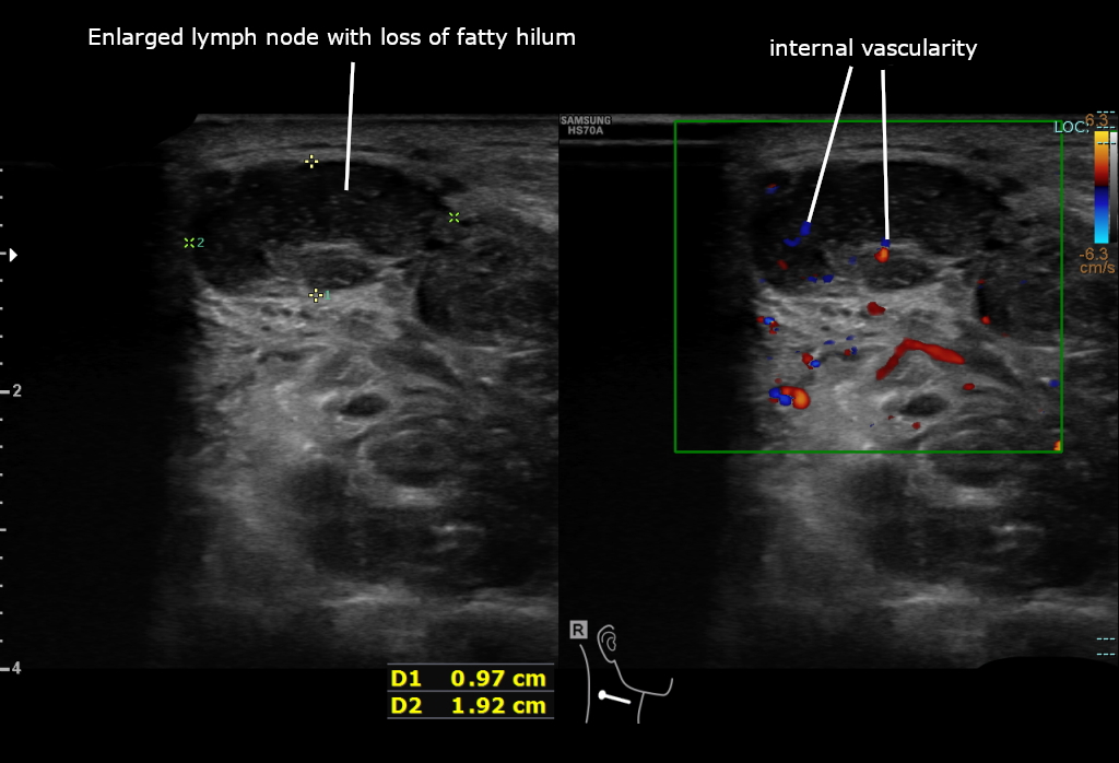
An enlarged neck node with the marks of malignancy. The grey panel shows a round node that has lost its bright fatty hilum, sized by the calipers. The colour panel shows the abnormal vessels growing inside it. The labels and arrows are the author’s.
Trouble shows when those friendly signs fall away.
The first thing to go is the shape. A node infiltrated by tumour swells against its own grain and turns round, its short axis growing to meet its long one. A reader measures the two and watches the ratio climb toward one, a round node carrying far more suspicion than an oval one of the same size. The next sign is the loss of the hilum. As tumour fills the node, the bright fatty core is squeezed thin, the streak narrowing and then vanishing, until the node reads as an even dark mass with no gateway left. A node that has shed its hilum has shed the clearest mark of a benign one. The absence ranks among the surest signs a scan can read. The flow changes last and tells the sharpest tale. A malignant node grows its own vessels in from the rim, the Doppler lighting up around the edge and through the capsule in a scattered, disorderly tangle. That peripheral flow, creeping in from the outside, is a major mark of a node gone bad. The eye is drawn to a thickened corner, a lost streak, a rind of colour, each a place the node has stopped reading like its calm neighbours. The eye returns to the hilum first on every node, its bright streak the quickest yes-or-no a neck offers. The ratio carries a rough rule of its own: a short axis grown past half the node’s length has rounded toward the malignant shape. The cortex gives its own warning, thickening on one side into a lump that bulges the outline, the uneven, eccentric swelling a tumour makes as it seeds one corner of the node first. A dark, structureless patch inside a node is necrosis, the dead centre of a node that has outgrown its blood supply, a sign that carries weight in a metastasis and in tuberculosis alike. A round, dark node with no hilum and a rind of peripheral flow carries the weight of three signs at once. A reader who sees the three together reaches for a needle with little doubt.
A node can carry two rarer marks, each one damning. Bright specks of calcium scattered through it point to a spread from a papillary thyroid cancer, the same dots that sit in the parent tumour. A node that turns part cystic, a dark pocket opening inside it, points to a spread from a papillary thyroid or a throat cancer, or to the breakdown at the heart of tuberculosis. Either sign, on its own, carries a node straight to a needle. A clear pocket inside a neck node points to a spread or a breakdown, never to nothing of concern. The calcium that scatters through a metastatic node shows as fine bright specks, the small dots of a papillary thyroid spread, each too small to throw a shadow of its own.
Size alone settles nothing. The shape, the hilum, and the flow weigh ahead of the bare measurement, the look of a node telling far more than its width. A reader logs the size to track a node across scans, watching whether it grows. The risk is read off the features themselves, the centimetres a record more than a verdict.
No single sign is the verdict. A node is read on the weight of all its features together, the worrying ones counted against the reassuring few. One worrying sign on its own may earn a watchful repeat scan. Three together earn a needle. The skill lies in reading the whole node, never one mark alone. A node read in doubt earns a scan in a few weeks to see which way it moves. A node that hardens or grows over that interval is sampled. The repeat scan is the cheapest test the neck offers. The reading is built like a verdict, each feature a piece of evidence weighed. A reader names the worrying signs aloud, counts them, and lets the count guide the call. A node can carry a worrying shape and a calm flow, or a lost hilum and a normal outline, the signs not always arriving together. The reader reads each on its own and adds the weights, the mixed picture the everyday reality of the neck.
What the cause looks like
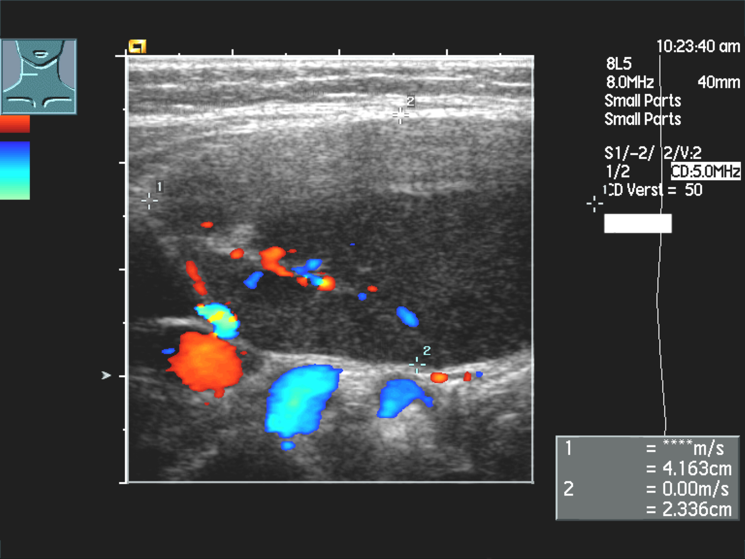
A large lymph node in the neck, its echoes dark, colour Doppler packing it with flow, the picture of a lymphoma node. Its size shows in the calipers at the right. A node this dark could pass for a cyst until the flow inside it marks it as solid.
The cause behind a malignant node often shows in its look. A node carrying a spread from a head-and-neck cancer reads as a firm, round, dark mass, sometimes with the cystic change or the calcium that betrays its origin. These nodes gather in the levels that drain the primary tumour, a map a surgeon reads to plan the neck. The neck drains the mouth, the throat, the voice box, and the thyroid. A spread there often arrives before the primary tumour is ever felt. A firm node found in the right level can be the first sign of a cancer hiding in the throat, the scan pointing the search toward its source. A spread climbs the chain in a known order, the cancer of one corner of the mouth seeding the nodes of one level first. A surgeon reads the level of an involved node as a clue to the primary and a guide to which nodes to clear. The cause read off the look is a guess to confirm, the needle telling which story a node holds. The first node to fill is the sentinel of the spread, the one nearest the tumour on the drain. Finding it early, before the others swell, can turn a wide cancer into a small one.
A lymphoma node looks different again. It runs large, its echoes strikingly dark, dark enough to pass for a cyst, the flow inside it the sign that the mass is solid. The nodes come in numbers, round and gathered in chains down the neck, the whole picture pointing away from a single spread toward a disease of the lymph system itself. A lymphoma node holds a fine, net-like grain inside its darkness, a reticular pattern a careful eye picks out. The flow runs through it in a branching tree, kept or even heavy, the sign that tells it from a simple cyst. Lymphoma asks for a core of tissue, not just a smear of cells, since the disease is named on the way a node is built inside. The scan that finds the chain of dark nodes guides the wider needle that takes that core.
Tuberculosis writes its own story in the nodes. The nodes mat together into a clump, their borders blurring into one another. Pockets of dark breakdown open inside them where the tissue has died. A cold abscess, a soft swelling with no heat over it, can track from such a node. The pattern, in the right patient, points to tuberculosis before any other test. Tuberculosis favours the nodes low in the neck and above the collarbone. A needle into such a node draws material sent for the tests that name the organism, the diagnosis made on the sample the scan guided. Tuberculosis of the neck runs in the patient the disease favours, the young, the recently arrived, the run-down. The scan reads the matted, broken nodes; the history and the test name the cause.
The everyday reactive node stands apart from all of these, swollen by a cold or a sore throat, its hilum bright and its flow central, settling on its own within weeks. A node that grows, hardens, or sheds its hilum across a few scans is the one that breaks from the benign crowd. The repeat scan is what tells the slow reactive node from the early malignant one. A reactive node answers a cause nearby: a sore throat, a dental infection, a cold. It grows fast, stays tender, and shrinks once the cause clears. Tenderness leans toward the benign. The commonest neck lump of all is a reactive node a child grows with every cold. A reader learns to leave these be, scanning to reassure, the skill as much in what goes unsampled as in what earns a needle.
Where the nodes sit
The neck is mapped into numbered levels, one through six, each draining its own territory. The level a node sits in is reported with it, the surgeon reading the scan told at once which region a spread has reached. The level of a worrying node points back toward the organ it came from, a guide to where the hidden primary may hide. The map a sonographer builds of the neck becomes the map a surgeon clears. Level one sits under the jaw and the chin, levels two through four run down the great vessels, level five fills the back triangle, and level six guards the midline around the thyroid. A node in level six, low against the midline, speaks of the thyroid above all. The level system is a shared language, the same numbers used by the radiologist, the surgeon, and the oncologist. A node reported in level three means the same thing to all three, the report a map they all read the same way. A scan that misses a level misses the nodes that drain there. A reader works every level in turn, the sweep a fixed routine the reader never skips. A node out of its expected level is a clue of its own, a spread that has jumped or a primary in an odd place.
Taking a sample
A node that reads suspect ends in a needle. An ultrasound-guided fine-needle aspiration draws cells from the node under the probe’s eye, the tip steered into the worrying part of it, clear of the vessels packed around the neck. The cells go to a pathologist, who names the spread or the lymphoma or the infection behind the swelling. The scan flags the node, the needle settles it. The aspiration is a quick clinic procedure under local anaesthetic, a fine needle passed two or three times into the node, the soreness gone by the next day. Bleeding is slight, the great vessels held clear under the probe’s eye the whole time. A clinician scans the node, numbs the skin, and guides the needle in one sitting, the cells smeared on a slide on the spot. A pathologist next door can say at once whether the sample holds enough to read. The needle for a node is fine, finer than the lump it samples, and it leaves nothing behind but a day’s soreness. A node already treated leaves a scar bed, a changed patch a reader learns to read apart from a true regrowth, the surgical change fading over the months that follow.
The probe is what makes the sample land true. A node a centimetre wide, buried beside the carotid, is a target a blind needle could never reach safely. Guided, the needle takes its cells from the exact spot the scan flagged and stays clear of the great vessels the whole way in. The scan that raised the alarm places the needle that answers it, in one sitting. The vessels of the neck make the guidance matter more than almost anywhere. The carotid and the jugular run close beside the nodes. A needle placed by feel courts a vessel the guided one slips past. A good sample is the gift of a steady hand and a clear picture, the needle landing where the worry lives.
A node can give more than cells. Fluid washed from the needle is tested for the proteins of a thyroid cancer, a trace that betrays a spread even when the cells are too few to read. A core of tissue, taken with a wider needle, gives the pathologist the architecture a lymphoma is named on. The needle is matched to the question the scan has raised. A node that comes back inconclusive earns a repeat, since a node that earns a needle earns a careful one. The pathologist and the sonographer work the same node, the picture and the cells read together toward one answer. A benign smear ends the worry. A malignant one starts the staging and the plan. The cells, not the picture, write the final line. A node in doubt sends the needle back or the patient to a core, the answer chased until it is found.
Where ultrasound stops short
The scan reads a node well. Naming the cells inside it is past the picture. A node with every worrying feature is still only a node under suspicion until a needle proves the cause. The picture narrows the odds and points the needle, the final word left to the pathologist.
The overlap is the hard part. An early spread can sit in a node that still looks calm, its hilum not yet lost. A florid reaction can swell a node into a round, angry lump for a few weeks. The honest read sits between two errors: calling every round node a cancer, and waving every oval through. The first frightens patients needlessly. The second misses the early spread that hides in plain sight.
The answer is the follow-up and the needle. A bolder read of the grey is no answer at all. A doubtful node is watched or sampled, never called on a single uncertain scan. The handheld earns its place by making that repeat easy, a quick scan the patient can have again and again at no cost but a few minutes. The grey picture has a floor it cannot pass. Two nodes can read alike on the screen, the different cells inside them past the picture to tell apart, and no rock of the probe parts them. The needle exists for exactly this, the test that reads what the picture only suspects. A confident read is its own danger, the features overlapping enough that certainty on the grey alone is a trap. The scan narrows, the needle decides, and the good operator knows the line between the two. The picture and the needle are partners, the one pointing, the other proving, neither whole on its own.
On a handheld
The neck is made for a pocket probe. The nodes sit shallow, a centimetre under the skin, square in the sharp range of a small linear head. A clinician who feels a lump in a neck reads it on the spot, the shape and the hilum and the flow all there in a two-minute scan, the patient’s question answered in the same visit. The scan asks no preparation of the patient, no fasting, no dye. A neck is bared, gel laid on, and the nodes read in the time it takes to describe them. A pocket probe reads the neck as a cart reads it, the nodes shallow enough that the small head gives up nothing in detail. The shape, the hilum, and the flow read the same on the phone as on the larger machine. The probe goes to the patient in the bed, the chair, the cot, no move to a scanning room asked of a sick or a small one. The neck is read where the patient lies. The tool fits the question a neck lump asks, quick, shallow, and answered on the spot.
The reach carries the scan to where the lump is found. A swollen neck node met in a clinic, a camp, or a rural ward is read at once, the worrying ones marked for a needle. The image saves to the phone, the node measured and its flow filmed, ready for a specialist to read from afar. A node followed over months is scanned again with the old images in hand, its growth or its settling plain across the series. A neck scanned today and again in a month tells its story in the change, the old clip on the phone laid beside the new. A node that has held its shape and its hilum is a node settling. A clip saved of a worrying node teaches the next reader the look of trouble, the round dark mass and the rind of colour there to study away from the patient. The cost has fallen far enough that an ENT office or a rural post can own the probe that reads its own nodes. The whole work-up of a neck lump rides in a coat pocket.
Common questions about cervical lymph node ultrasound
How does ultrasound tell a benign neck node from a malignant one?
It reads the shape, the core, and the flow. A reactive node is oval, with a bright fatty hilum at its centre and flow spraying out from there. A node that turns round, loses the bright hilum, and grows vessels in from its rim carries the marks of malignancy. No single sign decides it; the features are weighed together.
What is the fatty hilum?
The bright streak of fat at the centre of a node, where the vessels enter and leave. Its presence is the surest reassuring sign a node can show, the mark of a reactive or benign node. As a tumour fills a node, the hilum is squeezed out and vanishes. The loss of it is one of the surest signs of malignancy.
Does the size of a node decide whether it is cancer?
No. A large node is often a harmless reaction to a sore throat. The shape, the hilum, and the flow weigh far more than the size. Size is logged to track a node across scans, a record more than a verdict, the risk read from the features themselves.
What does a node need a needle?
When its features raise enough concern: a round shape, a lost hilum, peripheral flow, calcium, or cystic change, especially several together. An ultrasound-guided fine-needle aspiration then draws cells from the node to name the cause, the probe steering the needle clear of the vessels of the neck.
Can a handheld ultrasound assess neck lymph nodes?
Yes. The neck suits it well. The nodes lie shallow, within easy reach of a small linear probe. The whole assessment of shape, hilum, and flow takes a couple of minutes. A clinician reads a neck lump at the bedside and marks the worrying node for a needle in the same visit.
About the Author
Julien Mercier
Senior R&D Engineer · Medical Ultrasound Transducer Development
Senior R&D Engineer with an M.S. in Applied Physics and over 15 years of experience in medical ultrasound transducer development, specializing in the design verification and performance testing of high-frequency imaging transducers. Currently leading the development and verification of the company’s next-generation high-frequency linear-array transducer, responsible for imaging performance evaluation and reliability analysis in preclinical testing. Brings extensive hands-on experience in piezoelectric element tuning, beamforming parameter optimization, and system-level performance testing.