Breast ultrasound looks inside a lump that a hand can only feel. A high-frequency linear probe reads the tissue under the skin, sorting a fluid cyst from a solid growth, a harmless knot from a cancer. It is the test a clinician turns to when a woman finds a lump, when a mammogram leaves a doubt, or when dense tissue hides what an X-ray cannot show.
What the breast holds
A breast is built in layers, each of which ultrasound reads in turn. Skin caps it. A cushion of fat lies under the skin. Deeper down sits the glandular tissue that makes milk, laced with ducts and slung from the chest wall by fine bands called Cooper’s ligaments. On the screen these settle into stacked bands, the dark fat riding above the brighter, busier gland, the ducts drawn as thin dark threads that converge on the nipple. Behind the gland a thin layer of fat sets it off from the muscle of the chest wall. Under that the ribs arc past as bright curves trailing their own shadows. The skin draws the topmost line of all, a fine bright band the eye leans on to fix the depth of everything beneath it.
No two breasts read alike. A breast does not hold one form across a woman’s life. It runs dense and glandular in youth, softens toward fat in the later years, swells and firms across a pregnancy. The reader judges what is in front of her against the breast’s own pattern, the two sides held up to each other, a lump weighed against the tissue that surrounds it. Hormones stir that tissue on a monthly tide, the gland thickening and growing tender in the days before a period, then quieting. A reader keeps the rhythm in mind, since a vague fullness felt one week can be gone the next, the sort of change a single scan reads wrong.
A lump declares itself as a shape that fits none of the layers. To find it, the reader drags the probe across the breast in overlapping rows, circles the nipple, then carries the sweep up into the armpit. A few minutes covers the whole study. The woman lies back with her arm thrown overhead, the breast pulled thin and level across the chest wall, its deeper tissue brought within the probe’s short reach. A careful reader works the breast like the face of a clock, running the probe out from the nipple along each spoke the ducts follow, then back in toward it. A lump is logged by its hour and its distance from the nipple, a fixed address that lets the next scan land on the same spot exactly. The nipple itself casts a stubborn shadow, cleared only by tilting the beam in from one side.
Telling a lump apart
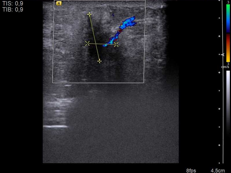
A breast cancer on a linear probe, measured with calipers and overlaid with color Doppler. The dotted yellow crosses mark the mass in two dimensions. The blue and red show the tangle of vessels feeding it, one of the signs a reader weighs. Image: Nevit Dilmen, CC BY-SA 3.0.
One question drives every breast scan. What is this lump?
The easiest answer is a cyst. A sealed bag of fluid reads as a clean black oval with a hair-thin wall, the tissue behind it brightened where the sound crossed the fluid unspent. Telling a benign lump from a malignant one often ends right here. A cyst is almost never a cancer, cleared at a single glance. A cyst that turns cloudy, thickens its wall, or fills with debris loses that easy pass and joins the queue for a closer reading, since the rare cancer now and then wears a cyst’s clothes.
A solid lump asks for the whole of the reader’s skill. The reassuring kind has a name and a shape every reader learns young: the fibroadenoma, the firm rubbery knot a woman so often carries, which sits on the screen as a smooth oval lying along the grain of the breast, its edges cleanly drawn, its substance even, sliding a little when the probe presses on it. A cancer betrays itself by breaking every one of those habits. It grows the wrong way, standing taller than it is wide, having forced out through the planes of the breast that a benign lump keeps within. The margin is the loudest tell, ragged or spiked or smudged into a bright halo where the tumour frays into the tissue it invades. Behind a hard cancer the sound frequently dies into a shadow, swallowed by the dense disordered growth. The lump sits anchored, gripping the tissue, refusing the easy glide of a benign one. Plenty of lumps decline to fall cleanly into either camp, part fluid and part solid, a complex mass that earns a closer look for the solid corner a pure cyst would never own. Size lends its own steady weight, a growth already past two centimetres carrying more menace than a five-millimetre speck wearing the same edges. No one of these signs convicts on its own. A reader who hangs a diagnosis on a single feature will be wrong often enough to do real harm, so the judgement stays cumulative, each feature a weight laid on one pan of a scale, the reading settling only once enough of them fall the same way. This is the work a finger cannot do. It is the reason a woman with a lump is sent for ultrasound ahead of almost anything else: the probe sees into the lump and reads its character in the room, a needle the only step left to prove what the picture already suspects.
If a single line had to carry the whole reading, it would be the orientation. A lump that lies flat, its long axis parallel to the skin, has grown along the natural planes of the breast, the gentle way of a benign thing. A lump that stands tall, deeper than it is wide, has shouldered across those planes in the manner of an invasion. The height set against the width sorts a remarkable share of lumps before any other feature is even weighed. The rule bends now and then, a few cancers lying flat and a few benign lumps standing tall, which is why orientation is where a reading starts, the other features carrying it on from there.
The hard cases live in the middle. A lump may carry one worrying feature among three reassuring ones, and there the reader leans on the strongest sign and on the company the lump keeps. Color Doppler adds a quiet vote, a knot of disordered vessels driven into a cancer counting against it. Elastography adds another, pressing the tissue to read its stiffness, a cancer giving back the hardness of a pebble in dough. Stiffness on its own proves nothing, a band of old scar reading just as hard, the measure only ever one more vote in the tally. When the picture still will not resolve, the needle settles it, the cost of a benign biopsy weighed against the cost of a cancer missed. Ultrasound serves as the second look as well. When a screening MRI lights up something the mammogram never showed, a targeted scan goes hunting for it. A lump the probe can find is a lump the probe can biopsy. That spares the patient the slower, costlier route of a biopsy taken inside the MRI machine.
Putting a number on it
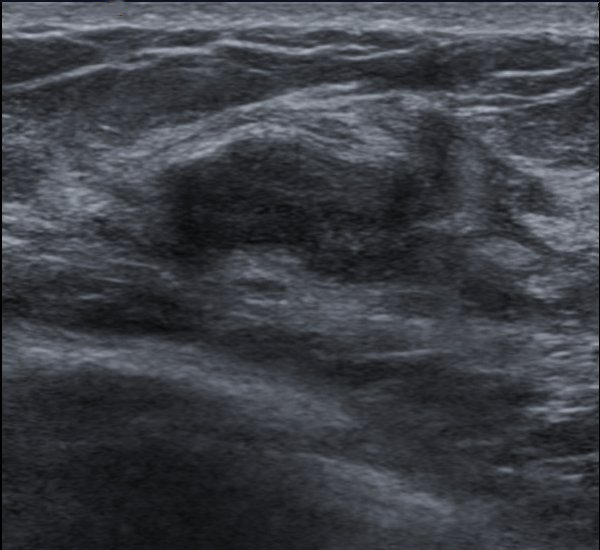
A fibroadenoma, the common benign breast lump, on a linear probe. It reads as a smooth dark oval lying wider than it is tall, along the grain of the breast, the reassuring shape a reader learns to know. Image: Nevit Dilmen, CC BY-SA 3.0.
A reader does not leave the verdict to instinct. The breast imaging reporting and data system, BI-RADS, gathers the features into a numbered category, a scale that runs from a plainly normal breast, through findings clearly and then probably benign, on up to the suspicious and the all-but-certain cancer. Two numbers stand off to the side: a zero when the study is not yet complete enough to score, a six for a cancer the biopsy already proved. The scale is the shared tongue every breast reader speaks. The middle rungs are where it earns its keep. A probably benign lump carries under one chance in fifty of being a cancer, low enough to watch by the calendar with no needle yet. A suspicious lump spans a wide band of risk, from a few percent up toward near certainty. Each step the reader climbs pulls the threshold for a biopsy lower.
Each rung carries an instruction, and that is the point of the whole apparatus. A benign reading returns the woman to ordinary screening. A probably benign one buys a short watch, a repeat scan in six months to prove the lump holds still. It spares a needle, the small residual risk kept in plain view. A suspicious reading sends her to biopsy. The number turns a grey picture into a plan a surgeon, an oncologist, and the woman herself all read the same way. The categories hold a clinic to account as well. A probably benign call ought to prove benign nearly every time it is made. A suspicious call ought to turn up cancer often enough to justify the needles it sends. A clinic that audits its own numbers keeps the scale honest, the same word meaning the same thing in every reader’s mouth.
The dense breast
Here ultrasound stops being an extra. Here it is the point.
Dense breast tissue glows white on a mammogram. So does a cancer. A tumour buried in dense tissue can vanish into that whiteness, and the denser the breast, the deeper a cancer can hide. Density carries a second sting, a dense breast running a higher baseline risk of cancer than a fatty one, so the woman hardest to read on a mammogram is also the one with more to find. The mammogram is not failing here. It has met its limit, a shadow-picture undone by tissue every bit as bright as the thing it tries to hide.
Ultrasound does not see by whiteness. It reads tissue by texture. Against the bright clutter of a dense breast, a dark cancer can step forward exactly where the X-ray went blind. The numbers bear this out. Across large studies, an ultrasound laid over a mammogram turns up two to four extra cancers in every thousand women with dense breasts. The greater part of those are small, many under a centimetre, caught early enough that the nodes are still clear. The cancers an ultrasound alone catches are heavily the invasive kind, the dangerous ones the mammogram had quietly let slip past.
The gain has a cost, and an honest account names it. The same scan that finds those cancers also lights up many harmless lumps, each one a fresh worry and now and then a biopsy that comes back benign. Scanning a whole dense breast well takes time and a patient, disciplined method, which is why some centres hand the sweep to an automated scanner. The handheld keeps the targeted job: the lump a woman can feel, the spot the mammogram flagged, the corner a reader wants one more angle on. Density has climbed out of the radiology report and into the conversation a woman has with her own doctor. Many places now print it where she can read it and weigh a supplemental scan for herself. A handheld carries that scan into the clinic visit, with no second appointment at a distant centre to arrange.
The flecks it struggles with
Ultrasound has one stubborn blind spot. The earliest cancers sometimes announce themselves only as microcalcifications, specks of calcium finer than a grain of salt, and a fleck that small, set in busy tissue, throws back too weak an echo for an ordinary probe to lift it out. A mammogram catches the same speck by the shadow it casts on the X-ray, the one corner of breast imaging the older test still owns outright. The reason sits in the physics. Ultrasound builds its picture from echoes. A calcium fleck a fraction of a millimetre wide, lost in the speckle of glandular tissue, does not throw back enough sound to register at all. An X-ray beam passes clean through the soft tissue and is stopped only by the dense fleck, which prints as a sharp white dot on the film. The calcium that hides from ultrasound is the calcium a mammogram reads best of all.
Ultra-high-frequency probes for microcalcifications have begun to close the gap. Pitched far above the everyday band, they render the outermost millimetres in a detail no standard head can match, and the brightest of the calcium has started to surface on them. The mammogram keeps the crown for now. The newest probes are quietly reaching for it. Where a probe does pick out calcium sitting inside a solid lump, those bright specks count toward the worry, one more line in the case against it. The faint, scattered calcium that fills a duct on its own, the earliest whisper of a cancer still trapped in the lining, stays the mammogram’s to find.
Guiding the needle
A suspicious lump ends at a needle, and ultrasound is what makes the needle trustworthy. An ultrasound-guided breast biopsy takes a thread of tissue from the lump while the probe holds both target and needle on the one screen, the bright tip steered into the worrying heart of the mass on the first pass. A lump too small or too deep for the fingers to pin sits in plain view on the monitor. A tiny metal clip dropped at the site marks the spot for a surgeon or a later scan, a small piece of foresight that pays off when chemotherapy shrinks a tumour to nothing. Someone still has to find where it stood. The whole thing runs in a clinic room under local anaesthetic, the soreness gone in a day, the diagnosis back within the week. The breast takes a few kinds of needle, each picked to fit the lump. A core needle, the workhorse, lifts a slim core of tissue a pathologist can read whole. A vacuum device draws a fuller sample when a single core leaves the question open. A fine needle, thinner again, suits a cyst that wants draining or a node that wants sampling. Whatever goes in, the reader holds the answer against the picture, since a benign result on a lump that looked like a cancer is a mismatch that sends the woman back for another core. That same clip guides her surveillance for years after, every follow-up scan finding the biopsy site without a search.
Reading the armpit
A breast cancer makes its first journey to the lymph nodes of the armpit, and so the same sweep that reads the breast carries on into the axilla. Reading the axillary lymph nodes is less about naming a node than about changing a plan, because what the probe finds there rewrites the surgery before it begins. The breast and the armpit are read as one problem, the size of the tumour and the state of the nodes together fixing how far the disease has travelled and how hard it has to be fought.
The tell is the rind. A healthy node keeps a thin shell of cortex wrapped around a bright fatty core; a node breeding cancer thickens that cortex past about three millimetres, swells toward a ball, and crowds out the fatty centre until it is gone. A node that reads that way takes a needle of its own there and then, a few cells drawn under the same probe to prove the spread. That one needle spares a woman a surprise on the operating table, the spread known and planned for long before she is ever put to sleep. The whole sweep of the axilla costs a reader only a minute past the breast itself.
The answer steers the whole operation. A clear axilla sends the surgeon to test a single sentinel node, the first that drains the tumour, and to leave the rest of the armpit undisturbed. A node already proven by needle sends her to clear the axilla outright, or sends the patient to chemotherapy before any blade is lifted. Found early on a handheld, that one node can reroute a woman’s entire treatment. Shape feeds the same decision. A node rounding up until its length barely beats its width joins the thick cortex and the lost hilum as a mark against it. When a proven node sends a woman to chemotherapy ahead of any surgery, a marker clip goes into that node, so the surgeon can find and lift the exact one once the drugs have shrunk the disease and the swelling has settled.
The painful or changing breast
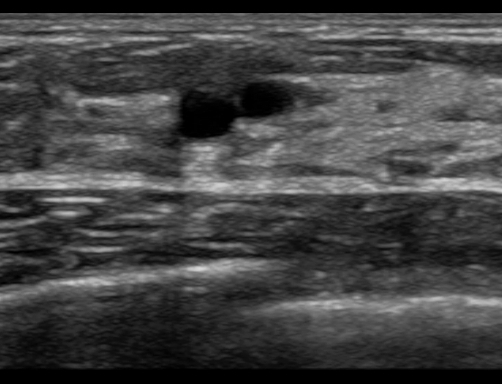
Two small breast cysts on a linear probe, each a clean black oval against the grey tissue, the simplest of all the calls a breast scan makes. A sealed bag of fluid like this is almost never a cancer. Image: schomynv, public domain.
Not every scan is hunting a cancer. A red, hot, tender breast points to infection, and the probe sorts a simple mastitis from a walled pocket of pus that needs a drain guided into it. A lump that comes and goes with the month is usually a cyst filling and emptying. A man’s swollen breast resolves into the disc of gynecomastia behind the nipple. The everyday and the alarming sit side by side under the same probe. The everyday wins, by a wide margin, on almost every list a clinic runs.
On a handheld
The breast is the easiest of all organs for a pocket scanner. It lies shallow, square in the linear probe’s sharpest range, and the whole exam is a surface job a handheld runs as well as any cart. A clinician feels a lump and reads it in the same visit, so a woman walks out with an answer the same day, no referral and no fortnight of dread. The study saves to the phone and travels to a radiologist or a surgeon in seconds. That speed changes the texture of the visit. A woman who feels a lump and fears the worst can be shown, then and there, the plain cyst behind it, the dread lifted before she is out of the chair. A lump that does need a needle gets booked before she leaves the room, the long gap between finding and knowing cut down to days.
The reach counts hardest where a mammogram is a day’s travel away. In much of the world a woman who finds a breast lump has no quick path to an X-ray. A handheld probe in a local clinic gives her a same-day look, a score, and a needle if she needs one. The gap in cost is steep. A mammography suite fills a shielded room with serviced equipment and needs a radiographer trained to drive it. A linear probe that reads a breast costs a small fraction of that, running from a phone the clinic already owns. Ultrasound cannot screen a whole population the way a mammogram can. The scan is too slow, too tied to the skill of the hand that holds it. For the woman who already has a lump, it is often the fastest true answer within her reach. A trained eye behind the probe counts for more than the price on it, the reading only ever as good as the sweep that built it.
Common questions about breast ultrasound
Can ultrasound tell if a breast lump is cancer?
It can tell which lumps need a biopsy. A benign lump and a cancer read differently on ultrasound, in their shape, their edges, and the shadow behind them. A reader scores the lump on those features. The biopsy gives the final word, since only the cells under a microscope confirm a cancer.
What is BI-RADS?
A scoring system for breast imaging. It files every finding into a numbered category, from a plainly normal breast up to one that all but names a cancer. Each number sets the next step: routine screening, a short recheck, or a biopsy. It gives every reader one rule for the same finding.
Why is ultrasound used for dense breasts?
Dense tissue and cancer both read white on a mammogram, so a cancer can hide inside the density. Ultrasound reads tissue by texture, picking a dark cancer out of the bright clutter. Added to a mammogram, it turns up a few more cancers per thousand women in dense breasts that the X-ray alone would miss.
Can a handheld ultrasound find breast cancer?
It finds the lumps that need a biopsy. A handheld linear probe reads the suspicious features of a breast mass to the standard of a cart, then guides the needle that gives the diagnosis. It works alongside a screening mammogram, since the two tests catch different cancers between them.
What does a breast biopsy under ultrasound involve?
A needle takes a small core of tissue from the lump. The probe shows the lump and the needle together on the screen, so the needle reaches the right spot on the first try. It is done under local anaesthetic in the clinic. The soreness fades in a day or two. The tissue gives the diagnosis the scan could only suspect.
About the Author
Julien Mercier
Senior R&D Engineer · Medical Ultrasound Transducer Development
Senior R&D Engineer with an M.S. in Applied Physics and over 15 years of experience in medical ultrasound transducer development, specializing in the design verification and performance testing of high-frequency imaging transducers. Currently leading the development and verification of the company’s next-generation high-frequency linear-array transducer, responsible for imaging performance evaluation and reliability analysis in preclinical testing. Brings extensive hands-on experience in piezoelectric element tuning, beamforming parameter optimization, and system-level performance testing.