Breast Biopsy Ultrasound Guidance Handheld Linear Probe Technique
Guiding a breast biopsy with ultrasound means watching the needle reach the lesion, with the tip and the target held on the screen together. The operator sees the moment the two meet and draws the sample from the spot on the screen, where a blind biopsy would be working from a guess. What comes back from that sample decides whether the lesion is left alone or treated.
When ultrasound guides the biopsy
Ultrasound guides a biopsy when the target shows on ultrasound. A solid mass, a complex cyst, or an area of distortion that the probe shows is a target the operator can reach under live imaging. The needle and the lesion appear together on the screen, so the operator steers the needle to the spot and watches it arrive.
Not every target shows on ultrasound. A cluster of microcalcifications with no mass is often invisible to the probe, since the flecks are too small to stand out. That kind of target goes to a stereotactic biopsy, guided by mammography. Ultrasound handles the masses it can see. Calcium with no mass takes the stereotactic route.
Ultrasound guidance has practical strengths for the targets it suits. It uses no radiation and shows the needle moving in real time. The patient lies on her back through the procedure, free of the compression a stereotactic table needs. The setup costs less to run. It fits in any room with an ultrasound machine. For a mass the probe can see, this is the method of choice.
The stereotactic route asks more of the patient. It puts her face down on a table with the breast through an opening, held under compression as the needle works. Radiation maps the spot. For the masses ultrasound can reach, the lighter path wins on comfort alone.
The core needle biopsy
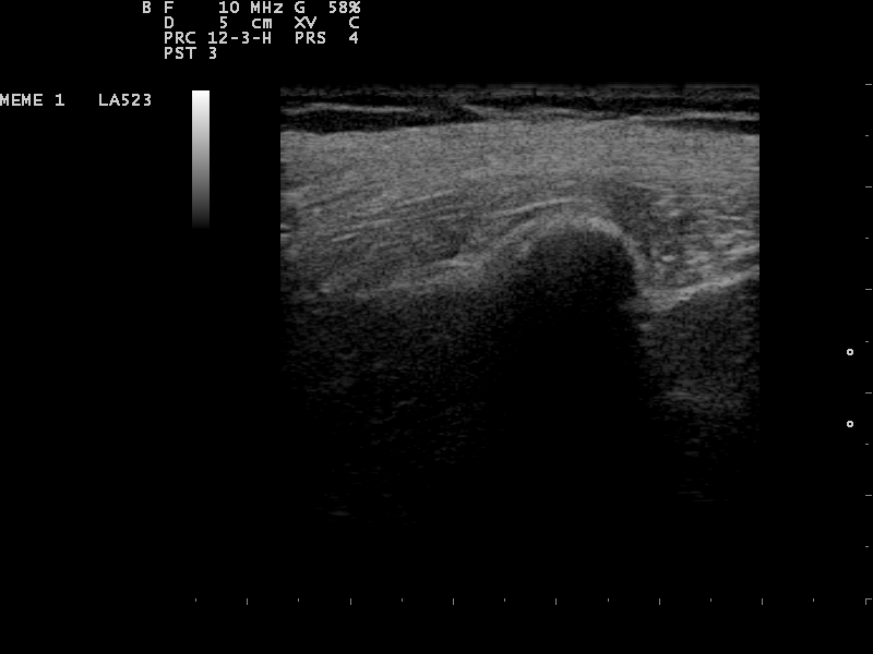
A hypoechoic breast mass on ultrasound. A core needle samples a target like this under live guidance, with the tip watched into the lesion.
The core needle biopsy is the workhorse of breast sampling. It takes a thin cylinder of tissue, a core, where a fine needle draws only loose cells. A pathologist can read the tissue architecture in a core, which tells an invasive cancer apart from a non-invasive one and types the tumor. The standard tool is a spring-loaded device with a 14-gauge needle. A local anesthetic numbs the skin and the track before the needle goes in, so the procedure is felt as pressure more than pain. The operator places the needle tip at the edge of the mass under ultrasound, then fires the spring. The needle shoots forward. A trough opens to catch a core. An outer sheath snaps across to cut it free. The throw takes a fraction of a second. The speed is the point, since a slow push would tear the soft tissue and crush the sample. A coaxial sheath can be left in the breast after the first pass, so the later cores travel down the same track without a fresh puncture each time. The operator pulls the device out, drops the core into fixative, then goes back in for the next. Three to six cores are taken from across the mass, so the sample covers more than one spot. The cores look like fine threads of tissue, a centimeter or so long. A dense core that sinks in the fixative can hint at cancer before the slides are read. Each pass is followed on the screen, the needle a bright line entering the dark mass. A radiologist or a trained operator runs the procedure, reading the screen and placing the needle at the same time. The 14-gauge size is the common choice, with 16-gauge and 18-gauge used for softer or trickier targets. The operator checks the throw length before each fire, since a long throw on a small breast could reach the chest wall. A firm mass can push the needle off its line as the spring fires, so the operator braces the lesion with the probe and corrects the angle when the tip meets resistance. The number of cores rises for a larger or mixed-looking mass, where one or two samples might miss the worst part. The cores give the pathologist enough tissue for a firm diagnosis in the large majority of cases, which is why it has replaced open surgical biopsy for the first look at a breast mass.
The vacuum-assisted biopsy
The vacuum-assisted biopsy takes larger samples through a single insertion. The needle is thicker, in the 8 to 12 gauge range. It stays in the breast through the procedure. Vacuum pressure pulls tissue into a side chamber in the needle. A blade cuts the sample. The needle rotates to a new position for the next, with no need to come out. Eight to ten samples can be taken in a fan around the lesion this way.
The larger samples are the point. A vacuum biopsy removes more tissue than a single core does. This raises the chance of a firm diagnosis. It also lowers the chance of an upgrade later, when surgery finds worse disease than the biopsy showed. The method suits a small or scattered target, where a few cores might miss.
The trade is a bigger needle. The vacuum device is wider, so it leaves more of a mark and a greater chance of bruising. For an everyday mass the core needle is enough. The vacuum method is held for the targets that need the extra tissue.
A vacuum device can do more than sample a lesion. For a small benign growth such as a fibroadenoma, the same tool can take it out piece by piece in one sitting, an option some women prefer to a surgical excision. The aim there shifts from diagnosis to removal.
Fine needle aspiration and cysts
Fine needle aspiration uses the thinnest needle of the three. A fine needle on a syringe draws fluid or a spray of loose cells. It gives no solid core. The sample is cytology, cells to study without the tissue architecture a core holds. The breast uses it less than the thyroid does, since a core of tissue answers more about a breast mass.
Where fine aspiration fits best is the cyst. A simple cyst is a fluid sac. Ultrasound shows it as a smooth black oval. If it causes symptoms, a fine needle can drain it in one pass, watched on the screen. The fluid relieves the pressure. A cyst that collapses cleanly and stays gone needs nothing more.
The fluid that comes out is read by eye. Clear or greenish fluid from a simple cyst carries no worry and is discarded. Bloody fluid is sent to the laboratory, since blood can come from a growth in the cyst wall. A cyst that fills again after draining calls for a second look.
A complex cyst is the one that calls for more. Thick walls, solid parts inside, or debris that does not move raise the question of something other than plain fluid. The solid part is the target. The operator cores it the way any solid mass is sampled.
The needle path in the breast
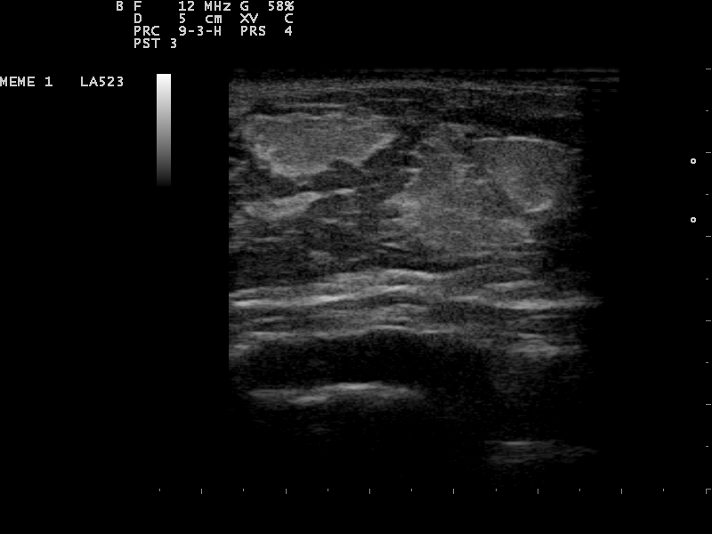
A breast mass on ultrasound. The needle path is planned to run flat, parallel to the chest wall, so the probe follows the needle along its length.
The path the needle takes through the breast is planned before the skin is broken. The operator picks an entry point and an angle so the needle runs nearly flat, parallel to the chest wall. A flat path keeps the needle inside the scan plane along its length, where the probe can follow it end to end.
The flat path has a safety reason as well. A needle aimed steeply down toward the chest wall risks the muscle and the lung beneath it. Running the needle parallel to the wall keeps it in the breast tissue and clear of harm, even if the spring throw carries it further than planned. The lung sits a short way under a thin breast, so the flat path is a habit the operator keeps on every pass.
A lesion sitting hard against the chest wall or close under the skin can be moved before the throw. The operator injects a little saline behind it or over it, which lifts the target into a safer window. This step, hydrodissection, opens room for the needle where the anatomy left none.
The tip is the part to keep in view.
A tip lost from the plane could sit deeper or to one side of where the screen suggests, so the rule is to keep the tip in sight and never assume where it is. Small angle changes bring a stray tip back into the plane. The target is approached until the tip rests at its near edge, ready for the throw.
The marker clip and aftercare
A tiny marker clip is often left at the biopsy site at the end. The clip is a speck of titanium or steel, placed through the biopsy needle into the spot that was sampled. It marks where the tissue came from, so a later mammogram or surgery can find the exact site even after the lesion is gone or has shrunk on treatment.
The clip counts when the biopsy removes a small lesion entirely, or when chemotherapy before surgery shrinks the tumor away. Without a marker, the surgeon would have no point to aim for. Clips come in several shapes, so more than one biopsy site in the same breast can be told apart on a later view. The metal is small enough to sit safely through a magnetic resonance scan. It sets off no alarm at airport security.
After the needle comes out, pressure is held on the site to stop bleeding. A cold pack and a snug dressing follow. The patient goes home the same day. Complications are uncommon, with bleeding, bruising, and infection together running under one and a half percent in large series.
Why the guidance is accurate
The accuracy of ultrasound-guided biopsy rests on seeing the needle reach the target. The operator does not infer that the sample came from the right spot. The screen shows the needle inside the lesion at the moment the core is taken. This direct sight is why the method reads so well against the alternatives.
The numbers back this up. For masses visible on the scan, ultrasound-guided core biopsy carries a false-negative rate near 1.7 percent, against around 8.9 percent for the stereotactic route. Sensitivity for core needle biopsy runs from 97 to 99 percent. One large series of 2,420 ultrasound-guided 14-gauge biopsies agreed with the final diagnosis in 96 percent of cases.
Accuracy still rests on good technique. A core taken from the rim of a mass, short of its center, can miss the cancer. A benign reading of a mass that looks malignant on the scan is a flag to sample again or to cut it out. The check on any biopsy is whether its answer matches the picture the imaging painted.
Every biopsy ends with one more check. The result is held up against the imaging that prompted it.
This agreement has a name, radiologic-pathologic concordance. A benign result on a benign-looking mass is concordant and reassuring. Where the mass looked malignant, that same benign result is discordant, a sign the needle may have missed, sending the case back for a repeat or for surgery.
Some results sit between benign and malignant. A lesion of uncertain potential, what pathologists call a B3, can hold a worse area nearby that the cores missed, so it often goes on to a wider excision to be sure. The biopsy starts the answer here without closing it. A clear result still rests on the cores having reached the right tissue, which the live scan is there to confirm. A doubtful case is always safer sampled again than left.
Guiding a biopsy on a handheld unit
A handheld unit brings biopsy guidance to the clinic room. The operator scans the mass, plans a flat path, then follows the needle on the same screen used for the diagnostic scan. The portable probe needs no fixed suite. A targeted biopsy can follow the scan that found the lesion, in one visit.
Common questions about ultrasound-guided breast biopsy
What is an ultrasound-guided breast biopsy?
It is a biopsy in which the operator watches the needle on an ultrasound screen as it enters the lesion. The probe shows the mass and the needle together, so the sample is taken under direct sight. It is used for lesions the ultrasound can see.
What is the difference between core needle and vacuum-assisted biopsy?
A core needle biopsy fires a 14-gauge needle to take three to six cylinders of tissue. A vacuum-assisted biopsy uses a thicker 8 to 12 gauge needle that stays in place and takes eight to ten larger samples through suction. The vacuum method removes more tissue.
When is ultrasound guidance used for a breast biopsy?
Ultrasound is used when the target shows on ultrasound, such as a solid mass or a complex cyst. A cluster of microcalcifications with no mass is often invisible to the probe and goes to a stereotactic biopsy guided by mammography instead.
Why is a marker clip placed?
A small titanium or steel clip marks the biopsy site. It lets a later mammogram or surgery find the exact spot, even after the lesion is removed by the biopsy or shrinks during chemotherapy. It is safe to leave in place.
How accurate is ultrasound-guided breast biopsy?
For masses the probe can see, ultrasound-guided core biopsy has a false-negative rate near 1.7 percent and a sensitivity of 97 to 99 percent. One series of 2,420 cases agreed with the final diagnosis in 96 percent. Accuracy rests on the needle being seen inside the target.
Can a handheld ultrasound guide a breast biopsy?
Yes. A handheld probe shows the mass and the needle in real time, so the operator can plan a flat path and watch the needle reach the target. A targeted biopsy can follow the diagnostic scan in one visit, with no fixed suite needed.
About the Author
Julien Mercier
Senior R&D Engineer · Medical Ultrasound Transducer Development
Senior R&D Engineer with an M.S. in Applied Physics and over 15 years of experience in medical ultrasound transducer development, specializing in the design verification and performance testing of high-frequency imaging transducers. Currently leading the development and verification of the company’s next-generation high-frequency linear-array transducer, responsible for imaging performance evaluation and reliability analysis in preclinical testing. Brings extensive hands-on experience in piezoelectric element tuning, beamforming parameter optimization, and system-level performance testing.